
Classification of necrotising fasciitis
necrotising fasciitis classification types microbiology
"necrotizing fasciitis" AND classification
necrotising fasciitis classification types skin infection
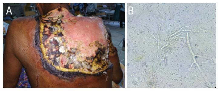
This composite educational image illustrates the clinical and laboratory findings of a severe fungal infection. Panel A is a clinical photograph showing an extensive necrotising wound on the upper back of a 60-year-old male. The lesion displays a heterogeneous wound bed with significant black necrotic eschar at the periphery, yellowish slough, and areas of central erythema and granulation tissue. The margins are irregular and appear undermined, consistent with creeping necrosis into subcutaneous tissues. Panel B is a diagnostic micrograph featuring a potassium hydroxide (KOH) mount of a biopsy from the lesion. It reveals hyaline, broad, aseptate fungal hyphae characterized by wide-angle (approximately 90-degree) branching. These morphological features are highly characteristic of mucormycosis, such as that caused by Apophysomyces species. The image demonstrates the clinical presentation of necrotising fasciitis and the essential microscopic diagnostic criteria for identifying invasive molds in a clinical dermatology or infectious disease context.
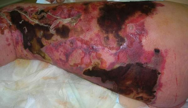
This clinical photograph displays a 43-year-old male's left lower extremity, demonstrating severe soft tissue infection consistent with necrotizing fasciitis. The image shows extensive, confluent areas of skin necrosis characterized by dark brown and black eschar formation, particularly on the lateral thigh and spreading toward the buttock and perineum. Surrounding these necrotic regions is marked, diffuse erythema and violaceous discoloration, indicating severe underlying inflammation. There is evidence of skin sloughing and epidermal detachment, with areas of pale, devitalized tissue exposed. The margins of the infection appear sharply demarcated in some areas while showing a spreading inflammatory border in others. Clinical signs of edema and hemorrhagic extravasation are visible throughout the affected limb. This image serves as a significant visual educational tool for recognizing the rapid progression of fulminant skin and soft tissue infections and the life-threatening necessity of surgical debridement.
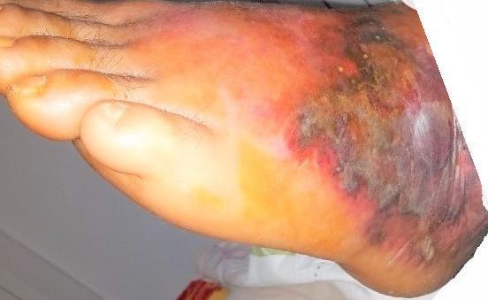
A clinical photograph of the dorsal and lateral aspect of a human foot demonstrating clinical signs of necrotizing fasciitis. The image shows a progressive cutaneous infection characterized by widespread, poorly demarcated erythema extending from the toes towards the midfoot and ankle. Central to the lesion, there is significant skin discoloration with shades of violaceous, dusky purple, and black, indicating deep tissue ischemia and evolving necrosis. The affected area exhibits visible swelling (edema) and irregular skin texture, including possible hemorrhagic bullae or desquamation. The visual findings represent a surgical emergency, illustrating the rapid progression from superficial inflammation to full-thickness soft tissue destruction. This content is intended for medical education regarding the identification of life-threatening soft tissue infections and distinguishing them from uncomplicated cellulitis.
Classification of Necrotising Fasciitis
1. Microbiological Classification (Most Widely Used)
Type I - Polymicrobial (Synergistic)
| Feature | Details |
|---|---|
| Organisms | Mixed aerobic + anaerobic flora: non-Group A Streptococci, Staphylococcus aureus, E. coli, Bacteroides fragilis, Clostridium spp., Pseudomonas, Klebsiella, Proteus |
| Frequency | Most common - accounts for 70-80% of all NF cases |
| Hosts affected | Immunocompromised patients: diabetics, malignancy, chronic renal/hepatic disease, elderly |
| Common sites | Perineum (Fournier's gangrene), abdominal wall (postoperative), trunk |
| Key mechanism | Synergistic virulence - aerobic bacteria consume local oxygen, creating conditions for anaerobic proliferation |
- Bailey and Love's Short Practice of Surgery, 28th Ed.
Type II - Monomicrobial (Haemolytic Streptococcal / Staphylococcal)
| Feature | Details |
|---|---|
| Organisms | Group A beta-haemolytic Streptococcus pyogenes (most common); also Staphylococcus aureus including MRSA strains |
| Frequency | Accounts for 20-30% of cases |
| Hosts affected | Often previously healthy individuals of any age group |
| Common sites | Extremities; often following minor or penetrating trauma |
| Key feature | This is the "flesh-eating disease" widely reported in media; may be associated with streptococcal toxic shock syndrome (STSS) |
Type III - Clostridial (Gas Gangrene) / Marine Organisms
| Feature | Details |
|---|---|
| Organisms | Clostridium perfringens, C. septicum; marine variants include Vibrio vulnificus, Aeromonas hydrophila |
| Frequency | Relatively uncommon |
| Hosts affected | Post-traumatic, post-surgical; intravenous drug users (black-tar heroin injection); marine exposure |
| Key feature | Produces gas in tissues - palpable crepitus, crackling sensation; rapid progression |
| Marine subtype | Vibrio vulnificus - saltwater/shellfish exposure; Aeromonas hydrophila - freshwater exposure |
Type IV - Fungal (Rare)
| Feature | Details |
|---|---|
| Organisms | Candida spp., Zygomycetes (Mucorales) - Apophysomyces, Rhizopus; Aspergillus |
| Frequency | Rare |
| Hosts affected | Severely immunocompromised patients (organ transplant, haematological malignancy) |
| Key feature | Broad aseptate hyphae on biopsy (mucormycosis); high mortality |
2. Anatomical/Site-Based Classification (Historical)
| Eponym/Term | Site | Notes |
|---|---|---|
| Fournier's gangrene | Perineum, scrotum, penis, vulva | Originally described as idiopathic; now known to be polymicrobial |
| Ludwig's angina | Submandibular/sublingual space | Rapidly spreading floor-of-mouth infection |
| Meleney's synergistic gangrene | Abdominal wall | Post-surgical; synergistic polymicrobial |
| Cervical NF | Neck and mediastinum | Can descend to cause mediastinitis |
3. Depth-of-Infection Classification
| Term | Depth |
|---|---|
| Necrotising fasciitis | Superficial fascia and subcutaneous fat (muscle relatively spared early) |
| Necrotising myositis / myonecrosis | Muscle layer involved (e.g., gas gangrene) |
| Necrotising adipositis | Primarily adipose tissue |
| Synergistic necrotising cellulitis | All soft tissue layers involved |
Clinical Correlation to Classification
- All types share the hallmark features: disproportionate pain, woody induration, skin discolouration (dusky blue/black), bullae formation, crepitus (especially Type III), rapid progression to septic shock
- Lymphangitis is typically absent - a useful differentiating sign from cellulitis
- Mortality ranges from 30-70% depending on type, extent, host factors, and time to debridement
- Surgical debridement is the cornerstone of treatment for all types
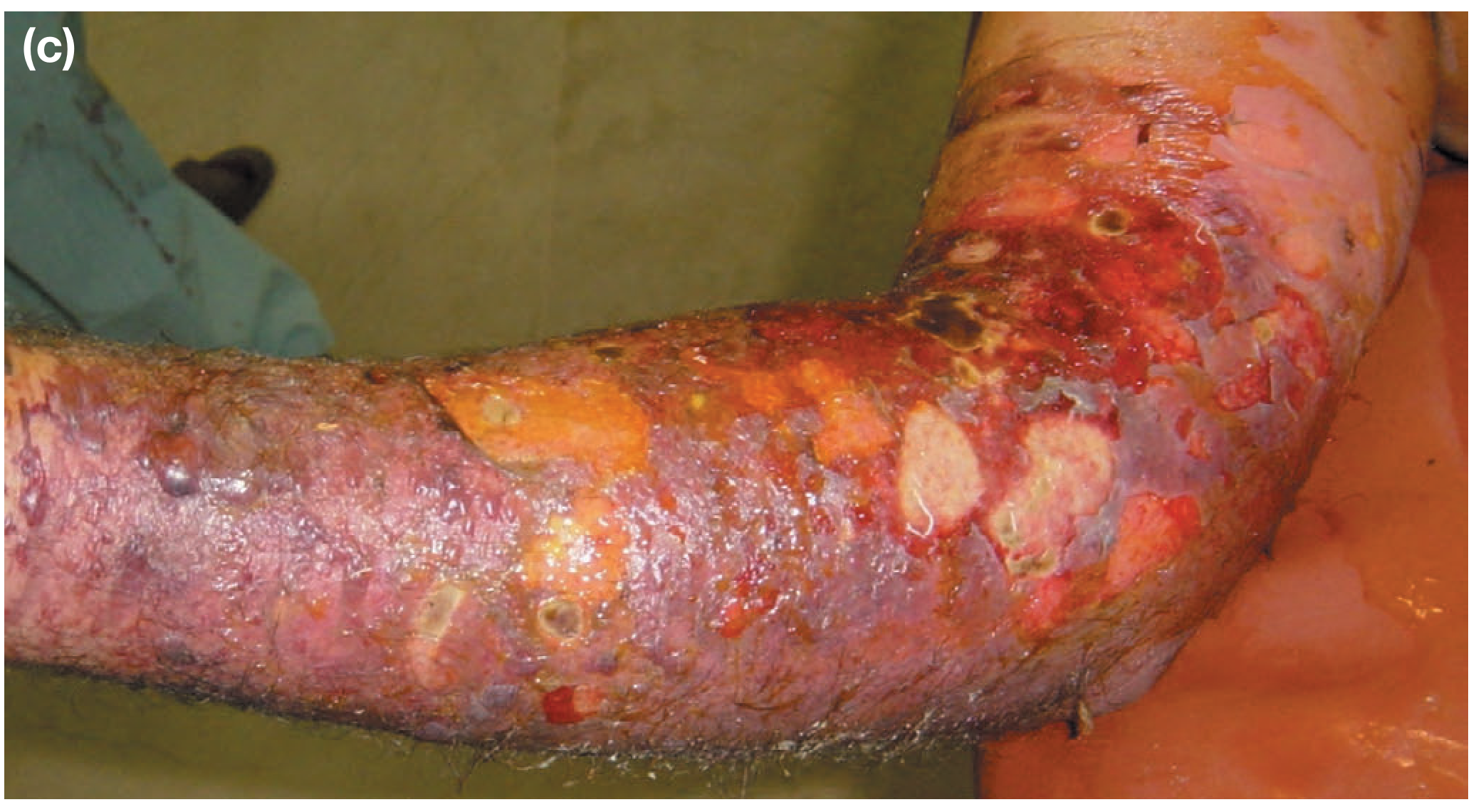
Clinical Images
Explore clinical diagnosis criteria and LRINEC score for necrotising fasciitis
"LRINEC score" AND "necrotizing fasciitis"
"LRINEC" AND "necrotizing fasciitis" AND diagnosis
LRINEC score necrotising fasciitis variables points sensitivity specificity validation 2024
PMID: 34657810
Clinical Diagnosis Criteria and LRINEC Score for Necrotising Fasciitis
A. Clinical Diagnosis
Early Clinical Signs
| Sign | Description |
|---|---|
| Disproportionate pain | Pain far exceeding the degree of visible skin inflammation - the most important early warning |
| Oedema beyond erythema | Swelling spreading beyond the visible erythematous margins |
| Tachycardia and fever | Systemic toxicity out of proportion to local appearance |
| Skin erythema and warmth | Early stage; may appear deceptively like cellulitis |
Late / Progressive Signs
| Sign | Description |
|---|---|
| Woody-hard induration | Subcutaneous tissues feel board-like; fascial planes cannot be distinguished on palpation |
| Skin discolouration | Progresses from dusky red → violaceous/purple → black (thrombosis + necrosis of microvasculature) |
| Haemorrhagic bullae | Fluid-filled blisters due to deep tissue destruction - a sinister sign |
| Crepitus | Crackling/gas in tissues; especially prominent in Type III (clostridial) NF |
| Cutaneous anaesthesia | Loss of skin sensation over affected area due to necrosis of cutaneous nerves |
| Lymphangitis absent | Notably absent - a distinguishing feature from cellulitis |
| Skip lesions | Non-contiguous spread that later coalesces |
Systemic Features
- Fever, rigors, tachycardia
- Rapid progression to septic shock and multiorgan failure
- Renal failure secondary to hypovolaemia
- Altered mental status
Intraoperative Diagnostic Criteria (Finger Test)
- A 2 cm incision is made down to the deep fascia under local anaesthesia
- Positive test: Lack of bleeding, grey necrotic fascia, absence of resistance to finger dissection along fascial planes ("lack of the normal resistance of healthy tissue"), and a characteristic "dishwater" or grey-brown fluid - all confirm NF
- Absence of resistance on blunt dissection along the fascial plane = fascial necrosis
Investigations Supporting Diagnosis
| Investigation | Findings in NF |
|---|---|
| X-ray (plain film) | Air/gas in soft tissues (especially Type III); should NOT delay surgery |
| CT scan | Best imaging modality; shows fascial plane thickening, gas tracking along fascial planes, fluid collections, loss of tissue planes |
| MRI | High sensitivity for fascial involvement; T2 hyperintensity along fascia; useful if diagnosis uncertain |
| Creatine kinase (CK) | Enormous elevation when muscle involved |
| Frozen section biopsy | Fascial necrosis with acute inflammatory infiltrate; thrombosis of microvasculature; confirms diagnosis histologically |
B. LRINEC Score (Laboratory Risk Indicator for Necrotising Fasciitis)
Variables and Scoring
| Parameter | Range | Points |
|---|---|---|
| C-Reactive Protein (CRP) | < 150 mg/L | 0 |
| ≥ 150 mg/L | 4 | |
| White Cell Count (WBC) | < 15 × 10⁹/L | 0 |
| 15-25 × 10⁹/L | 1 | |
| > 25 × 10⁹/L | 2 | |
| Haemoglobin (Hb) | > 13.5 g/dL | 0 |
| 11-13.5 g/dL | 1 | |
| < 11 g/dL | 2 | |
| Serum Sodium (Na⁺) | ≥ 135 mmol/L | 0 |
| < 135 mmol/L | 2 | |
| Serum Creatinine | ≤ 1.6 mg/dL (141 µmol/L) | 0 |
| > 1.6 mg/dL | 2 | |
| Blood Glucose | ≤ 180 mg/dL (10 mmol/L) | 0 |
| > 180 mg/dL | 1 |
Note: CRP alone contributes up to 4 points - it is the single most heavily weighted variable, reflecting the intense acute-phase response in NF.
Score Interpretation
| Score | Risk Category | Action |
|---|---|---|
| < 6 | Low risk | NF unlikely; consider alternative diagnosis |
| 6-7 | Intermediate risk | ≈ 50% probability; close monitoring, serial reassessment |
| ≥ 8 | High risk | Strong suspicion of NF; urgent surgical exploration |
Diagnostic Performance (Original vs. Validation Studies)
| Study | Cut-off | Sensitivity | Specificity | PPV | NPV |
|---|---|---|---|---|---|
| Wong et al. 2004 (derivation) | ≥ 6 | ~90% | ~95% | 92% | 96% |
| Prospective validation (Fernando et al.) | ≥ 6 | 43% | 83% | 25% | 92% |
| Prospective validation (Fernando et al.) | ≥ 8 | 27% | 93% | 33% | 91% |
| Meta-analysis - extremities (Tarricone et al. 2022) | ≥ 6 | 49% | 83% | 35% | 90% |
Limitations of LRINEC Score
- Poor sensitivity in validation studies - a score < 6 does NOT reliably rule out NF; the prospective extremity study (PMID 34657810) showed NPV of only 90% meaning 10% of NF cases are missed
- Not designed for head/neck NF - performance is worse outside extremities and perineum
- Cannot replace clinical judgement - a high clinical suspicion mandates surgical exploration even with a low LRINEC score
- Does not include imaging findings - purely laboratory-based; ignores gas on X-ray or CT findings
- Variables confounded by comorbidities - diabetics may have baseline hyperglycaemia and elevated creatinine, inflating scores
Key Clinical Rule
"A low LRINEC score cannot be used to rule out NF. Clinical suspicion + surgical exploration remain the gold standard."
Newer Competing Scores
- SIARI score (Site Other Than Lower Limb, Immunosuppression, Age, Renal Impairment, Inflammatory markers) - prospectively validated against LRINEC by R K et al. 2023 (PMID 36462382)
- J-LRINEC score (2025, Japanese population) - logistic regression-derived, C-statistic 0.97, sensitivity 91%, specificity 85% - outperforms original LRINEC in that population
Clinical Images
Find recent clinical guidelines for necrotising fasciitis management 2024-2025
"necrotizing fasciitis" AND management
necrotising fasciitis management guidelines 2024 2025 surgical debridement antibiotics
PMID: 41404821
"necrotizing fasciitis" AND treatment AND antibiotics
https://www.cdc.gov/group-a-strep/hcp/clinical-guidance/necr…
PMID: 40052618
PMID: 42202366
IDSA WSES guidelines necrotizing fasciitis 2024 IVIG hyperbaric oxygen antibiotic regimen
https://pmc.ncbi.nlm.nih.gov/articles/PMC12512284
"necrotizing fasciitis" AND "antibiotic" AND "surgical debridement"
Recent Clinical Guidelines for Necrotising Fasciitis Management (2024-2026)
1. Resuscitation and ICU Care (Immediate Priority)
| Action | Detail |
|---|---|
| Airway | Endotracheal intubation if airway threatened (especially cervical/head-neck NF); early anaesthetic involvement |
| Fluid resuscitation | Aggressive IV fluid replacement; correct hypovolaemia before and during surgery |
| Haemodynamic monitoring | ICU-level monitoring; vasopressors for refractory septic shock |
| Organ support | Early renal replacement therapy if acute kidney injury develops |
| Blood cultures | Taken immediately before antibiotics, but must NOT delay surgical or antibiotic treatment |
2. Surgical Management - Cornerstone of Treatment
Principles of Debridement
- Urgency: Operate as soon as diagnosis is suspected. Every hour of delay increases mortality. Do NOT wait for imaging if clinical diagnosis is clear.
- Extent: Incise well beyond the visible margins of erythema and induration. Remove all grey/necrotic fascia until healthy, bleeding tissue is encountered.
- Wound management: Wound left open - never primarily closed. Lightly packed with saline-soaked gauze or negative-pressure (VAC) dressings.
- Re-look at 24-48 hours: Mandatory re-exploration at 24-48 hours. Further debridement is almost always required. Repeat until no further progression.
- Amputation: May be necessary for limb NF with uncontrolled spread or compartment syndrome; life takes priority over limb.
Reconstruction
- Vacuum-assisted closure (VAC) dressings for wound bed preparation
- Split-thickness skin grafting once wound bed is clean and granulating
- Early grafting reduces protein and fluid losses from large open wounds - Bailey & Love's Short Practice of Surgery, 28th Ed.
3. Antibiotic Therapy
3a. Empirical Broad-Spectrum Cover (Initiated Immediately)
| Regimen | Components | Indication |
|---|---|---|
| Regimen 1 | Piperacillin-tazobactam 4 g/6h (prolonged infusion) + Clindamycin 600 mg/6h + Vancomycin | First-line polymicrobial/unknown; if MRSA risk |
| Regimen 2 | Meropenem 2 g loading then 2 g/8h (prolonged infusion) + Clindamycin + Vancomycin | Severe polymicrobial; carbapenem-resistant concern |
| Regimen 3 | Ceftriaxone 2 g/24h + Metronidazole 500 mg/8h + Clindamycin | Moderate severity; community-acquired |
| For penicillin allergy | Ciprofloxacin 400 mg/8h IV + Metronidazole 500 mg/8h + Clindamycin ± Vancomycin/Daptomycin |
Key principle: Clindamycin is added to ALL regimens regardless of other cover because it inhibits bacterial toxin and superantigen production via ribosomal protein synthesis blockade - particularly important in Group A Streptococcal NF and toxic shock syndrome. - CDC GAS Clinical Guidance
3b. Pathogen-Directed Therapy (After Culture Results)
| Type | Organism | Antibiotic of Choice |
|---|---|---|
| Type I (polymicrobial) | Mixed aerobic/anaerobic | Continue broad-spectrum; narrow per sensitivities |
| Type II - GAS | Group A Streptococcus pyogenes | High-dose Benzylpenicillin 2.4 g/4h IV + Clindamycin 1.2 g/6h IV |
| Type II - MRSA | S. aureus MRSA | Vancomycin (30 mg/kg loading, then continuous infusion 30 mg/kg/24h) OR Daptomycin 10 mg/kg/24h OR Linezolid 600 mg/12h |
| Type III - Clostridial | C. perfringens | High-dose Penicillin G 4 MU/4h IV + Clindamycin 900 mg/8h IV |
| Type III - Marine (Vibrio) | Vibrio vulnificus | Doxycycline 100 mg/12h + Ceftriaxone 2 g/24h |
| Perineal/Abdominal (Type I) | Mixed including anaerobes | Add Metronidazole 500 mg/8h or Ciprofloxacin 400 mg/8h |
3c. Duration
- IV antibiotics continued until all debridements complete, patient haemodynamically stable, and systemic sepsis resolved
- Most regimens: 7-14 days total
- Joint involvement: up to 6 weeks IV
- Step-down to oral therapy guided by clinical response and sensitivities
4. Adjunct Therapies
Intravenous Immunoglobulin (IVIG)
| Aspect | Current Guidance |
|---|---|
| Rationale | Neutralises streptococcal superantigens and exotoxins; may blunt immune dysregulation in toxic shock |
| Indication | Consider in Type II NF with streptococcal toxic shock syndrome (STSS) unresponsive to surgery + antibiotics |
| Dosing | 1-2 g/kg as single dose or divided over 2-3 days |
| Evidence level | Not proven in RCTs. IDSA and CDC both state "may be considered" but efficacy not established. - CDC Guidance |
| 2024 update | Routine use not recommended; reserve for severe STSS cases with deterioration despite optimal surgical and antibiotic management |
Hyperbaric Oxygen (HBO) Therapy
| Aspect | Current Guidance |
|---|---|
| Rationale | Raises tissue pO₂, kills obligate anaerobes, inhibits alpha-toxin in clostridial infections, improves leucocyte function |
| Indication | May be considered as adjunct (not primary) - especially Type III clostridial NF |
| Current guideline position | Data conflicting; not consistently recommended. Must NEVER delay surgical debridement - PMC 2025 review |
| Protocol | If available: 100% O₂ at 2-3 atmospheres, 90-120 min sessions, 1-2 times daily |
Negative-Pressure Wound Therapy (NPWT/VAC)
- Strongly recommended post-debridement to accelerate wound bed preparation, reduce oedema, and facilitate grafting
- Early surgical review with NPWT reduces need for repeated debridement in selected cases - Bailey & Love's, 28th Ed.
Hypochlorous Acid Irrigation (Emerging, 2024-2025)
- Increasing evidence from periorbital NF literature: dilute hypochlorous acid irrigation reduces infection burden and may lower exenteration rates - AAO 2024 evidence report
- Not yet in mainstream guidelines; considered in select ophthalmic/facial cases
5. Multidisciplinary Team
- General/plastic/reconstructive surgeon
- Intensivist/critical care physician
- Infectious disease specialist (antibiotic stewardship)
- Anaesthetist (airway in cervicofacial NF)
- Nephrologist (renal support)
- Microbiologist (culture-directed therapy)
6. Mortality Risk Stratification (2025 Meta-Analysis)
| Risk Factor | Odds Ratio (95% CI) |
|---|---|
| Clostridium infection | 6.42 (2.17-19.0) |
| Cardiopathy | 2.21 (1.27-3.83) |
| Elevated creatinine | 2.20 (1.13-4.29) |
| Advanced age | 1.16 per year (1.10-1.22) |
- Overall mortality: 15-20% for streptococcal NF (Type II); up to 30-50% for polymicrobial/fulminant cases; higher with delayed debridement - CDC
Summary of Key 2024-2026 Guideline Points
| Domain | Current Standard |
|---|---|
| Diagnosis | Clinical; LRINEC supplementary; CT if uncertain, but never delays OR |
| Surgery | Immediate aggressive debridement; re-look 24-48h; VAC dressing |
| Antibiotics | Piperacillin-tazobactam or carbapenem + clindamycin ± vancomycin empirically; narrow per culture |
| GAS Type II | Penicillin + clindamycin (toxin inhibition) |
| IVIG | Consider in STSS; not routinely recommended |
| HBO | Adjunct only; never delays surgery; most useful in clostridial NF |
| MDT | Mandatory for all cases |
| Duration | 7-14 days IV antibiotics minimum |
The 2025 Expert Consensus (Zhou et al., Burns Trauma 2025; doi: 10.1093/burnst/tkaf031) represents the most recent dedicated NF management document, though its full text was not accessible. It covers adult NF diagnosis and treatment algorithm.