
Eye anatomy and physiology
eye anatomy cross section diagram cornea retina lens
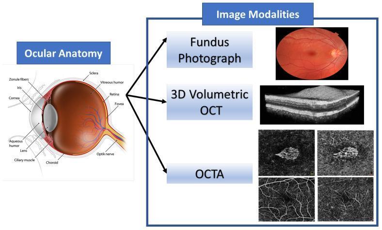
This educational graphic combines an anatomical diagram of the human eye with corresponding ophthalmic imaging modalities. The 'Ocular Anatomy' section features a detailed sagittal cross-section identifying key structures: the cornea, aqueous humor, iris, pupil, biconvex lens, zonule fibers, and ciliary muscle in the anterior segment; and the vitreous humor, retina, fovea, choroid, sclera, and optic nerve in the posterior segment. The 'Image Modalities' section illustrates diagnostic tools used to visualize these structures. A color Fundus Photograph provides a surface view of the retina, optic disc, and vasculature. A 3D Volumetric Optical Coherence Tomography (OCT) scan displays a high-resolution, grayscale cross-sectional view of retinal layers, highlighting tissue stratification. Finally, Optical Coherence Tomography Angiography (OCTA) panels show non-invasive, depth-resolved images of the retinal and choroidal microvasculature. This composite serves as a reference for correlating anatomical landmarks with clinical diagnostic imaging used in the management of retinal diseases such as Age-related Macular Degeneration (AMD).
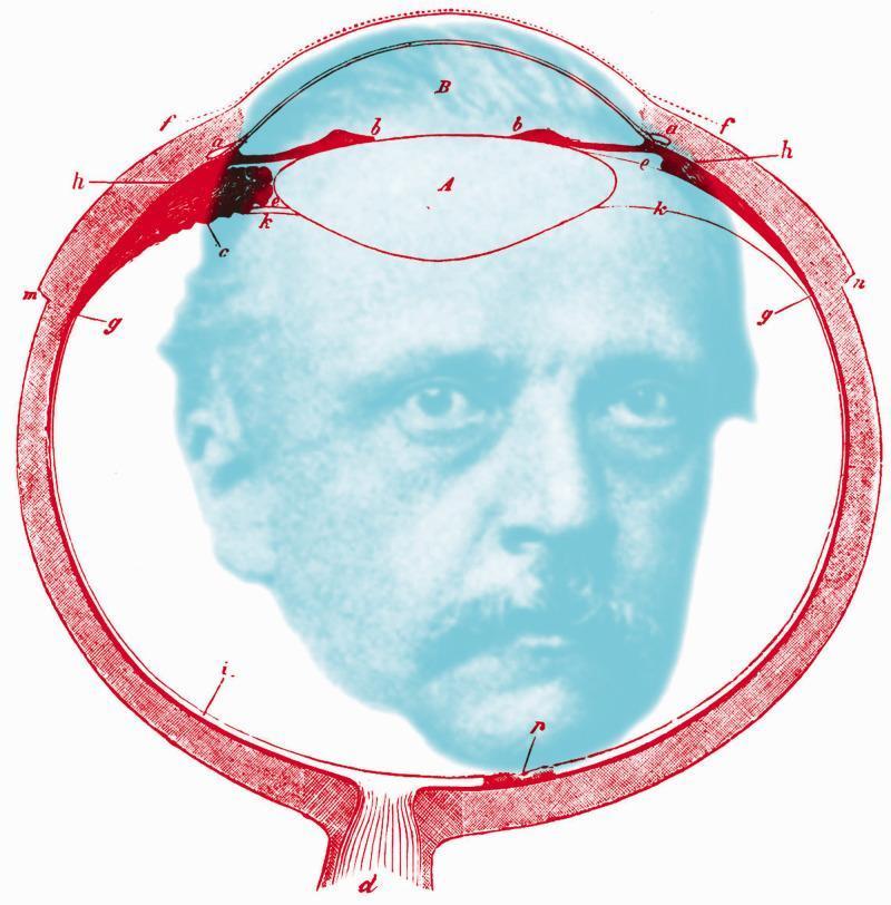
This educational illustration is an anatomical diagram of the human eye in sagittal cross-section, overlaid with a portrait of Hermann von Helmholtz. The diagram, rendered in red linework, depicts essential ophthalmic structures including the cornea (labeled B), the crystalline lens (labeled A), and the ciliary body (b). Various alphabetical annotations (c, d, e, f, g, h, i, k, m, n) indicate specific histological and anatomical regions, such as the sclera, retina, choroid layers, and the optic nerve exit (d). The lens is shown in its anatomical position behind the anterior chamber and iris. The image serves as a conceptual tribute to physiological optics, bridging the anatomy of vision with the historical figure responsible for major advancements in the field, such as the invention of the ophthalmoscope and the primary treatise on physiological optics. The spatial arrangement emphasizes the internal ocular environment, with the retinal lining and posterior segment framing the central portrait.
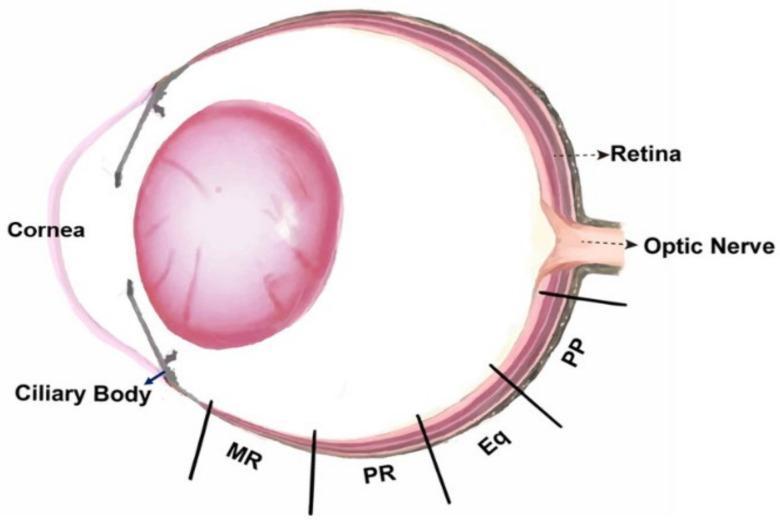
This medical anatomical diagram illustrates a sagittal cross-section of the eye, specifically designed to show sampling locations for retinal analysis. The illustration depicts key anterior structures including the cornea, ciliary body, and a prominent crystalline lens. The posterior segment highlights the multi-layered retina and the exit point of the optic nerve. The diagram serves as a map for topographical retinal research, marking four specific regions along the inferior hemisphere: 1) the Posterior Pole (PP) located adjacent to the optic nerve; 2) the Equator (Eq) representing the mid-peripheral region; 3) the Peripheral Retina (PR) continuing anteriorly; and 4) the Marginal Retina (MR) situated most anteriorly near the ciliary body. This visual provides clinical and research context for studying regional variations in retinal cell density, morphology, and pathophysiology, such as changes in rod and cone distribution or responses to form deprivation.
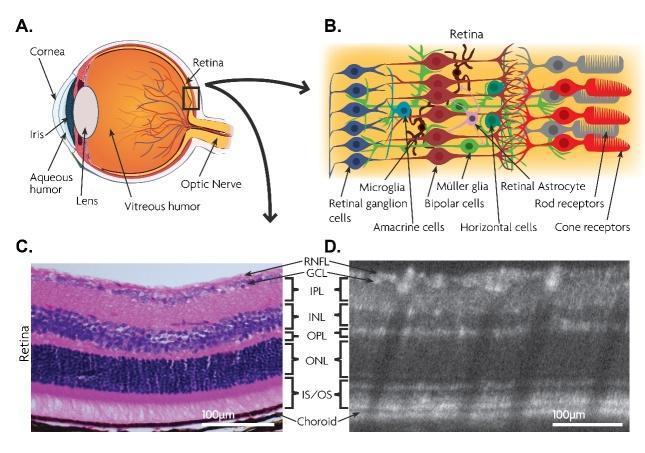
This composite educational illustration presents the multi-layered anatomy of the retina through schematic diagrams and comparative diagnostic imaging. Panel A shows a sagittal anatomical diagram of the eye, highlighting key structures including the cornea, iris, lens, vitreous humor, and optic nerve. Panel B provides a high-magnification schematic of the retinal cellular architecture, illustrating the relationship between photoreceptors (rods and cones), bipolar cells, horizontal cells, amacrine cells, Müller glia, and retinal ganglion cells (RGCs). Panels C and D provide a side-by-side comparison of a histological cross-section (H&E stain) and an in vivo spectral-domain optical coherence tomography (SD-OCT) scan. Both panels are annotated to delineate the discrete retinal laminations: Retinal Nerve Fiber Layer (RNFL), Ganglion Cell Layer (GCL), Inner Plexiform Layer (IPL), Inner Nuclear Layer (INL), Outer Plexiform Layer (OPL), Outer Nuclear Layer (ONL), and the Inner/Outer Segment (IS/OS) junction, terminating at the choroid. Scale bars represent 100 μm, emphasizing the correlation between histological cellularity and OCT reflectivity bands for ophthalmic diagnostic training.
Eye Anatomy and Physiology

1. Gross Anatomy - Layers of the Eyeball
Outer Fibrous Layer
- Sclera - the tough, opaque white outer shell that gives the eye its shape and protects internal structures. Light cannot pass through it.
- Cornea - the transparent anterior modification of the sclera. It is the primary refracting surface of the eye, accounting for ~75% of total refractive power. It is avascular and nourished by the aqueous humor and tear film. The lateral margin of the cornea is continuous with the conjunctiva, a clear mucous membrane covering the sclera.
Middle Vascular Layer (Uvea)
- Choroid - highly vascular layer just inside the sclera; supplies oxygen and nutrients to the outer retina.
- Ciliary body - contains circular and longitudinal smooth muscle fibers that control lens shape (accommodation). It also produces aqueous humor by active transport from plasma.
- Iris - the pigmented, opaque diaphragm in front of the lens. It has two muscles:
- Sphincter pupillae (parasympathetic) - constricts the pupil (miosis)
- Dilator pupillae (sympathetic) - dilates the pupil (mydriasis)
- Variations in pupil diameter can produce up to a 16-fold change in the amount of light reaching the retina.
Inner Neural Layer
- Retina - the light-sensitive neural tissue lining the posterior two-thirds of the choroid. Contains photoreceptors and multiple neural processing layers (see Section 4 below).
2. Internal Compartments and Fluids
| Compartment | Contents | Notes |
|---|---|---|
| Anterior chamber | Aqueous humor | Between cornea and iris |
| Posterior chamber | Aqueous humor | Between iris, zonule, and lens |
| Vitreous chamber | Vitreous humor | Between lens and retina |
- Aqueous humor - a clear, protein-free fluid produced by the ciliary body. It nourishes the avascular cornea and iris, then drains through the trabecular meshwork into the canal of Schlemm at the filtration angle. Obstruction of this canal raises intraocular pressure (IOP), the primary risk factor for glaucoma.
- Vitreous humor - a viscous, gel-like fluid that fills the posterior chamber and keeps the eyeball spherical.
3. The Lens and Image Formation
- The crystalline lens is a transparent biconvex structure held in place by zonule fibers (suspensory ligament) attached to the ciliary body.
- At rest (distant vision), ciliary muscle is relaxed, zonules are taut, and the lens is flattened (reduced refractive power).
- For near vision, the ciliary muscle contracts, zonule tension reduces, and the lens becomes more convex (increased refractive power) - this is called accommodation. In young individuals, this can add up to 12 diopters of refractive power.
Refractive errors:
- Myopia (nearsightedness) - eyeball too long; image focuses in front of the retina. Corrected with biconcave (diverging) lens.
- Hyperopia (farsightedness) - eyeball too short; image focuses behind the retina. Corrected with biconvex (converging) lens.
- Presbyopia - age-related hardening of the lens reducing accommodative ability.
4. The Retina - Structure and Cell Layers
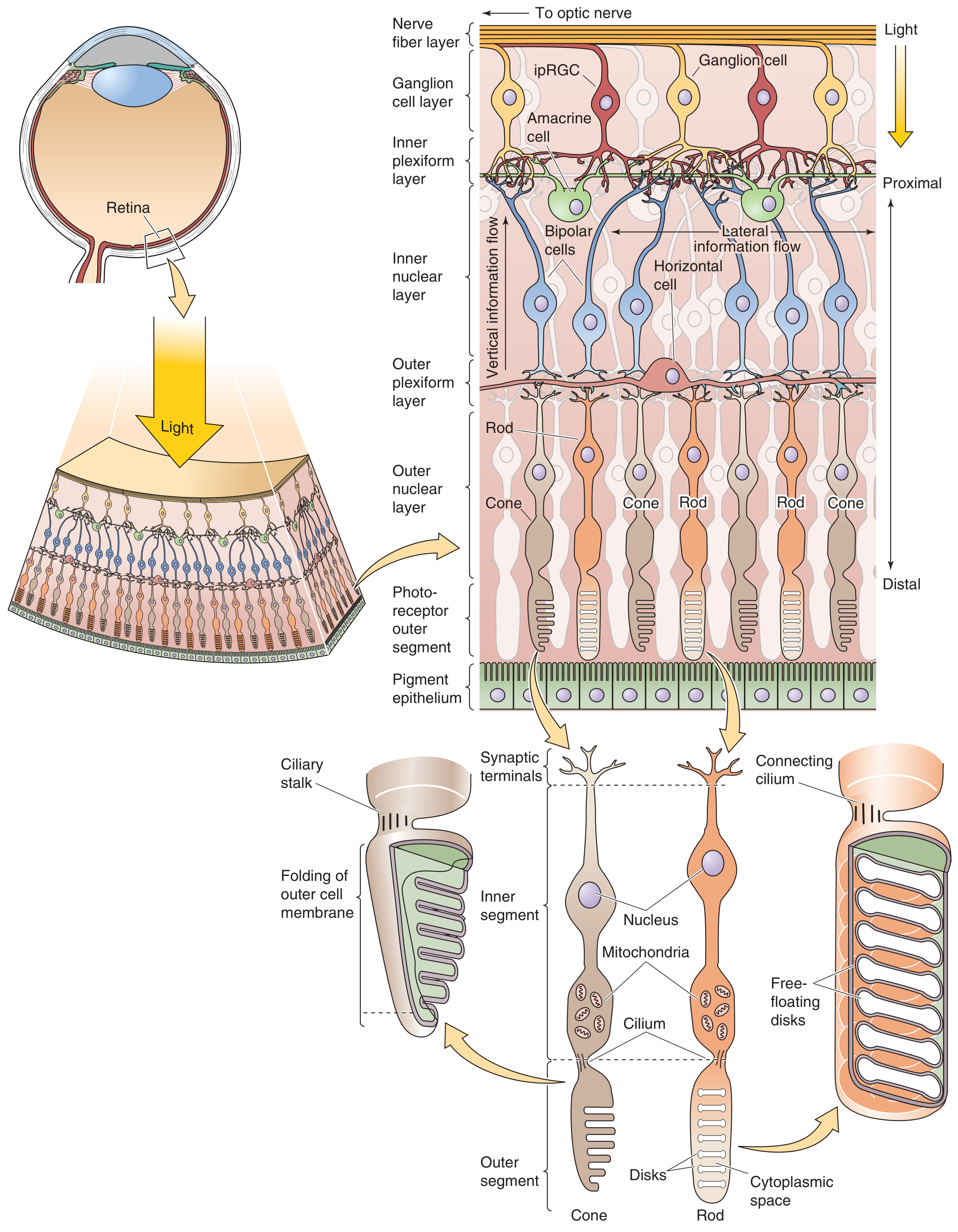
- Nerve fiber layer - axons of ganglion cells traveling to optic nerve
- Ganglion cell layer - output neurons of the retina
- Inner plexiform layer - synapses between bipolar/amacrine cells and ganglion cells
- Inner nuclear layer - bipolar, horizontal, and amacrine cells
- Outer plexiform layer - synapses between photoreceptors and bipolar/horizontal cells
- Outer nuclear layer - photoreceptor cell bodies (nuclei of rods and cones)
- Photoreceptor outer segments - the transduction compartment
- Retinal pigment epithelium (RPE) - absorbs stray light, regenerates photopigments
Key retinal landmarks:
- Fovea centralis - a small pit (~300-700 µm) at the center of the macula where visual acuity is highest. Contains only cones packed at maximal density (~0.05 µm center-to-center). Inner neural layers are displaced laterally to minimize light scattering.
- Optic disc (blind spot) - where ganglion cell axons exit as the optic nerve. Contains no photoreceptors, hence a physiological blind spot.
- Macula - the central area surrounding the fovea, responsible for fine-detail and color vision.
5. Photoreceptors - Rods vs. Cones
| Feature | Rods | Cones |
|---|---|---|
| Number | ~120 million | ~6-7 million |
| Distribution | Peripheral retina | Concentrated in fovea |
| Sensitivity | Extremely sensitive (1 photon sufficient) | Less sensitive |
| Vision type | Scotopic (dim light, monochromatic) | Photopic (bright light, color) |
| Photopigment | Rhodopsin | S, M, or L opsins (blue, green, red) |
| Outer segment discs | Free-floating discs | Infolded, continuous with outer membrane |
| Convergence | High (many→1 ganglion cell) | Low at fovea (1:1:1) |
6. Phototransduction - Converting Light to Neural Signals
- Synaptic terminal - releases glutamate onto bipolar cells
- Inner segment - contains nucleus, mitochondria, and synthesizes photopigments
- Outer segment - the transduction site; a highly modified cilium with stacked disc membranes packed with photopigment
The Dark Current
Light Response - The Cascade
- Photon absorption - light strikes rhodopsin (rod) or cone opsin, converting retinal from 11-cis to all-trans configuration.
- Transducin activation - activated rhodopsin (metarhodopsin II) activates the G-protein transducin.
- Phosphodiesterase (PDE) activation - transducin activates PDE, which hydrolyzes cGMP → GMP.
- Channel closure - falling cGMP closes the outer-segment cation channels.
- Hyperpolarization - the cell hyperpolarizes (to ~-70 mV), reducing glutamate release.
- Signal to bipolar cells - decreased glutamate signals downstream neurons.
This is counterintuitive: photoreceptors are most active in the dark and reduce activity (hyperpolarize) in response to light.
7. Visual Pathway - From Retina to Cortex
Photoreceptors
    ↓
Bipolar cells (+ Horizontal cells for lateral inhibition)
    ↓
Ganglion cells (+ Amacrine cells)
    ↓
Optic nerve (CN II)
    ↓
Optic chiasm (nasal fibers cross, temporal fibers stay ipsilateral)
    ↓
Optic tract
    ↓
Lateral Geniculate Nucleus (LGN) of thalamus
    ↓
Optic radiation
    ↓
Primary visual cortex (V1) - occipital lobe (calcarine sulcus)
8. Protective and Accessory Structures
- Orbit - bony socket of the skull providing mechanical protection
- Eyelids and blinking - keep the cornea moist and protected
- Lacrimal gland - produces tears that cleanse and moisturize the corneal surface; tears drain via the nasolacrimal duct into the nose
- Extraocular muscles - three pairs (superior/inferior rectus, medial/lateral rectus, superior/inferior oblique) innervated by CN III, IV, and VI; move the eyeball in the orbit
- Conjunctiva - mucous membrane lining eyelids and covering anterior sclera
Summary Table - Key Structures at a Glance
| Structure | Function |
|---|---|
| Cornea | Primary refraction (~43 diopters), light transmission |
| Iris/pupil | Regulates light entry (up to 16-fold range) |
| Lens | Fine-tune focus, accommodation |
| Aqueous humor | Nourishes avascular structures, maintains IOP |
| Vitreous humor | Maintains eye shape |
| Retina | Transduces light to neural signals |
| Fovea | Highest visual acuity, color vision |
| Optic nerve | Transmits visual signals to brain |
| Choroid | Vascular supply to outer retina |
| RPE | Photopigment recycling, light absorption |