
Chalazoin
I'll look up chalazion from the medical library for you.
Reading File
Searching sources...
Finding Sources
Finding Sources
Good sources found. Now reading the most comprehensive entries in parallel.
Reading File
Reading File
Reading File
Now let me get the Wills Eye Manual treatment details and fetch the key clinical images.
Reading File
Reading File
Loading Image
Loading Image
Here is a thorough overview of chalazion based on multiple ophthalmology and primary care textbooks.
Chalazion (Meibomian Cyst)
Definition
A chalazion is a sterile, chronic granulomatous inflammatory lesion (lipogranuloma) of the meibomian gland (or occasionally the gland of Zeis), caused by obstruction and retention of lipid-rich sebaceous secretions within the eyelid. It is distinct from a hordeolum (stye), which is an acute bacterial abscess.
- Goldman-Cecil Medicine, p. 302
- Wills Eye Manual, p. 381
Pathogenesis
The retained lipid-rich material acts as a foreign body, triggering a lipogranulomatous foreign-body inflammatory reaction. Histopathology shows:
- Extracellular fat deposits (appear as clear spaces after processing dissolves the fat)
- Lipid-laden epithelioid cells and multinucleated giant cells
- Surrounding lymphocytes
Histopathology - lipogranuloma:
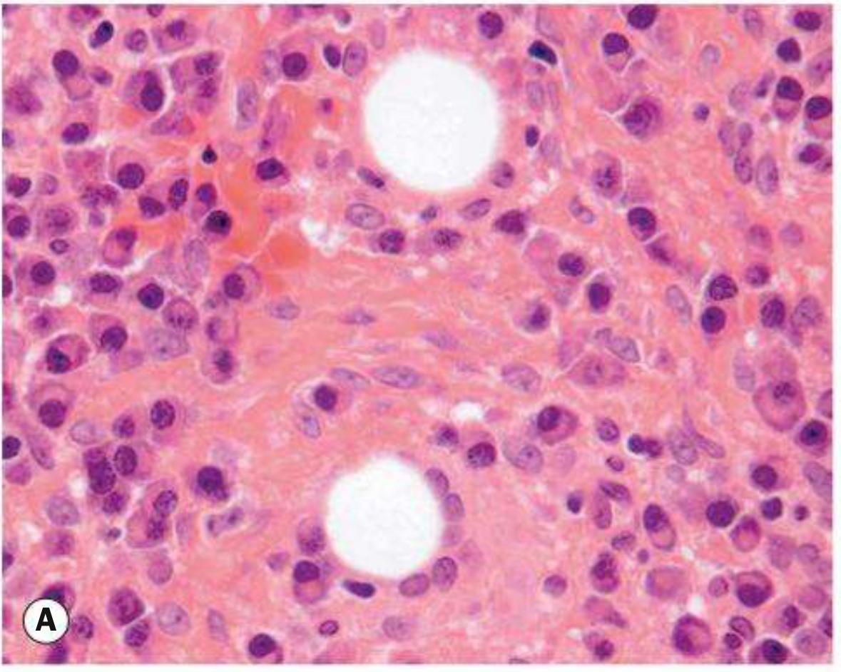
- Kanski's Clinical Ophthalmology 10th ed., p. 54
Associated conditions:
- Blepharitis (commonly present)
- Acne rosacea (associated with multiple/recurrent chalazia)
- Bortezomib (proteasome inhibitor used in myeloma) can predispose to chalazia within 3 months of starting treatment
Key warning: A recurrent chalazion in the same lid position - especially in older patients - must be biopsied to exclude sebaceous carcinoma masquerading as a chalazion.
Clinical Features
Bilateral chalazia in upper eyelids (clinical photo):
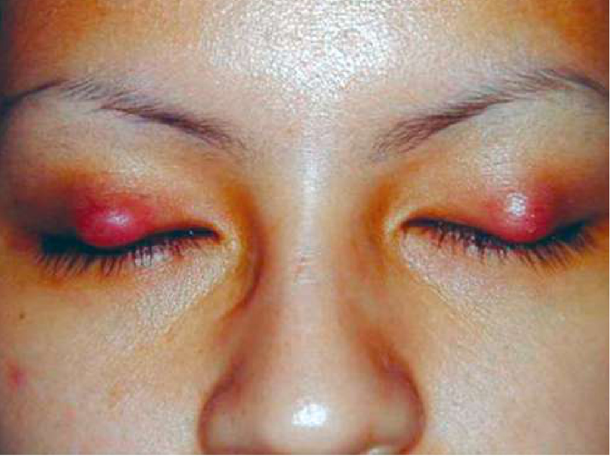
Symptoms
- Gradual onset of a painless or mildly tender eyelid lump (subacute/chronic form)
- Acute form: localized cellulitis and sterile inflammation; if secondarily infected, becomes an internal hordeolum
Signs
| Feature | Description |
|---|---|
| Critical | Well-defined, palpable subcutaneous nodule within the tarsal plate |
| Meibomian gland | Blocked orifice, inspissated secretions may be visible |
| Associated | Eyelid swelling, erythema, focal tenderness, blepharitis, acne rosacea |
| Possible | Conjunctival granuloma, or lesion pointing/draining mucopurulent material |
Differential Diagnosis
| Condition | Key distinguishing feature |
|---|---|
| Hordeolum (stye) | Acute, painful, bacterial; external (gland of Zeis) or internal (meibomian gland) |
| Sebaceous carcinoma | Recurrent chalazion same spot; madarosis, eyelid thickening, chronic unilateral blepharitis in older patient |
| Preseptal cellulitis | Diffuse eyelid/periorbital erythema, edema, warmth |
| Pyogenic granuloma | Deep-red, pedunculated conjunctival lesion, often post-chalazion/surgery |
| Forniceal foreign body | Eyelid swelling, history of contact lens use or trauma |
- Wills Eye Manual, p. 382
Workup
- History - prior chalazia, ocular surgery, trauma, medications (bortezomib)
- External examination - palpate eyelid for nodule; inspect for madarosis or ulceration
- Slit lamp - evert eyelid, evaluate meibomian gland inspissation; assess for signs suggesting malignancy
- Screen for rosacea (look at skin, check for telangiectasias)
Treatment
Step 1 - Conservative (first-line)
- Warm compresses for at least 10 minutes 4x daily with gentle massage over the lesion
- At least 1/3 resolve spontaneously - observation alone is appropriate if improving
- Expression: compression between two cotton-tipped applicators can express contents of a fresh, superficial lesion
Step 2 - Medications
- Topical antibiotic/steroid (e.g., neomycin/polymyxin B/dexamethasone ointment b.i.d. for 1-2 weeks)
- Doxycycline 20-50 mg orally daily to b.i.d. (low-dose, for its anti-inflammatory effect) - indicated for multiple/recurrent chalazia or associated ocular rosacea
Step 3 - If no resolution after 3-4 weeks: Choose one
A. Intralesional steroid injection
- Triamcinolone acetonide 40 mg/mL mixed 1:1 with 2% lidocaine with epinephrine (0.2-1.0 mL)
- Alternatives: betamethasone or dexamethasone formulations
- ~80% success rate with one injection; can repeat 1-2 weeks later
- Preferred for marginal lesions or lesions near the lacrimal punctum
- Risks: skin depigmentation and fat atrophy (less common via conjunctival approach); rare retinal vascular occlusion from retrograde intra-arterial infiltration - discuss with patient before use
B. Incision and curettage (I&C) - surgical
- Administer local anesthesia; evert the eyelid with a specialized chalazion clamp
- Incise vertically through the tarsal plate
- Curette the cyst contents; excise solid inflammatory material with fine scissors if needed
- No suture required
- Apply topical antibiotic 3x daily for 5 days post-procedure
- All excised tissue (especially recurrent or atypical chalazia) should be sent for histopathology
Follow-Up
- If medical therapy is used: review only if lesion persists beyond 3-4 weeks
- After I&C: re-examine as needed
- Recurrent chalazion at the same site = biopsy mandatory to rule out malignancy (sebaceous carcinoma)
Sources: Kanski's Clinical Ophthalmology 10th ed. | Wills Eye Manual | Goldman-Cecil Medicine | Textbook of Family Medicine 9e
This is a shared conversation. Sign in to Orris to start your own chat.