
65 years old male came to the A& E with aphasia & B/L hemiparesis. O/E his BP 178/9 4 mmHg, PR 80,Rhythem is irregular. what is the most appropriate next step of management? a) Mechanical thrombolectomy b) Intra arterial tPA c) IV labetalol d) IV tPA e) Low dose aspirin
Answer: (d) IV tPA
Clinical Reasoning
- Aphasia (dominant hemisphere involvement)
- Bilateral hemiparesis (large vessel or bilateral territory involvement)
- Irregular pulse - strongly suggests atrial fibrillation as the embolic source
- BP 178/94 mmHg - elevated but does NOT exceed the thrombolysis threshold
Why IV tPA (d) is Correct
"Acute ischemic stroke treated with tissue plasminogen activator (tPA) has an improved neurologic outcome when treatment is given within 4.5 h of onset of symptoms, with likely increased benefit associated with earlier administration."
- Systolic >185 mmHg OR diastolic >110 mmHg = contraindication to thrombolytics
- This patient's BP (178/94) is within the acceptable range - tPA can proceed
Why the Other Options Are Wrong
| Option | Reason Incorrect |
|---|---|
| (a) Mechanical thrombectomy | Requires large vessel occlusion confirmed on CT angiography/neuroimaging. It is NOT the first step - IV tPA is given first (if eligible), and thrombectomy is considered as an additional or alternative step if large vessel occlusion is confirmed. "Drip and ship" (give IV tPA, then transfer for thrombectomy) is the standard approach. |
| (b) Intra-arterial tPA | Not first-line. Reserved for selected patients who are ineligible for IV tPA or in the 3-6 hour window for specific arterial territories. More invasive, requires catheter lab. |
| (c) IV labetalol | Antihypertensive treatment is not indicated at BP 178/94 in acute ischemic stroke. The AHA/ASA guidelines recommend permissive hypertension (up to 220/130 mmHg) in non-thrombolysis candidates to maintain cerebral perfusion. Abrupt BP reduction can worsen ischemia. Labetalol is only used if BP exceeds 185/110 before giving tPA - and even then, the goal is to lower it just enough to allow tPA, not to aggressively treat it. |
| (e) Low-dose aspirin | Antiplatelet therapy is appropriate in the long-term secondary prevention of ischemic stroke. In the acute setting within the tPA window, IV tPA takes priority. Aspirin is deferred for 24 hours after thrombolysis. |
Key Management Points
- Before giving tPA: Check BP - if >185/110, give labetalol/nicardipine to bring it down to <185/110, then give tPA
- BP here is 178/94: Already below the threshold - tPA can be given immediately without antihypertensive pretreatment
- Atrial fibrillation: Explains the cardioembolic mechanism; anticoagulation (not tPA) is for prevention, not acute treatment
- Thrombectomy consideration: If CT angiography reveals a large vessel occlusion (e.g., MCA, basilar), mechanical thrombectomy may be added, but IV tPA remains the first step
Sources: Harrison's Principles of Internal Medicine 22E; Tintinalli's Emergency Medicine - "Blood Pressure Control in Stroke" (Table 167-9); Bradley & Daroff's Neurology in Clinical Practice - "General Management of Acute Ischemic Stroke"
60 years old man was admitted to the hospital with the sudden onset of severe vertigo, hiccups and vomiting. On examination, the soft palate was drawn up to the left side when patient is saying 'Ah', associated dysarthria, ptosis, constricted right pupil. There is an impairment of the pain and temperature sensation in the left side of the trunk. Upper limb and lower limb motor examination is normal. What is the most likely reason for this presentation? a) Anterior cerebral artery territory Infarction b) Anterior inferior cerebellar artery infarction c) Anterior spinal Artery infarction d) Posterior Inferior cerebellar artery infarction e) Posterior cerebral artery infarction
lateral medullary Wallenberg syndrome PICA infarction brainstem anatomy diagram
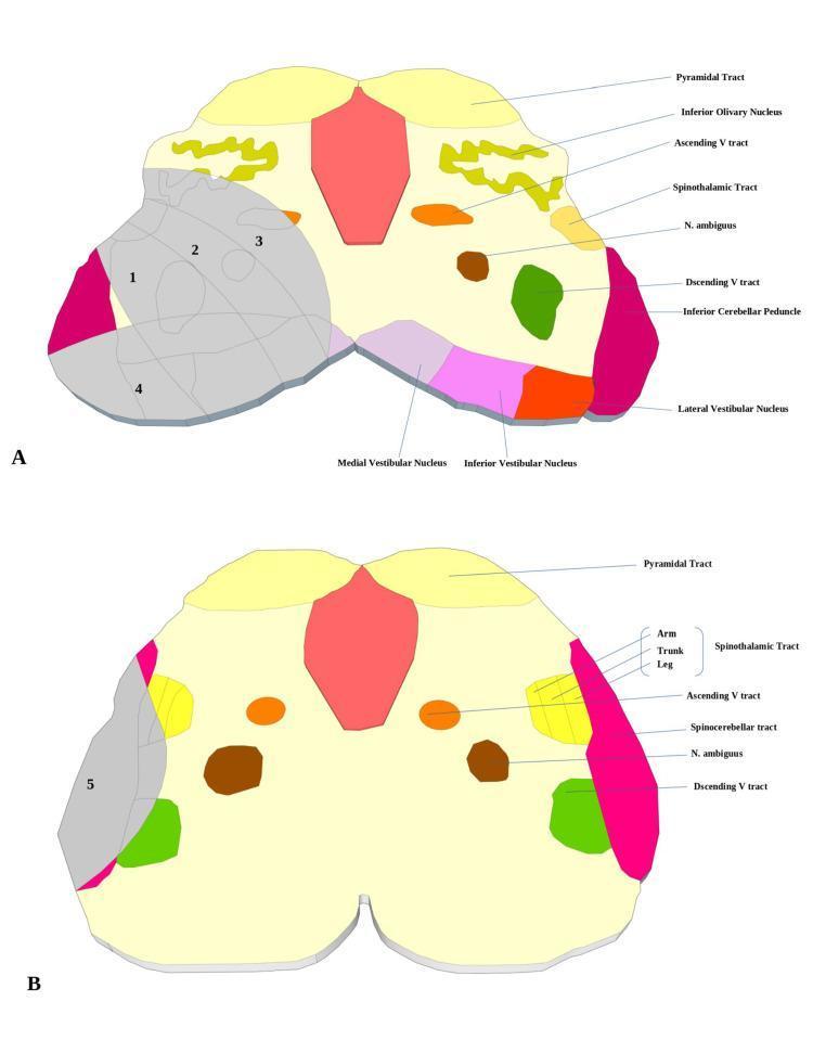
This medical diagram illustrates the cross-sectional neuroanatomy and clinical pathology of the brainstem at the rostral (A) and caudal (B) medullary levels. The illustrations use color-coding and numerical labeling (1-5) to demonstrate common patterns of Lateral Medullary Infarction (Wallenberg Syndrome). In the rostral level (A), labeled structures include the Pyramidal tract, Inferior Olivary Nucleus, Ascending V tract, Spinothalamic tract, Nucleus ambiguus, Descending V tract, and various vestibular nuclei (Medial, Inferior, and Lateral). In the caudal level (B), the diagram adds detail to the Spinothalamic tract topography (Arm, Trunk, Leg) and shows the Spinocerebellar tract. Shaded gray regions indicate specific infarction subtypes: Typical (1+2), Ventral (2+3), Large (1+2+3), Dorsal (4), and Lateral (5). These sections highlight the vascular distribution of the Posterior Inferior Cerebellar Artery (PICA) and its clinical relevance in localizing brainstem strokes. The diagrams serve as an educational tool for correlating visual neuroanatomical landmarks with specific neurological deficits observed in medullary syndromes.
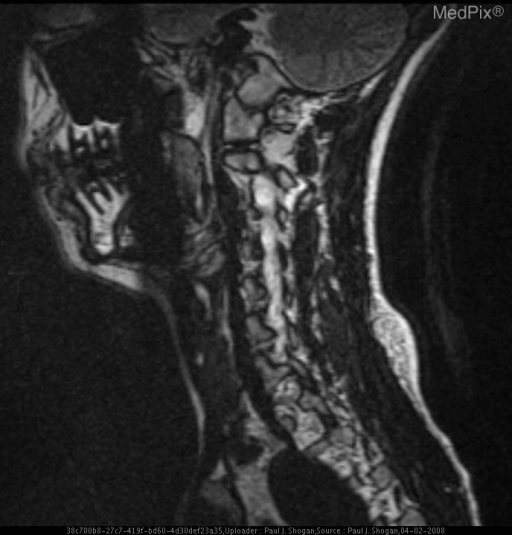
**Imaging Modality:** Sagittal Magnetic Resonance Imaging (MRI), T2-weighted sequence. **Anatomical Region:** Cervical spine and posterior fossa, including the brainstem (medulla oblongata), cerebellum, and upper spinal cord. **Observed Pathology:** The image demonstrates the neuroanatomical structures associated with Lateral Medullary Syndrome (Wallenberg Syndrome). While the acute infarct may be subtle on this specific sagittal view, the focus is on the regional anatomy of the medulla oblongata and its proximity to the cerebellum and vertebral-basilar arterial distribution. **Characteristic Visual Features:** - **Sagittal View:** Clear visualization of the craniovertebral junction and the brainstem. - **Brainstem:** The medulla oblongata is visible anterior to the fourth ventricle and cerebellum. - **Spinal Cord:** Intact signal intensity of the cervical spinal cord. - **Soft Tissue:** Visual representation of the posterior neck musculature and cervical vertebrae. **Relevant Clinical Context:** Lateral medullary infarction typically results from occlusion of the Posterior Inferior Cerebellar Artery (PICA) or the vertebral artery. Key diagnostic cues involve identifying ischemia within the retro-olivary (lateral) portion of the medulla, which houses the vestibular nuclei, spinothalamic tract, and descending sympathetic fibers.
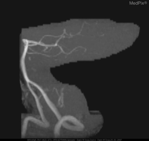
**Imaging Modality:** Magnetic Resonance Angiography (MRA), Maximum Intensity Projection (MIP) reconstruction. **Anatomical Region:** Posterior cranial fossa vasculature, specifically the vertebrobasilar system. **Observed Pathology:** Lateral medullary infarction (Wallenberg Syndrome) context. The image demonstrates the arterial anatomy of the posterior circulation. There is a notable absence or significant attenuation of the distal segments of the posterior inferior cerebellar artery (PICA) on the affected side compared to expected vascular distribution. The vertebral artery and basilar artery are visualized, but the lateral branching patterns relevant to the lateral medulla appear compromised. **Characteristic Visual Features:** - High-signal vascular reconstruction against a suppressed background. - Visualization of the bilateral vertebral arteries merging into the basilar artery. - Evaluation of the PICA origin and course; vascular dropout in the distribution of the lateral medulla and inferior cerebellum is the primary diagnostic cue. **Clinical Context:** This neuroimaging is used to identify vascular occlusion or dissection in patients presenting with acute lateral medullary syndrome, characterized clinically by ipsilateral ataxia, Horner's syndrome, and contralateral loss of pain/temperature sensation. **Key Diagnostic Features:** Focal vascular truncation or absence of the PICA, correlating with acute ischemic territory in the lateral medulla.
Answer: (d) Posterior Inferior Cerebellar Artery (PICA) Infarction
This is the Classic Lateral Medullary (Wallenberg) Syndrome
Symptom-by-Symptom Anatomical Mapping
| Patient's Finding | Structure Damaged | Side |
|---|---|---|
| Severe vertigo, nausea, vomiting | Vestibular nuclei | Ipsilateral (Right) |
| Hiccups | Reticular formation / respiratory centers | Ipsilateral |
| Soft palate drawn up to the LEFT on saying "Ah" | Nucleus ambiguus (CN IX/X) - RIGHT sided paralysis causes deviation to LEFT | Ipsilateral right |
| Dysarthria | Nucleus ambiguus (CN X) + cerebellar peduncle | Ipsilateral |
| Ptosis + constricted right pupil (Horner's syndrome) | Descending sympathetic fibers in lateral tegmentum | Ipsilateral (Right) |
| Pain & temperature loss in left trunk (contralateral) | Spinothalamic tract (already crossed) | Contralateral (Left) |
| Normal motor power in limbs | Pyramidal tract (ventral medulla) - SPARED | - |
"Because the syndrome affects the lateral tegmentum, motor involvement is usually not prominent, and prognosis is generally good... Involvement of the descending sympathetic fibers causes an ipsilateral Horner's syndrome (ptosis, miosis). Infarction of the nucleus ambiguus and exiting fascicles of CN X causes breathy hoarseness and dysphagia."
Key Localizing Clue - The "Crossed" Pattern
- Ipsilateral face - loss of pain/temperature (trigeminal spinal nucleus - NOTE: not prominent here, but ipsilateral Horner's and palatal palsy confirm RIGHT-sided lesion)
- Contralateral body (left trunk) - loss of pain/temperature (spinothalamic tract, already crossed before entering the medulla)
- Normal motor function (pyramids are ventral - spared in lateral lesions)
Why the Other Options Are Wrong
| Option | Structure | Why it Doesn't Fit |
|---|---|---|
| (a) Anterior cerebral artery | Medial frontal lobe, paracentral lobule | Causes contralateral leg weakness and frontal lobe signs - no cranial nerve or Horner's features |
| (b) AICA | Lateral pons + inner ear | Would cause ipsilateral hearing loss (labyrinthine artery from AICA) and facial (CN VII) palsy - absent here |
| (c) Anterior spinal artery | Medial medulla (pyramid + medial lemniscus + CN XII) | Causes contralateral hemiplegia (pyramidal tract) + contralateral loss of vibration/proprioception (medial lemniscus) + ipsilateral tongue deviation - no Horner's, no vertigo |
| (e) Posterior cerebral artery | Occipital lobe, thalamus, midbrain | Causes hemianopia, thalamic sensory loss, or CN III palsy - no Horner's, no medullary syndrome |
Vascular Anatomy Note
- In ~80% of cases, Wallenberg syndrome is actually caused by vertebral artery occlusion (not isolated PICA occlusion), because the PICA typically branches from the vertebral artery
- Isolated PICA occlusion accounts for the remaining ~20%
- In the question context, PICA territory infarction is the accepted answer as it defines the syndrome
Sources: Neuroanatomy through Clinical Cases 3rd Ed., pp. 684-685; Localization in Clinical Neurology 8e, pp. 919-920; Harrison's Principles of Internal Medicine 22E; K.J. Lee's Essential Otolaryngology