
Alt surgery
What aspect of ALT (Anterolateral Thigh) flap surgery are you interested in?
anterolateral thigh flap complications management
PMID: 40318362
anterolateral thigh flap anatomy perforator diagram
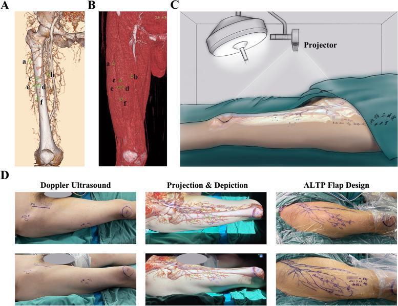
This composite educational image illustrates the multi-step process of using Augmented Reality (AR) for preoperative Anterolateral Thigh Perforator (ALTP) flap design. (A) Three-dimensional CT angiography (CTA) reconstruction of the thigh showing the skeletal framework and vascular network, with six specific perforator emergence points (a-f) marked. (B) Volume-rendered CTA focusing on the muscular anatomy, showing the same markings relative to the muscle surface. (C) A clinical diagram depicting a portable projector mapping the reconstructed vascular map onto a patient’s thigh in the preoperative setting. (D) A clinical photograph sequence demonstrating the practical application: initial Doppler ultrasound marking of perforators, followed by 'Projection & Depiction' where the AR map is overlaid on the skin to align with anatomical landmarks like the patella and ASIS, and finally the 'ALTP Flap Design' where the surgical incision lines are drawn based on the projected vascular course and perforator locations. This demonstrates an advanced surgical planning technique in reconstructive microsurgery to increase the accuracy of flap harvesting.
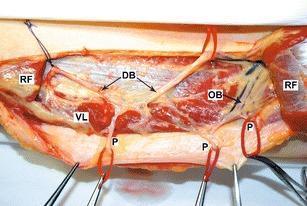
This clinical photograph of a cadaveric dissection illustrates the vascular anatomy of the anterolateral thigh, specifically relating to the design and elevation of an anterolateral thigh (ALT) flap. The image identifies the vastus lateralis (VL) and the rectus femoris (RF) muscles, with the RF being partially cut to expose the underlying pedicle. Labeled anatomical structures include the descending branch (DB) and the oblique branch (OB) of the lateral circumflex femoral artery. Multiple perforators (P) are shown originating from both the descending and oblique branches, which are critical for providing the blood supply to the overlying skin and fascia in flap surgery. The vessels are highlighted with red vessel loops for clarity. This anatomical study demonstrates variation in perforator origin, serving as an educational resource for plastic and reconstructive surgeons to understand the different vascular patterns encountered during flap harvest to ensure tissue viability.
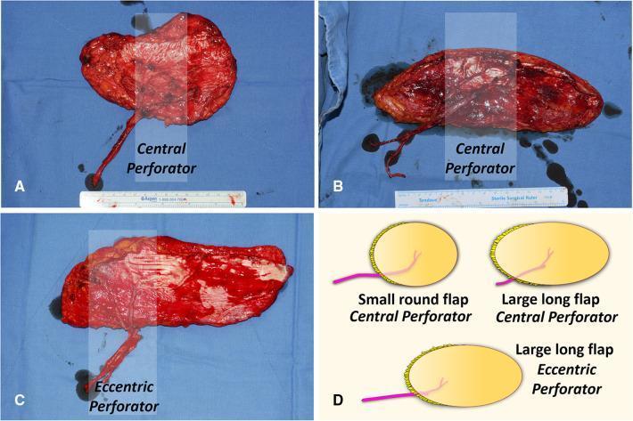
This composite educational graphic details variations in the design of Anterolateral Thigh Perforator (ALTP) flaps for soft tissue reconstruction. The image consists of clinical photographs of harvested specimens (A, B, C) and a corresponding schematic diagram (D). Panels A and B demonstrate Cen-ALTP flaps, where the vascular pedicle enters the skin paddle centrally. Image A shows a small, round paddle with a shorter pedicle, while Image B displays a significantly elongated, larger paddle. Panel C illustrates an Ecc-ALTP flap, featuring a large, long skin paddle with the perforator vessel located eccentrically toward one end. The clinical photographs show the raw, vascularized deep surface of the flaps and the length of the dissected vascular pedicles. Panel D provides an illustrative summary of these three configurations, emphasizing how perforator location (central vs. eccentric) and paddle dimensions (small round vs. large long) are varied to accommodate different recipient site requirements and pedicle reach. These flaps are commonly used in reconstructive surgery for large defects such as those following debridement for chronic osteomyelitis in the lower limb.
ALT Flap Surgery: Complications & Management
Overview
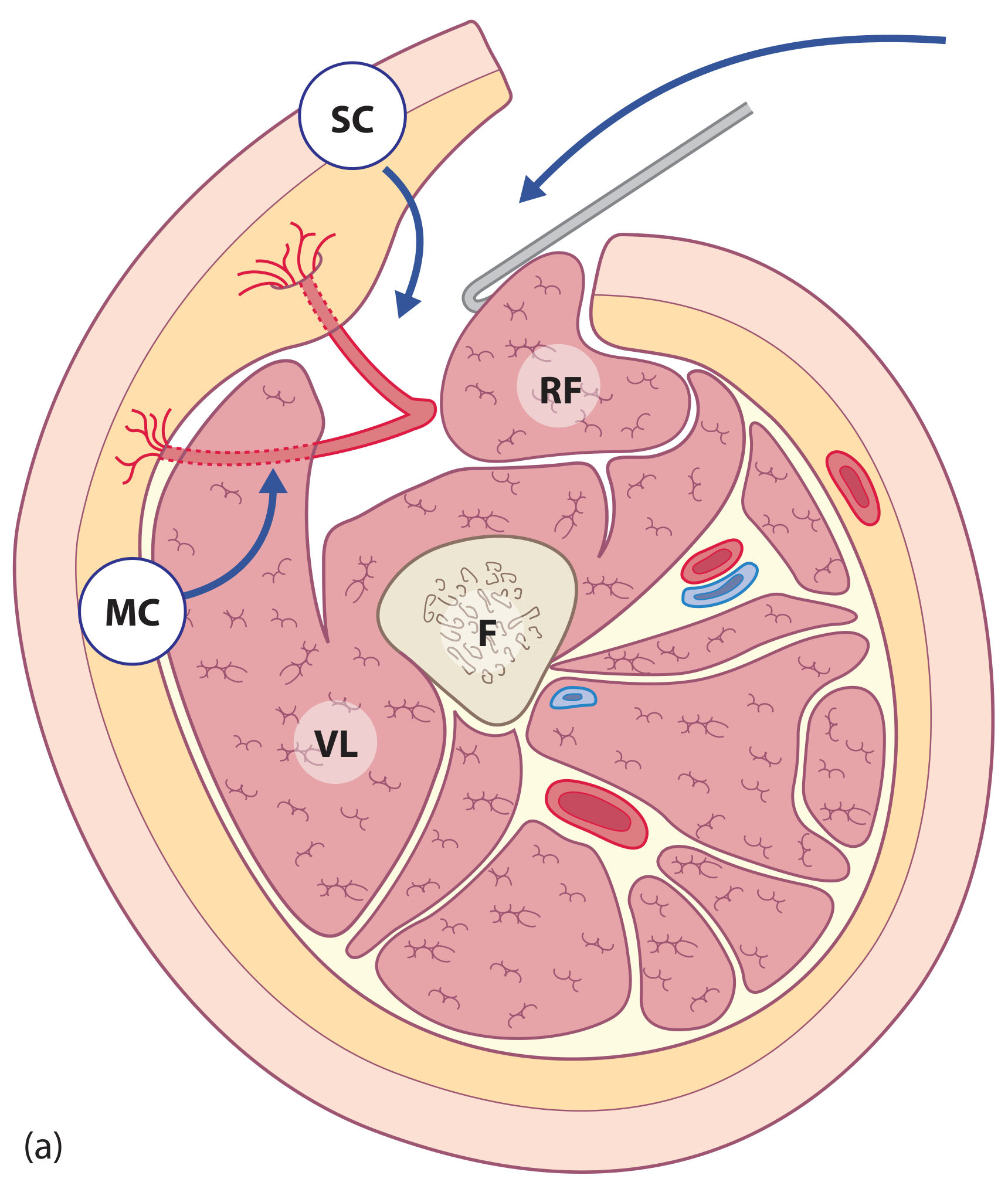
Recipient Site Complications
1. Flap Failure (Partial or Total)
- Cause: Vascular compromise - arterial thrombosis, venous congestion, or pedicle kinking/compression
- Key vulnerability: The ALT is usually perfused by a single perforator. Proximal venous occlusions are particularly difficult to salvage because of the small calibre of the perforating vessels - once compromised, there is no redundancy
- The small perforator calibre also means the pedicle is easily compromised by external compression, making ALT a poor choice when pedicle tunnelling is required
- Management: Close monitoring in the first 48-72 h; urgent re-exploration for venous congestion or arterial insufficiency; salvage with pedicled pectoralis major or local flap if free flap fails
2. Pedicle Anatomy Variability
- Musculocutaneous perforators are present in ~81.9% of cases; absent perforators occur in ~5.4% - the surgeon must be prepared for intraoperative surprises
- Variable perforator number, location and course is the single biggest technical hazard
- Management: Pre-operative Doppler (8-MHz) or CT/MR angiography mapping; Augmented Reality CTA overlay is an emerging pre-operative technique; always design flap with the principal perforator at centre; be ready to extend to an oblique branch perforator
3. Haematoma / Seroma
- Inadequate haemostasis during perforator dissection through vastus lateralis is a common source
- Management: Meticulous haemostasis when ligating muscular side-branches; suction drains at donor and recipient sites
4. Wound Dehiscence / Infection
- Common in contaminated wounds (trauma, oncological resections)
- Management: Debridement, antibiotic coverage, negative pressure wound therapy (NPWT), secondary closure
5. Flap Bulk / Poor Contouring
- The ALT tends to be thick, especially in obese patients and women with greater thigh adiposity
- In Caucasian patients, skin colour mismatch for facial/neck reconstruction is also noted
- Management:
- Thin the flap at the time of harvest (suprafascial dissection) or use it as a fascial-only flap with skin grafting to reduce bulk
- Staged debulking procedures post-operatively
- Consider alternative flap (e.g., radial forearm) when thin, pliable tissue is essential in non-Asian populations
- The flap is generally considered unsuitable for obese patients and men with extremely hairy thighs (Campbell's Operative Orthopaedics 15th Ed 2026)
6. Motor Nerve Injury to Vastus Lateralis
- The descending branch is accompanied by motor nerve branches to the vastus lateralis
- Management: These nerves must be identified and preserved during pedicle dissection - failure to do so causes quadriceps weakness at the donor thigh
7. Sensory Loss
- Injury to the lateral femoral cutaneous nerve (which can optionally be included to make the flap sensate) results in lateral thigh numbness if sacrificed unintentionally
- Management: Identify and protect or formally include the nerve for a neurosensory flap
Donor Site Complications
1. Wound Closure Problems
| FW/TCR Ratio | Recommended Closure |
|---|---|
| < 16% | Direct primary closure (tension-free) |
| 16-30% | Locoregional flap (avoids graft morbidity) |
| > 30% | Skin grafting or "kiss" (split skin paddle) technique |
- Widths > 8-13 cm cannot usually be closed primarily and require skin grafting (Rockwood & Green's 10th Ed 2025)
- Harvesting deep fascia allows muscle to bulge, further hindering closure
- Management: Advance skin edges and suture to muscle to reduce defect; NPWT, acellular dermal matrices, tissue expansion as adjuncts
2. Seroma / Haematoma at Donor Site
- Particularly common when large paddles are harvested and skin grafts are placed
- Management: Suction drains, compressive dressings, NPWT over graft
3. Skin Graft Failure
- When primary closure is not feasible, skin graft take may be partial
- Management: NPWT to improve graft adherence; re-grafting if necessary
4. Functional Deficit
- Provided the nerve to vastus lateralis is preserved, functional disability is limited
- If substantial muscle cuff is taken with musculocutaneous perforators, some quadriceps bulk is lost
- Management: Preserve nerve; minimise muscle cuff around perforator
5. Cosmetic Scar
- The donor scar on the lateral thigh is generally considered acceptable and "relatively inconspicuous," but may be a concern in younger or female patients who are concerned about visible thigh scarring
- Primary closure leaves a vertical scar; skin-grafted donor sites have greater cosmetic morbidity
Summary: Patient Selection Pitfalls
| Situation | Problem | Action |
|---|---|---|
| Obese patient | Excessive flap bulk; unreliable perfusion | Choose alternative flap |
| Women (higher thigh fat) | Poor contouring; bulk mismatch | Consider debulking or alternative |
| Caucasian patients | Poor colour match for head/neck | Forearm or other flap preferred |
| Hairy thigh | Intraoral reconstruction | Contraindicated |
| Pedicle tunnelling needed | Compression risk on small perforator | Poor choice; use other flap |
Key Takeaways
- Vascular anatomy variability is the defining challenge - pre-operative Doppler or CTA mapping is standard
- Flap bulk is the most common functional/aesthetic problem at the recipient site - plan for thinning or staged debulking
- Donor site closure should be planned based on the FW/TCR ratio; primary closure is only reliable when width is < ~16% of thigh circumference
- Preserve the motor nerve to vastus lateralis and identify the lateral femoral cutaneous nerve at every harvest
- The flap is generally avoided in obese patients and when pedicle compression is likely
What is this surgery about explain as you explain a donkey
ALT Flap Surgery — Explained Simply 🫏
You steal from Peter to pay Paul
But skin needs blood to survive
Why the thigh?
- It's a big, meaty area with lots of skin to spare
- The blood supply there is fairly predictable
- Taking skin from the thigh doesn't cripple you — you can still walk fine
- The scar it leaves behind is hidden under clothes
What can go wrong (in plain language)
| Problem | What it means in plain English |
|---|---|
| Blood pipe blocks off | The patch doesn't get blood, it dies — needs emergency re-operation |
| Patch too thick | Like putting a big puffy duvet where you need a thin sheet — doesn't fit well |
| Can't close the thigh | The hole left on the thigh is too big to zip shut — needs a skin graft |
| Weird blood pipe location | Every person's thigh pipes are in slightly different spots — surgeon gets a surprise mid-operation |
| Nerve gets cut | The thigh goes numb, or leg muscle gets slightly weaker |
The bottom line
Surgeons borrow living tissue from your thigh, reconnect its blood supply in a new location, and use it to rebuild a missing piece of your body somewhere else.
What are the complications and management
ALT Flap Complications & Management — Still Explained Simply 🫏
GROUP 1: Problems at the PATCH SITE (where the skin was moved TO)
💀 1. The Patch Dies (Flap Failure)
- Watch the flap every hour for the first 2-3 days (colour, temperature, capillary refill)
- If it goes wrong → rush back to operating theatre to unblock or redo the vessel connection
- If the patch is truly dead → use a different flap (e.g. pectoralis muscle) as salvage
🔀 2. Blood Pipe Kinking or Squashing
- Never tunnel the pedicle under tight tissue — bad idea for ALT
- Position the patient carefully post-op
- Use a monitoring device (Doppler probe) over the pedicle site
🎯 3. Surprise Blood Pipe Location (Anatomy Variability)
- Pre-op Doppler scan (or CT angiography) to map the perforators before cutting
- Always have a backup flap plan before you start
- Experienced surgeons adjust intraoperatively — extend the search or use oblique branch perforators
🏋️ 4. Patch Too Bulky / Too Thick
- Thin the flap during harvest (shave off fat from the underside)
- Use a fascial-only version + skin graft on top (less bulk)
- Plan a staged debulking operation months later
- In obese patients — just avoid this flap altogether and pick a thinner one (e.g. radial forearm flap)
🩸 5. Bleeding / Haematoma Under the Patch
- Meticulous haemostasis during surgery
- Suction drains left in post-op
- If haematoma forms → take back to theatre, wash out, re-drain
🦠 6. Infection / Wound Breakdown
- Antibiotics
- Wound debridement (clean it out)
- Negative pressure wound therapy (NPWT — a vacuum dressing that sucks the wound clean)
- Secondary closure once clean
🎨 7. Colour Mismatch (Cosmetic Problem)
- Accept it (functional > cosmetic in most cases)
- In face/neck reconstruction where appearance matters a lot → consider using radial forearm or other flaps with better colour match
- Laser or tattooing can be used later in selected cases
GROUP 2: Problems at the DONOR SITE (where skin was taken FROM — the thigh)
🧵 8. Can't Close the Thigh Wound
| Flap Width vs Thigh Size | What to do |
|---|---|
| Small (< ~16% of thigh circumference) | Stitch it closed directly |
| Medium (16–30%) | Borrow a local flap from nearby thigh tissue |
| Large (> 30%) | Skin graft the hole |
- Direct stitching (best result, least scarring)
- Locoregional flap (borrow nearby tissue to cover)
- Skin graft (take thin skin from elsewhere and patch the thigh)
- NPWT dressings to help the wound heal down before grafting
😴 9. Thigh Numbness
- Identify and protect the nerve during harvest
- OR deliberately include it in the flap to make the patch sensate (a feature, not a bug)
- If cut accidentally — numbness may partially recover over months; no great fix exists
🦵 10. Leg Muscle Weakness
- These nerve branches must be identified and preserved at every single operation
- No excuse for cutting them — careful dissection avoids this entirely
- If injured → physiotherapy; recovery is variable
💧 11. Seroma / Fluid Collection at Donor Site
- Suction drains for a few days post-op
- Compressive bandaging
- Aspiration in clinic if persistent
Who should NOT have an ALT flap?
| Patient | Why it's risky |
|---|---|
| Very obese | Flap too thick; unreliable blood supply through fat |
| Women with large thighs | Same bulk problem |
| Caucasian patient needing face/neck work | Colour mismatch looks bad |
| Man with very hairy thighs | Hair in the mouth/wound = not ideal |
| Any patient needing pedicle tunnelled | Tiny vessel compresses too easily |
One-line summary
The patch can die, get infected, or be too thick at the new site — and the thigh it came from can be hard to close, go numb, or get weak. Most complications are preventable with good planning and surgical technique.
Any picture of procedure
anterolateral thigh flap harvest surgical procedure
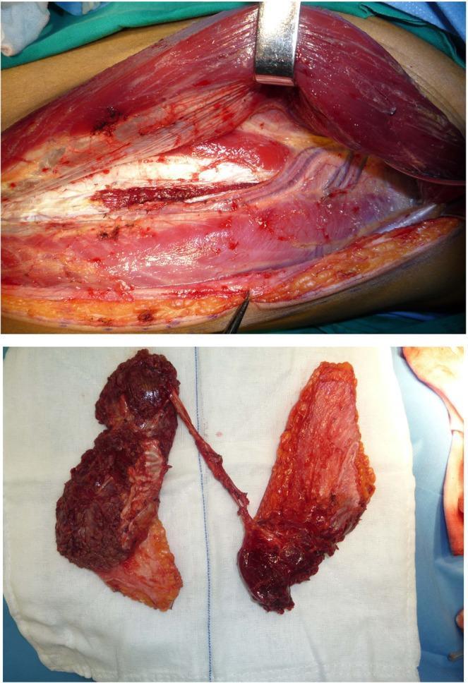
Clinical photograph of a surgical procedure involving the harvest of a branch-based anterolateral thigh (ALT) flap for reconstructive surgery. The top panel shows the donor site in the thigh under surgical retraction. Key anatomical structures visible include the vastus lateralis muscle being retracted, underlying vascular structures, and deep fascia. Dissection of the subcutaneous adipose tissue and muscle is evident, exposing the descending and oblique branches of the lateral circumflex femoral artery. The bottom panel displays the harvested chimeric flap on a white sterile surgical towel. The flap is composed of two distinct tissue blocks: one primarily muscular and thicker in appearance, and the other consisting of a skin paddle with underlying adipose and muscle components. Both blocks remain connected via a common vascular pedicle, demonstrating the flap's versatility for reconstructing complex, multi-site or 'Type B' soft tissue defects. The image illustrates advanced microsurgical techniques in head and neck reconstruction using chimeric free flaps.
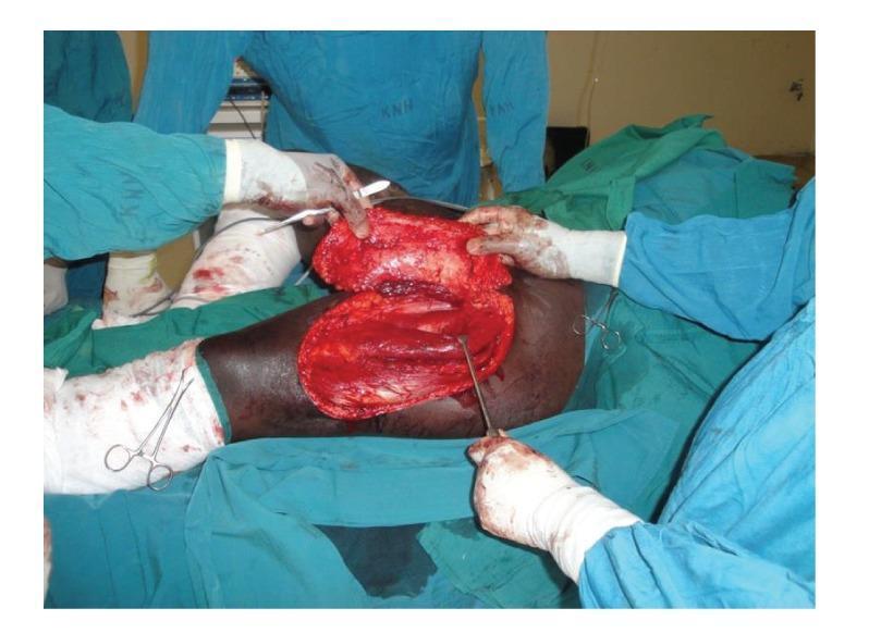
This clinical intraoperative photograph documents the elevation of a pedicled anterolateral thigh (ALT) flap during a reconstructive surgical procedure. The image depicts a large, rectangular fasciocutaneous flap that has been meticulously raised in a subfascial plane, exposing the underlying vastus lateralis and rectus femoris muscles of the donor thigh. The flap's undersurface reveals a highly vascularized appearance with visible subcutaneous fat and fascial tissue, maintained by a central perforating vessel. Surgical personnel in sterile green scrubs and gloves are seen manipulating the flap with both hands and instruments, such as a surgical dissector or retractor, to assess mobility and pedicle length. The surgical field is surrounded by green drapes, and peripheral instruments including hemostats are visible. This visual serves as an educational example of plastic and reconstructive surgery techniques, specifically illustrating the harvest of a locoregional flap for the management of extensive soft tissue defects or contractures.
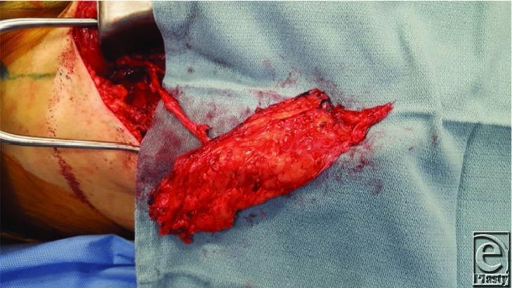
This clinical photograph captures a surgical procedure involving a fascia-only anterolateral thigh (ALT) flap during its elevation. The flap is a rectangular, erythematous tissue mass with a textured, granular surface, resting on blue surgical drapes. A key anatomical feature shown is the vascular pedicle, appearing as a thin, tubular structure extending from the flap back into the donor site in the thigh. The surgical site is visible to the left, showing dissected subcutaneous tissue and exposed muscle, held open by a metal retractor. The surrounding skin shows surgical markings and preparation for harvesting. This image serves as a detailed educational example of reconstructive surgery techniques, specifically illustrating the harvest of a microvascular free flap and the preservation of the vascular pedicle before division. It is highly relevant for plastic and reconstructive surgery training, focusing on flap anatomy, donor site management, and microsurgical preparation.
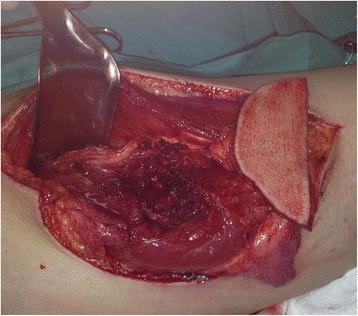
An intraoperative clinical photograph showing the harvest of an anterolateral thigh (ALT) free flap for reconstructive surgery. The image depicts the donor site on the lateral thigh, where a wide surgical incision has exposed the deep anatomical structures. A large metallic retractor is positioned superiorly to provide visualization of the surgical field. The harvested flap is visible, consisting of a paddle of skin and subcutaneous tissue attached to underlying muscle fibers. The donor bed reveals exposed vastus lateralis muscle tissue with evidence of meticulous dissection to isolate the vascular pedicle, likely derived from the descending branch of the lateral circumflex femoral artery. The surgical field shows typical signs of manipulation, including minor localized hemorrhage and exposed fascia. This image demonstrates the technical stage of microvascular flap elevation used in complex head and neck reconstructions, such as glossectomy defects.
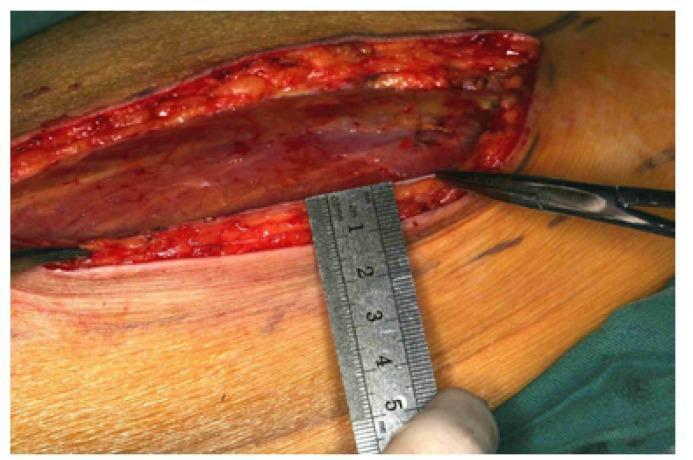
This clinical photograph shows an intraoperative view of an anterolateral thigh (ALT) flap during harvest for reconstructive surgery. An elongated surgical incision is visible along the medial margin of the flap, extending down to the deep fascia. Within the wound bed, the striated texture of the underlying muscle and adipose tissue is evident. A metallic surgical ruler is held perpendicularly against the elevated skin flap to measure its vertical thickness. The ruler shows that the flap currently measures approximately 10 mm in thickness before the thinning process. Surgical instruments, including a pair of forceps, are positioned within the field to assist in tissue retraction and dissection. The image illustrates the anatomical layers of a typical fasciocutaneous flap and demonstrates the clinical technique of verifying flap thickness prior to microvascular transplantation. This material is intended for surgical education, specifically focusing on the harvest of thin ALT flaps for procedures such as tongue reconstruction following hemiglossectomy.
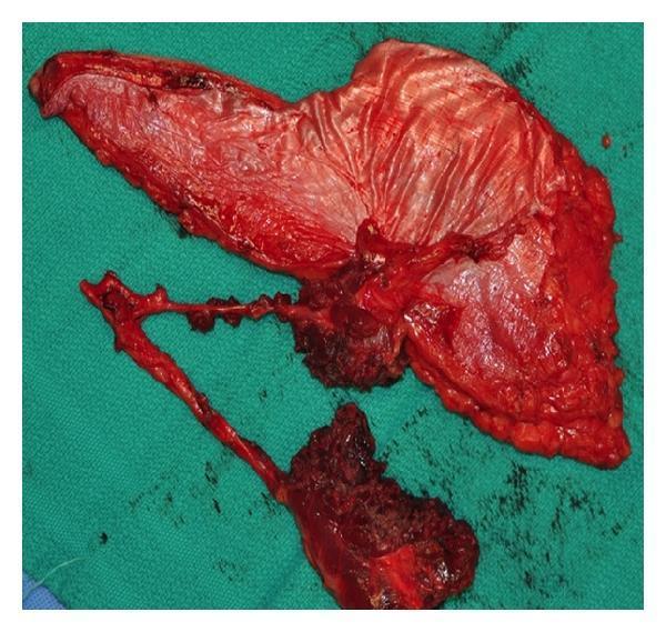
This clinical photograph shows a harvested anterolateral thigh (ALT) free flap, a versatile fasciocutaneous flap used in reconstructive surgery. The specimen is displayed against a green surgical drape immediately following harvest. The flap is composed of a large cutaneous portion and a complex vascular pedicle. Notably, the tissue has been divided into separate skin islands based on multiple perforators branching from the main descending branch of the lateral circumflex femoral artery. The vascular pedicle is visible as a long, branching structure with patent vessels and associated cuff of connective tissue. The deep surface of the flap shows the fascia and subcutaneous adipose tissue, which appears highly vascularized with a reddish-pink hue. This image illustrates the anatomical basis for creating chimera or multi-paddle flaps, allowing a single donor site to reconstruct non-contiguous defects, such as combined intraoral and extraoral maxillofacial defects. This is a key concept in advanced plastic and reconstructive surgery, highlighting the flap's adaptability in complex head and neck reconstruction.
📸 Step 1 — Flap Elevation (Raising the patch off the thigh)
The surgeon lifts the skin and fat off the thigh. You can see the underlying vastus lateralis muscle exposed. The flap is being held up to check how far it can reach.
📸 Step 2 — Measuring Flap Thickness (Is it too thick?)
A ruler measures how thick the flap is (~10 mm here). If it's too thick, the surgeon shaves off excess fat before transplanting it.
📸 Step 3 — The Vascular Pedicle (The life-line blood pipe)
The thin tube you see is the blood vessel pedicle - the artery and vein keeping the flap alive. It will be cut and re-connected (under a microscope) at the new location.
📸 Step 4 — Fully Harvested Flap (Ready to transplant)
The flap is now completely free from the body, sitting on a surgical drape. The branching blood vessel stalk is clearly visible. The clock is now ticking - this tissue must be connected to new blood supply quickly.
📸 Bonus — Chimeric / Two-Paddle Flap (For complex defects)
Sometimes surgeons harvest two separate tissue blocks from the same thigh on a shared blood pipe - one for filling a deep hole, one for covering the surface. One donor site, two problems solved.
Vascular pedicle is artery or vein?
The Vascular Pedicle — Both! 🫏
What's inside the pedicle:
| Component | What it does |
|---|---|
| 1 Artery | Carries fresh blood WITH oxygen INTO the flap |
| 1 or 2 Veins (venae comitantes) | Carries used blood WITHOUT oxygen OUT of the flap |
Think of it like a two-lane road:
- One lane going IN (artery) 🚗
- One lane going OUT (vein) 🚙
- Both lanes are needed — block either one, the patch dies
Specifically in the ALT flap:
- Artery: Descending branch of the Lateral Circumflex Femoral Artery (LCFA)
- Veins: One or two venae comitantes (companion veins that run alongside the artery)
- Internal diameter of the artery: > 3 mm — small but workable under a microscope
Which one causes more trouble when it blocks?
| Blockage | What happens | How quickly? |
|---|---|---|
| Artery blocked | No blood in — flap goes pale and cold | Fast (minutes to hours) |
| Vein blocked | Blood in but can't get out — flap swells, turns purple/blue | Slightly slower but still an emergency |
Vein blockage (venous congestion) is actually more common and in the ALT it is harder to fix because the perforator veins are tiny — surgeons say it is difficult to salvage once it happens.
Simple analogy 🫏
- Artery = the pipe bringing water IN 💧
- Vein = the drain taking water OUT 🕳️
- If the drain blocks, the pot overflows and the roots rot
- If the supply pipe blocks, the plant dries up and dies
What happens if a blood clot forms in the pedicle?
Blood Clot in the Pedicle — A Race Against Time ⏱️
First — What causes the clot?
| Cause | Plain explanation |
|---|---|
| Vessel wall trauma during surgery | Cutting and stitching irritates the inner lining → clot forms at the join |
| Tension on the pedicle | Stretched vessel → wall cracks → clot |
| Kinking / twisting | Like bending a hose → flow slows → clot forms |
| External compression | Tight dressing, haematoma pressing on pedicle → flow stops |
| Patient's blood is too "sticky" | High clotting tendency (hypercoagulable state) |
| Vasospasm | Vessel goes into spasm → no flow → clot forms in stagnant blood |
What happens inside the flap
Clot blocks the vessel
        ↓
Blood flow stops
        ↓
Oxygen delivery to the flap cells stops
        ↓
Cells start dying (ischaemia)
        ↓
After ~6 hours → irreversible cell death
        ↓
Flap turns black and dies (necrosis)
The window to save the flap is roughly 4–6 hours from when the clot forms. After that, the damage is permanent.
How do you KNOW a clot has formed?
| Sign | What it means |
|---|---|
| Flap goes pale / white / cold | Artery blocked — no blood coming IN |
| Flap goes purple / blue / swollen | Vein blocked — blood coming in but can't get OUT |
| Capillary refill > 2 seconds | Blood flow sluggish |
| Handheld Doppler signal disappears | Blood flow in vessel has stopped |
| Flap feels hard / turgid | Venous congestion — blood pooling inside |
Management — Step by Step
🚨 Step 1 — Immediately back to theatre
🔬 Step 2 — Find the problem
| Finding | Action |
|---|---|
| Pedicle kinked | Straighten it, re-route it |
| Haematoma compressing the vessel | Evacuate the blood clot pressing on it |
| Clot at the anastomosis (join) | Open the join, flush it out |
| Vasospasm | Apply warm saline, topical papaverine (a drug that relaxes vessels) |
🧵 Step 3 — Redo the anastomosis (re-sew the blood pipe join)
- Cut out the clotted segment
- Flush the vessel with heparin solution (anti-clot drug)
- Re-sew the vessel under the microscope
- Watch for blood flowing freely again ("pinking up")
💊 Step 4 — Drugs to prevent re-clotting
| Drug | What it does |
|---|---|
| Heparin (IV or low molecular weight) | Thins the blood, prevents new clots |
| Aspirin | Stops platelets sticking together |
| Dextran 40 (IV drip) | Reduces blood "stickiness", improves flow in small vessels |
| Papaverine (applied directly) | Relaxes vessel spasm |
🩸 Step 5 — If venous clot won't resolve: Leeches
When the vein is clotted and the flap is congested, medicinal leeches are applied to the flap surface. They suck out the pooled blood AND release a natural anti-clotting chemical called hirudin into the tissue. This keeps the flap alive while new small veins grow in over 5–7 days.
What if re-exploration fails?
- Partial flap loss — just the edges die, the centre survives → edges trimmed, wound closed
- Total flap loss — entire patch dies → surgeon must start over with a different flap (e.g. pectoralis major muscle flap as salvage)
Prevention is better than cure
| Prevention strategy | How it helps |
|---|---|
| Careful microsurgical technique | Clean, tension-free vessel join = less clot risk |
| No tight dressings over pedicle | Prevents compression |
| Keep patient warm and well-hydrated | Cold + dehydration = sticky blood + vessel spasm |
| Avoid vasoconstrictors (e.g. adrenaline) post-op | These cause vessels to spasm |
| Hourly flap observations | Catch the clot EARLY before 6-hour window closes |
Bottom line 🫏
A clot in the pedicle is a surgical emergency. The flap has about 4–6 hours before it dies permanently. The answer is always the same — get back to theatre fast, find the clot, remove it, re-sew the vessel, and give blood-thinning drugs. Leeches for venous clots. And if all else fails — use a different flap.