
Recurrent pregnancy loss
Reading File
Now let me search the medical library and PubMed in parallel.
Finding Sources
Searching PubMed
"recurrent pregnancy loss" AND management
Searching the Web
recurrent pregnancy loss causes investigation management 2024 guidelines
Good results. Now let me read the key textbook sections in parallel.
Reading File
Reading File
Good content coming in. Let me read more of the Berek & Novak chapter and the Creasy & Resnik evaluation section.
Reading File
Reading File
Excellent content. Let me now read more of the Berek chapter covering etiology details and management.
Reading File
Reading File
I have rich content. Let me read the management/therapy section from Berek & Novak and the APS/immunologic section from Creasy & Resnik.
Reading File
Reading File
I now have comprehensive content from both textbooks. Let me compile the full answer.
Recurrent Pregnancy Loss (RPL)
Definition
RPL is traditionally defined as 3 or more consecutive pregnancy losses before 20-24 weeks gestation. Many guidelines (ASRM, ESHRE) now justify evaluation after 2 losses, given that the prevalence of abnormal findings is similar after 2 or 3 miscarriages. RPL affects approximately 1% of couples attempting pregnancy. Sporadic (isolated) miscarriage is far more common, occurring in 15-25% of recognized pregnancies.
Etiology
1. Genetic Factors (most common cause of first-trimester loss)
- Fetal aneuploidy - the single most common cause of sporadic miscarriage. As the number of losses increases, the proportion with a chromosomally abnormal conceptus decreases (from ~78% with 1 loss to ~18% with 7+ losses), meaning other etiologies become proportionally more important.
- Parental chromosomal abnormalities - found in 3-5% of RPL couples vs. 0.2-0.7% of the general population. The most common is a balanced translocation (reciprocal 60%, Robertsonian 40%).
- Polyploidy and uniparental disomy - detectable by SNP array but may be missed by standard G-banding or aCGH.
2. Anatomic Abnormalities (~15% of RPL)
Congenital Mullerian anomalies arise from failure of duct development, abnormal fusion, or failure of septal absorption:
- Septate uterus - most common anomaly associated with RPL and most amenable to surgical correction (hysteroscopic metroplasty)
- Bicornuate, unicornuate, and didelphic uteri
- Acquired defects: submucosal fibroids, intrauterine adhesions (Asherman syndrome), endometrial polyps
Imaging: 3D transvaginal ultrasound is preferred (ESHRE); alternatives include sonohysterography, HSG, and MRI.
3. Antiphospholipid Syndrome (APS) - most treatable cause
APS is an acquired thrombophilia and the only immunologic cause with proven treatment. Diagnostic criteria require:
- Clinical criteria: 1+ pregnancy loss at ≥10 weeks, 3+ losses before 10 weeks, or thrombosis
- Laboratory criteria (must be positive on 2 occasions ≥12 weeks apart):
- Lupus anticoagulant (LA)
- Anticardiolipin antibodies (aCL) IgG/IgM
- Anti-β2-glycoprotein I (aβ2GPI) IgG/IgM
4. Endocrine Abnormalities
| Disorder | Mechanism | Management |
|---|---|---|
| Hypothyroidism / subclinical hypothyroidism | Ovulatory and luteal phase dysfunction | Levothyroxine (especially if TSH >2.5 mIU/mL in pregnancy or TPO-Ab positive) |
| Thyroid peroxidase antibodies (euthyroid) | Marker of generalized autoimmunity; impairs thyroid response to pregnancy demands | Levothyroxine shown to reduce miscarriage in IVF patients |
| Hyperprolactinemia | Alters folliculogenesis, luteal phase; shortened luteal phase | Dopamine agonist (bromocriptine/cabergoline) |
| PCOS / insulin resistance | Elevated LH, androgen excess, insulin resistance; miscarriage rate ~50% in PCOS vs 15-20% general | Metformin may reduce miscarriage in insulin-resistant RPL women (small studies) |
| Diabetes mellitus | Hyperglycemia directly damages embryo; vascular compromise | Tight glycemic control |
5. Thrombophilias (Inherited)
- Factor V Leiden, Prothrombin gene mutation (G20210A), MTHFR mutations, Protein C/S deficiency, Antithrombin III deficiency
- Evidence for routine screening is weak - inherited thrombophilias have a less established link to early RPL than to late fetal loss. ASRM does not recommend routine thrombophilia screening beyond APS.
6. Immunologic Factors
- Cellular immunity: Altered NK cell function and Th1/Th2 imbalance have been proposed. A 2023 meta-analysis (PMID 37236061) confirmed elevated peripheral NK cell cytotoxicity in recurrent miscarriage.
- Leukocyte immunization, IVIG, and paternal lymphocyte immunotherapy remain unproven and experimental.
7. Male Factors
- Sperm DNA fragmentation and chromosomal abnormalities in sperm contribute to embryo aneuploidy and RPL.
8. Other/Environmental Factors
- Obesity (BMI >25-30 independently increases risk)
- Smoking, alcohol, caffeine (>200 mg/day)
- Advanced maternal age (due to increased aneuploidy)
- Uterine blood flow abnormalities
9. Unexplained RPL
Despite thorough evaluation, at least 50% of couples have no identifiable cause. This is not necessarily discouraging - even after several losses, the chance of a subsequent live birth exceeds the chance of another miscarriage in most age groups.
Evaluation
When to evaluate: After 2 or more losses (ASRM, ESHRE, and most current guidelines agree).
History
- Number, timing (trimester), and characteristics of each loss
- Subfertility, menstrual pattern, DES exposure, infections
- Personal/family history of thrombosis, autoimmune disease
- Medications, environmental/substance exposures
- Consanguinity
Physical Examination
- BMI, signs of PCOS (hirsutism, acanthosis nigricans)
- Thyroid palpation
- Galactorrhea
- Pelvic exam (anatomy, estrogenization)
Recommended Laboratory and Imaging Tests
| Category | Tests |
|---|---|
| Genetic | Parental peripheral blood karyotype (both partners); chromosomal microarray (CMA) on products of conception |
| Uterine | 3D transvaginal ultrasound (preferred), sonohysterography, HSG, or MRI |
| APS/Autoimmune | Lupus anticoagulant; aCL IgG/IgM; aβ2GPI IgG/IgM |
| Endocrine | TSH (with TPO antibodies per ESHRE); prolactin; HbA1c; consider fasting glucose/insulin if PCOS suspected |
| Thrombophilia | Only if personal/family thrombotic history; not routine |
Tests NOT routinely recommended
- ANA, celiac markers, other autoantibodies
- Ovarian reserve testing (AMH, FSH/E2) - not part of ASRM standard RPL work-up, though it may guide prognosis
- Routine thrombophilia panel beyond APS
Management
Genetic Abnormalities
- Parental balanced translocation: Genetic counseling; IVF with preimplantation genetic testing for structural rearrangements (PGT-SR) can improve live birth rates. A 2025 meta-analysis (PMID 39151684) found PGT for aneuploidy in unexplained RPL does not clearly improve live birth rates over expectant management in unselected patients.
- Karyotype products of conception from subsequent losses to determine whether it is a "random" aneuploid event (somewhat reassuring) or euploid (indicating another etiology).
Anatomic Correction
- Septate uterus: Hysteroscopic metroplasty is first-line; live birth rates improve significantly.
- Submucosal fibroids / polyps: Hysteroscopic removal.
- Intrauterine adhesions: Hysteroscopic lysis.
APS Treatment
- Low-dose aspirin (75-81 mg/day) + unfractionated or low-molecular-weight heparin throughout pregnancy - this is the only treatment for RPL with strong evidence of benefit. Live birth rates improve from ~10-20% (no treatment) to 70-80%.
Endocrine Treatment
- Hypothyroidism / subclinical hypothyroidism: Levothyroxine to maintain TSH <2.5 mIU/mL in first trimester.
- Hyperprolactinemia: Bromocriptine or cabergoline to normalize levels.
- PCOS/insulin resistance: Metformin may be considered; evidence insufficient for routine use.
- Strict glycemic control in women with diabetes.
Progesterone Supplementation
- Vaginal progesterone is widely used in clinical practice. The PROMISE trial showed no benefit in unexplained RPL; however, the PRISM trial (n>4000) suggested possible benefit in women with early pregnancy bleeding. Current consensus: reasonable to use in clinical practice but not strongly evidence-based for unexplained RPL.
Psychological Support
- Strong evidence that dedicated support programs with early ultrasound monitoring (the "tender loving care" model) significantly improve live birth rates in unexplained RPL - independent of any specific treatment.
- Close monitoring with serial beta-hCG and ultrasound in subsequent pregnancy.
Interventions of Unproven/No Benefit
- Leukocyte immunization therapy
- Paternal lymphocyte immunization
- IVIG alone
- Routine anticoagulation for inherited thrombophilias without thrombotic history
Prognosis
Even without treatment, prognosis is better than many patients believe:
- After 3 losses: ~55-75% chance of next live birth (age-dependent)
- After 5 losses: ~40-55%
- Prognosis worsens significantly with advanced maternal age and increasing number of losses
Recent Guidelines (2024-2025)
- Australasian ACCEPT 2024 (PMID 38934293) - two-part guideline covering investigation and management
- SOGC Guideline No. 464, 2025 (PMID 41176277) - Canadian practice guideline updated
- APS in pregnancy, 2025 (PMID 40128683) - comprehensive review confirming aspirin + LMWH as standard of care
Sources: Berek & Novak's Gynecology, Ch. 33 (Recurrent Pregnancy Loss); Creasy & Resnik's Maternal-Fetal Medicine, Ch. 41; Australasian ACCEPT Guidelines 2024; SOGC Guideline No. 464, 2025.
Phecomelia causes
Phocomelia is a rare limb reduction defect where the proximal limb segments (upper arm/thigh) are absent or severely shortened, causing the hands or feet to attach directly to the trunk (like a seal's flipper). The most notorious cause is thalidomide (a sedative/antiemetic used in the late 1950s-early 1960s). Other causes include genetic syndromes and other teratogens.
Searching sources...
Reading File
Finding Sources
Searching the Web
phocomelia causes thalidomide genetic syndromes teratogen mechanism
Good hits. Now let me read the key textbook sections in parallel.
Reading File
Reading File
Reading File
Let me also get the radiology section and fetch the teratogen timeline image.
Reading File
Loading Image
I also want the phocomelia X-ray image and the thalidomide mechanism section.
Loading Image
Finding Sources
I now have comprehensive information to give a complete answer. Here it is:
Phocomelia - Causes
The term phocomelia (from Greek phoke = seal, melos = limb) describes a congenital malformation in which the proximal limb segments (humerus, femur) are absent or severely shortened, so that the hand or foot appears to attach directly at the shoulder or hip - resembling a seal's flipper.
Here is an X-ray demonstrating thalidomide-induced phocomelia of the right upper limb (hand attached near the shoulder, no humerus or forearm):
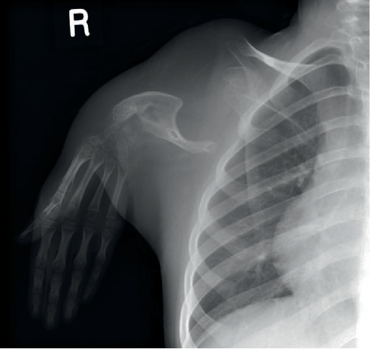
Fig. 73.46 Phocomelia - upper limb anomalies secondary to toxic effects of thalidomide. (Grainger & Allison's Diagnostic Radiology)
Causes
1. Thalidomide (most notorious/classical cause)
Thalidomide was a sedative and antiemetic prescribed to pregnant women in the late 1950s-early 1960s, primarily in West Germany and the UK. It caused a catastrophic epidemic of phocomelia:
- 60% of infants born to mothers who took thalidomide between days 38 and 54 after conception developed this deformity.
- The critical window is weeks 4-7 of gestation - exactly when the arms and legs are forming (see developmental timeline below).
- The anomaly was previously extremely rare (0.8% of all congenital hand malformations) but became common during the thalidomide epidemic.
- Thalidomide was withdrawn from the market in 1961 precisely because of these effects.
Mechanism of thalidomide teratogenicity (multifactorial):
- Anti-angiogenic effect - thalidomide inhibits new blood vessel formation (VEGF-mediated angiogenesis), destroying the immature capillary network supplying the developing limb bud
- Oxidative stress - generates reactive oxygen species that damage DNA in limb bud cells
- Cereblon binding - thalidomide binds the E3 ubiquitin ligase substrate receptor CRBN, disrupting the ubiquitination and degradation of transcription factors (SALL4, IKZF1/3) critical for limb development
- FGF/Wnt signaling disruption - interferes with fibroblast growth factor signaling essential for limb outgrowth
The critical teratogenic window for limb development (weeks 4-7):
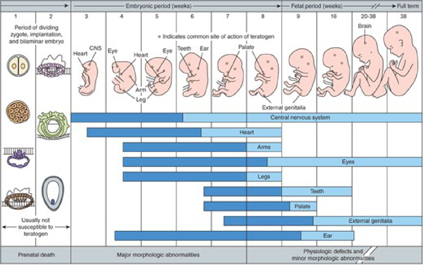
Schematic of critical periods of human development - arms and legs are maximally sensitive to teratogens during weeks 4-7. (Katzung's Basic & Clinical Pharmacology, 16th Ed.)
2. Genetic Syndromes
Several genetic syndromes list phocomelia as a feature. The key ones include:
| Syndrome | Inheritance | Key Features |
|---|---|---|
| Roberts syndrome | Autosomal recessive | Phocomelia of all 4 limbs, cleft lip/palate, prenatal growth retardation, chromosomal heterochromatin repulsion (ESCO2 gene mutation) |
| SC phocomelia syndrome | Autosomal recessive | Variant of Roberts syndrome; milder limb reduction, corneal clouding, hemangiomas |
| Al-Awadi/Raas-Rothschild/Schinzel phocomelia syndrome | Autosomal recessive | Severe limb reduction + genitourinary/cardiac defects (WNT7A gene) |
| Grebe syndrome | Autosomal recessive | Severe acromesomelic shortening of limbs (CDMP1/GDF5 gene) |
| Cornelia de Lange syndrome | Autosomal dominant (NIPBL mutations) | Limb reduction defects including phocomelia in severe forms, distinctive facies, growth retardation |
| Fuhrmann syndrome | Autosomal recessive | Aplasia/hypoplasia of fibula, femoral bowing, poly/oligodactyly |
| Holt-Oram syndrome | Autosomal dominant (TBX5 gene) | Radial ray defects, atrial/ventricular septal defects |
| Fanconi anemia | Autosomal recessive | Radial ray hypoplasia, aplastic anemia, cancer predisposition |
Also associated with chromosomal anomalies: trisomy 18, trisomy 13, and other aneuploidies.
3. Other Teratogens (rare reports)
- Misoprostol (prostaglandin E1 analog used as an abortifacient) - associated with limb reduction defects including transverse deficiencies
- Cocaine - vasoconstriction causing limb ischemia
- Maternal diabetes - associated with various limb defects (caudal regression, limb reduction)
- Warfarin embryopathy - stippled epiphyses and nasal hypoplasia; occasionally limb defects
- Organic solvents / environmental exposures - weak evidence
4. Vascular Disruption Sequence
Isolated (sporadic) phocomelia may result from a vascular disruption event in utero - an interruption of blood supply to the developing limb bud leading to tissue infarction and resorption. This can occur without a teratogen or identifiable genetic cause.
Associated Features (non-thalidomide phocomelia)
Half of all patients with phocomelia have additional anomalies:
- Radial ray deficiencies
- Cleft lip and palate (especially Roberts syndrome)
- Scoliosis
- Cardiac defects
- Skin and calcification aberrations
- Chromosomal abnormalities
Anatomically, the clavicle and scapula are always present. There is no true elbow joint, and the hand typically has only 3-4 digits with an absent thumb.
Classification (Frantz & O'Rahilly)
| Type | Description |
|---|---|
| Complete phocomelia | All proximal and intermediate limb bones absent - hand/foot directly on trunk |
| Proximal phocomelia | Proximal bone (humerus/femur) absent - forearm/leg + hand/foot present |
| Distal phocomelia | Intermediate bone (radius-ulna/tibia-fibula) absent - hand/foot attached to humerus/femur |
Summary of causes - mnemonic TGVI:
- Thalidomide (classic teratogen, critical window weeks 4-7)
- Genetic syndromes (Roberts, SC phocomelia, Al-Awadi, Cornelia de Lange, etc.)
- Vascular disruption (sporadic in utero ischemia)
- Idiopathic / chromosomal (trisomies, unclassified)
Sources: Campbell's Operative Orthopaedics 15th Ed 2026, Ch. 85; Katzung's Basic & Clinical Pharmacology 16th Ed, Ch. 59; Grainger & Allison's Diagnostic Radiology, Ch. 73
Placenta and cord disorders
Placental and cord disorders are a major category in obstetrics and perinatal pathology. Key placental disorders include placenta previa, placental abruption, placenta accreta spectrum, and molar pregnancy. Cord disorders include velamentous insertion, vasa previa, nuchal cord, and true/false knots. Let me pull comprehensive textbook content on all of these.
Searching sources...
Reading File
Finding Sources
Finding Sources
Searching the Web
placenta and cord disorders obstetrics overview classification 2024
Let me search more specifically in the library.
Finding Sources
Finding Sources
Finding Sources
Finding Sources
Finding Sources
Now let me read the main content sections in parallel.
Reading File
Reading File
Reading File
Excellent content. Now let me read the accreta/percreta section and the cord pathology section in parallel.
Reading File
Reading File
Reading File
Excellent rich content. Let me also read the accreta/abruption from the radiology book and Creasy's abruption section, plus fetch the PAS ultrasound image.
Reading File
Reading File
Loading Image
I now have comprehensive, well-sourced content across all major categories. Let me compile the full answer.
Placenta and Cord Disorders
PART A: PLACENTAL DISORDERS
1. Placenta Previa
Definition: Implantation of the placenta over or adjacent to the internal cervical os.
Classification (current terminology - "partial/marginal/complete" are no longer standard):
- Low-lying placenta - placental edge is within 2 cm of the internal os but does not cover it
- Placenta previa - placenta covers the internal os (formerly called "complete" or "total" previa)
- If the placental edge is ≤1 cm from the os or covers it, caesarean section is generally recommended
Incidence: ~1 in 200-250 pregnancies
Risk factors:
- Advanced maternal age
- Multiparity
- Prior caesarean section / uterine surgery
- Prior placenta previa
- Uterine abnormalities
- Multiple gestation
- Tobacco use
Clinical features:
- Painless, bright-red vaginal bleeding in the second/third trimester - classic presentation
- Average first bleed at 27-32 weeks
- ~20% have some uterine irritability
- Abnormal fetal lie (transverse, breech) due to lower uterine placenta
- Bleeding is initially self-limited but recurs; central previa can cause exsanguination
Important: Digital or instrumental cervical examination is absolutely contraindicated until placenta previa is excluded by ultrasound - it can precipitate catastrophic hemorrhage.
"Placental migration": 90% of second-trimester previas diagnosed on US resolve by term as the lower uterine segment elongates, drawing the placenta away from the os. Only ~20% of previas reaching term are central/complete.
Diagnosis:
- Transvaginal ultrasound (TVUS) - preferred, safe, most accurate
- Transabdominal US - 93-98% accurate; empty bladder required (full bladder over-diagnoses previa)
- MRI - useful in equivocal cases or when PAS is suspected
Management:
- Immediate obstetric consultation; two large-bore IV lines, fluid resuscitation
- Continuous fetal monitoring
- CBC, coagulation studies (fibrinogen <300 mg/dL = significant coagulopathy), type and crossmatch
- Rh-negative patients: Rh immune globulin 300 µg within 72 hours
- Expectant management if not in labor and hemodynamically stable
- Caesarean delivery if persistent, bleeding, or at term
2. Placental Abruption (Abruptio Placentae)
Definition: Premature separation of a normally implanted placenta from the uterine wall before delivery of the fetus.
Incidence: ~1% of pregnancies; most frequent between 24-26 weeks
Risk factors:
- Hypertension (most common)
- Trauma (including domestic violence)
- Advanced maternal age
- Cocaine/tobacco use
- Prior abruption
- Preterm premature rupture of membranes
Types by bleeding pattern:
| Type | Description |
|---|---|
| Revealed (external) | Blood escapes through the cervix - visible vaginal bleeding |
| Concealed | Blood collects behind the placenta, no external bleeding; may be more severe |
| Mixed | Elements of both |
Clinical features:
- Painful vaginal bleeding (contrast with previa = painless)
- Uterine tenderness and rigidity ("woody" uterus)
- Fetal distress or intrauterine fetal death
- Coagulopathy/DIC (especially concealed abruption with large retroplacental clot)
- Couvelaire uterus (uterine apoplexy) - hemorrhage dissects into myometrium
Diagnosis:
- Primarily clinical - USS has only 25% sensitivity for abruption; ultrasound cannot exclude abruption
- US may show retroplacental hematoma (if zone >2 cm, suspicious)
- CT: full-thickness areas of low attenuation making an acute angle with the myometrium
- MRI: more accurate than US, limited routine use
Management:
- Hemodynamic stabilization
- Emergency caesarean if fetal distress or maternal instability
- Coagulopathy correction (FFP, cryoprecipitate, platelets)
- Monitor for DIC
- Kleihauer-Betke test for feto-maternal hemorrhage
3. Placenta Accreta Spectrum (PAS)
Abnormal invasion of placental villi into the myometrium due to deficient decidua basalis. PAS is not homogeneous - foci of accreta, increta, and percreta can coexist in the same placenta.
Graded spectrum:
| Type | Definition | Histology |
|---|---|---|
| Placenta accreta | Placenta and myometrium in direct contact (no decidua) | Villi attached directly to superficial myometrium |
| Placenta increta | Placenta invades into the myometrium | Villi penetrate myometrial fibres |
| Placenta percreta | Placenta breaches uterine serosa; may invade bladder, bowel | Villi penetrate through serosa |
Risk factors:
- Previous caesarean section (most important - risk rises with each CS)
- Placenta previa overlying a uterine scar
- Prior myomectomy or uterine curettage
- Advanced maternal age
Mortality: Overall 3%; rises to 20% if bladder is involved (percreta). Leading causes of death: massive hemorrhage, DIC.
Ultrasound features (sensitivity 90.7%, specificity 96.9%):
| PAS Type | Key US Feature |
|---|---|
| Accreta | Bridging vessels (71%), "tornado vessels" with multidirectional flow |
| Increta | Loss of retroplacental clear zone (62-85%) |
| Percreta | Placental lacunae (82%) ± turbulent flow; subplacental hypervascularity; bladder-uterine interface irregularity |
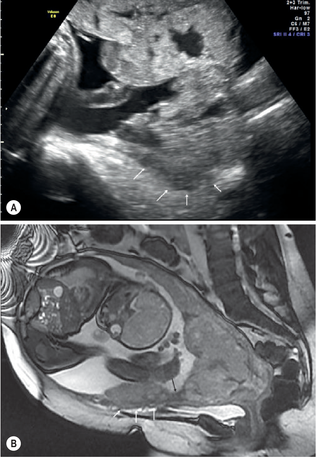
Fig. 35.53 - (A) Transvaginal US showing irregularity of the uterine-placental interface (white arrows) indicating PAS. (B) Sagittal T2 MRI showing low-lying placenta with low T2 placental bands (black arrow) and myometrial irregularity consistent with placenta increta (white arrow). (Grainger & Allison's Diagnostic Radiology)
MRI features: Heterogeneous placental signal; uterine bulge with myometrial thinning <1 mm or focal interruption; low T2 intraplacental bands; disorganized placental vasculature; bladder tenting or invasion. MRI is preferred when posterior placenta or US is inconclusive.
Management:
- Pre-delivery diagnosis allows planned caesarean hysterectomy (reduces morbidity/mortality)
- Temporary aortic or iliac artery balloon occlusion during surgery
- Multidisciplinary team: maternal-fetal medicine, urology, interventional radiology, blood bank
- Minimally invasive accreta may be removed manually/by curettage; increta/percreta usually require hysterectomy
4. Vasa Previa
Definition: Fetal blood vessels unsupported by the umbilical cord or placental tissue traverse the fetal membranes over the internal cervical os, lying below the presenting fetal part.
Association: Almost always coexists with velamentous cord insertion; also associated with bilobed or succenturiate-lobed placentas where vessels run between lobes.
Risk: When membranes rupture (spontaneously or artificially), these unprotected vessels tear → acute fetal exsanguination. Fetal mortality 33-100% if undiagnosed.
Diagnosis:
- Color Doppler US: hypoechoic vessels overlying internal os with flow on Doppler; they do not change position with maternal repositioning (differentiates from a cord loop)
- Clinically: pulsatile vessels palpated ahead of presenting part on digital exam; sinusoidal FHR pattern after membrane rupture; APH with sudden fetal bradycardia
Management:
- Elective caesarean section before labor at 35-36 weeks if diagnosed antenatally
- Emergency caesarean if membranes rupture with vasa previa - immediate neonatal resuscitation and transfusion
5. Molar Pregnancy / Gestational Trophoblastic Disease
(Covered separately - involves abnormal placental villous development)
6. Retained Products of Conception (RPOC)
Residual placental tissue remaining in the uterine cavity after miscarriage, termination, or delivery - especially with abnormal placental morphology or unsuspected PAS.
- Complications: Postpartum hemorrhage, infection, Asherman syndrome
- US: Heterogeneous echogenic mass with vascularity on Doppler (vs. intrauterine hematoma which is avascular)
- CT: Heterogeneous enhancing mass in uterine cavity
- Management: Surgical evacuation (suction curettage) or medical management (misoprostol)
PART B: UMBILICAL CORD DISORDERS
Cord abnormalities account for 10% of stillbirths.
1. Abnormal Cord Insertion
| Type | Definition | Incidence | Clinical Significance |
|---|---|---|---|
| Normal (central) | Inserts on the central portion of the placenta | ~80% | - |
| Marginal (battledore) | Inserts within 2 cm of placental edge | <10% singletons; 25-33% twins | Generally benign; FGR risk |
| Velamentous | Cord inserts on the fetal membranes; vessels travel unprotected through membranes before entering placenta | 1% singletons; 15% monochorionic twins | Hemorrhage, FGR, vasa previa |
| Furcate | Vessels lose Wharton's jelly protection before entering chorionic plate; they splay widely | Rare | Exposed vessels susceptible to trauma, rupture, torsion, and stillbirth |
Velamentous insertion is the most clinically dangerous cord insertion anomaly - the unprotected membrane vessels are susceptible to compression, rupture, and vasa previa formation.
2. Cord Prolapse
Definition: The umbilical cord presents in advance of the fetal presenting part after rupture of membranes (overt prolapse) or lies alongside the presenting part (occult prolapse).
Risk factors:
- Abnormal fetal presentation (breech, transverse)
- Prematurity
- Multiparity
- Polyhydramnios
- Obstetric manipulation (amniotomy, version)
- Abnormally long umbilical cord
Clinical features: Cord visible/palpable at or outside the cervix; sudden severe fetal bradycardia after membrane rupture
Management: Obstetric emergency - relieve cord compression by manually elevating the presenting part, position mother in knee-chest or Trendelenburg, urgent caesarean delivery.
3. Nuchal Cord
- Cord wrapped around the fetal neck
- Occurs in up to 30% of uncomplicated deliveries (single nuchal cord in 23.6%, multiple cords in 3.7%)
- Generally not an independent cause of stillbirth - isolated nuchal cord at birth without histopathologic evidence of cord occlusion is insufficient to attribute adverse outcome to it
- Tight nuchal cord with evidence of cord occlusion, fetal hypoxia, and histopathologic thrombosis is a recognized cause of perinatal death
4. True Knots
- Occur when the fetus passes through a loop of cord
- More common with long cords, polyhydramnios, active fetuses
- Common incidentally at birth; a "tight" knot causing stillbirth should show grooving of the cord, constriction of vessels, edema, congestion, or thrombosis to be considered causative
- Isolated finding of a true knot at birth is insufficient to diagnose cord accident as cause of stillbirth without pathologic evidence of occlusion
5. Cord Torsion
- Most often at the fetal end of the cord
- Pathological torsion: congested, edematous cord, often with thrombosis of cord vessels
- Can cause fetal death
6. Cord Hematoma, Stricture, and Rupture
- Hematoma: Usually at fetal end; spontaneous or traumatic; associated with FGR and fetal death
- Stricture: Focal narrowing of cord; rare; associated with torsion and fetal death
- Rupture: Extremely rare; associated with abnormally short/long cords or varices; can cause rapid fetal exsanguination
7. Single Umbilical Artery (Two-Vessel Cord)
- Normal cord has 2 arteries + 1 vein
- Single umbilical artery occurs in ~1% of pregnancies
- Associated with congenital anomalies (renal, cardiac, chromosomal), FGR
- Isolated finding (no other anomalies on detailed scan) carries low risk
Summary Table
| Condition | Key Feature | Classic Presentation | Main Risk |
|---|---|---|---|
| Placenta previa | Placenta over os | Painless antepartum bleeding | Hemorrhage, emergency CS |
| Placental abruption | Premature separation | Painful APH + uterine rigidity | DIC, fetal death |
| Placenta accreta | Absent decidua, myometrial attachment | Retained placenta at delivery | Catastrophic PPH |
| Placenta increta | Myometrial invasion | As above | PPH, hysterectomy |
| Placenta percreta | Serosal/bladder breach | As above + organ invasion | 20% maternal mortality if bladder |
| Vasa previa | Vessels over os in membranes | APH + sudden fetal bradycardia at ROM | 33-100% fetal mortality |
| Velamentous cord | Vessels in membranes | FGR, vasa previa | Hemorrhage, stillbirth |
| Cord prolapse | Cord ahead of presenting part | Bradycardia after ROM | Fetal asphyxia |
| True knot (tight) | Cord knot with vessel constriction | Usually found at delivery | Stillbirth |
| Single umbilical artery | 1 artery + 1 vein | Incidental on US | Fetal anomalies, FGR |
Sources: Rosen's Emergency Medicine 10th Ed, Ch. on Antepartum Hemorrhage; Creasy & Resnik's Maternal-Fetal Medicine, Ch. 41 (Umbilical Cord Pathology); Grainger & Allison's Diagnostic Radiology, Ch. 35 (Placenta & Cord); Textbook of Family Medicine 9th Ed, Ch. 20
Battledore placenta
Battledore placenta refers to a marginal cord insertion - where the umbilical cord inserts at the very edge (margin) of the placenta, resembling a battledore (an old paddle-shaped racket used in badminton). It occurs in about 7-9% of singleton pregnancies. It is generally considered a benign variant but may be associated with FGR and preterm delivery.
Searching sources...
Finding Sources
Searching the Web
battledore placenta marginal cord insertion clinical significance complications
Finding Sources
Finding Sources
Good - I have image URLs and file locations. Let me read the key sections and fetch the ultrasound images in parallel.
Reading File
Reading File
Loading Image
Loading Image
Now let me read the detailed Creasy & Resnik chapter on cord insertion types and clinical management.
Reading File
Reading File
I now have all the content needed. Let me also read the key clinical findings section just before management.
Reading File
I have thorough information. Here is the complete answer:
Battledore Placenta
Definition and Etymology
Battledore placenta is a placenta with marginal cord insertion - the umbilical cord inserts at or within the margin (edge) of the placenta, rather than in the center. The name comes from the resemblance to a battledore - the old paddle-shaped racket used in early badminton - where the placenta is the paddle and the cord is the handle attached at its edge.
The term marginal insertion is formally defined as cord insertion within 2 cm of the placental edge (but the cord still enters the placental disc, not the membranes beyond it - which would be velamentous insertion).
Comparison of Cord Insertion Types
| Type | Definition | Incidence (Singleton) | Clinical Risk |
|---|---|---|---|
| Central | Inserts into central portion of disc | ~80% | Lowest |
| Eccentric | Inserts off-center but >2 cm from edge | ~10% | Minimal |
| Marginal (Battledore) | Inserts within 2 cm of placental edge | 7-10% | Mild |
| Velamentous | Cord inserts into membranes beyond placental edge | 1-3% | High (vasa previa risk) |
Ultrasound Appearance
The cord insertion site is identified using color Doppler and gray-scale imaging in two right-angle planes. In battledore placenta, the vessels are seen arriving at the very margin of the placental disc.
Sagittal view - marginal cord insertion (battledore):
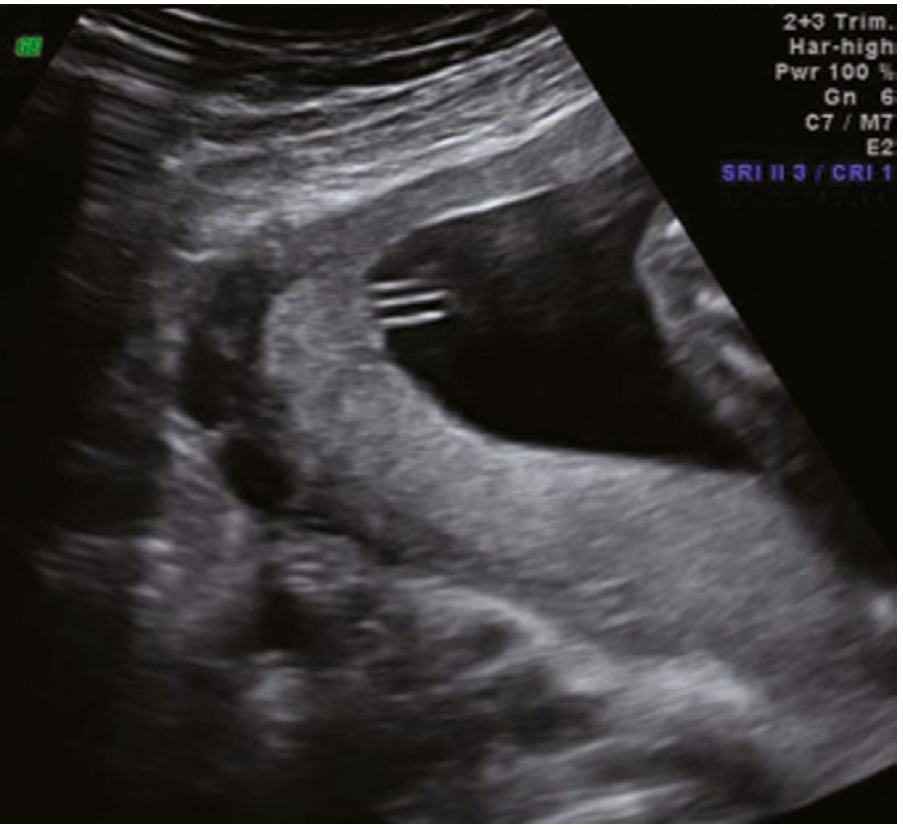
Figure 27.3 - Sagittal view of the posterior placenta showing a vascular structure near the upper placental margin. (Creasy & Resnik's Maternal-Fetal Medicine)
Color Doppler - marginal cord insertion:
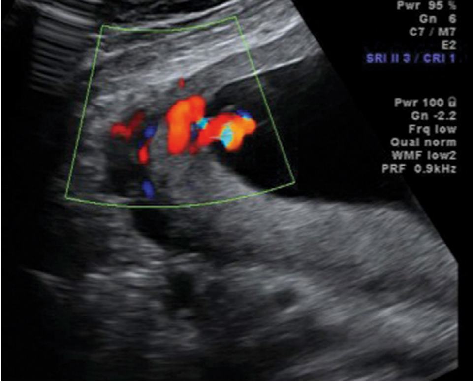
Figure 27.4 - Color Doppler image demonstrating umbilical cord insertion at the placental margin. (Creasy & Resnik's Maternal-Fetal Medicine)
Key differential from velamentous insertion: To confirm marginal (not velamentous) insertion, there should be no segment of umbilical vessels traveling through the fetal membranes beyond the placental edge. If at least 1 cm of vessels are seen traveling in the membranes beyond the edge, the diagnosis is velamentous, not marginal.
Incidence
- Singletons: <10% (approximately 7-9%)
- Twin gestations: 20-33% - significantly higher, especially in the smaller of the twins (the smaller twin typically has the peripheral - marginal or velamentous - insertion)
- More common when there is placenta previa
Pathogenesis
The developmental dynamics governing where the umbilical vessels insert as the placental disc expands are poorly understood. Based on first-trimester ultrasound observations, the cord insertion position appears to be established very early in pregnancy. Differential growth of the placental disc may result in a cord that was originally more central becoming relatively marginal over time.
Clinical Significance
Generally Favorable Prognosis
Central and marginal (battledore) cord insertion have no established association with abnormal fetal outcome when occurring in isolation in singleton pregnancies. Creasy & Resnik's states directly: "Central and marginal PCI have no associations with abnormal fetal outcome."
Potential Associations (mild/context-dependent)
- Fetal growth restriction (FGR) - marginal insertion is associated with slightly reduced placental efficiency; the smaller twin with peripheral cord insertion in discordant twins shows this most clearly
- Preterm premature rupture of membranes (PPROM) and preterm birth - velamentous and, to a lesser extent, marginal cord insertions are associated with increased risk of PROM, preterm PROM, and preterm birth
- Postpartum hemorrhage - marginal umbilical cord insertion is listed as a risk factor for PPH (Tintinalli's Emergency Medicine)
- Fetal distress in labor - some association reported, though weak in isolation
- Cord prolapse - theoretically higher risk if cord is eccentric/marginal with less secure anchoring
NOT associated with:
- Fetal structural anomalies
- Vasa previa (this is a risk of velamentous insertion, not marginal - an important distinction)
Differentiating Battledore from Velamentous Insertion
This distinction is clinically important because velamentous insertion carries a much higher risk:
| Feature | Battledore (Marginal) | Velamentous |
|---|---|---|
| Cord entry point | At the placental disc edge | Into the fetal membranes, beyond disc edge |
| Vessels in membranes | No (or <1 cm) | Yes - vessels travel unprotected in membranes |
| Wharton's jelly protection | Present up to the placenta | Lost in the membrane segment |
| Vasa previa risk | No | Yes (if vessels overlie internal os) |
| FGR risk | Mild | Moderate |
| Fetal mortality risk | Minimal | High if vasa previa ruptures |
Management
- Battledore placenta in isolation (singleton): No specific obstetric intervention required beyond standard care. Normal mode of delivery is appropriate.
- Serial growth ultrasounds are indicated when marginal insertion occurs in twin gestations (where growth discordance is more likely).
- Exclude vasa previa: When marginal or peripheral cord insertion is found, color Doppler should confirm that no vessels course across the internal cervical os.
- At delivery: Be aware of potential cord avulsion during placental delivery (applies more to velamentous than marginal, but relevant with any peripheral insertion).
Summary
Battledore placenta (marginal cord insertion) is a common, usually benign anatomical variant where the umbilical cord attaches at the edge of the placenta. It is clinically significant mainly in the context of twin pregnancies (contributing to growth discordance) and as a risk factor for postpartum hemorrhage and preterm birth. It does not carry the high-risk implications of velamentous insertion or vasa previa. The diagnosis is made on ultrasound using color Doppler, and the key clinical task is to distinguish it from velamentous insertion and confirm the absence of vasa previa.
Sources: Creasy & Resnik's Maternal-Fetal Medicine, Ch. 27 (Marginal and Velamentous Umbilical Cord Insertion); Grainger & Allison's Diagnostic Radiology, Ch. 35 (Table 35.4 - Anomalous Cord Insertions); Tintinalli's Emergency Medicine (Table 101-7 - PPH Risk Factors)
This is a shared conversation. Sign in to Orris to start your own chat.