I now have sufficient content from the textbooks to write a comprehensive, well-structured 10-mark exam answer on the cerebrum. Let me compile it:
THE CEREBRUM
Sources: Neuroanatomy through Clinical Cases 3rd Ed. | Guyton & Hall Textbook of Medical Physiology | Neuroscience: Exploring the Brain 5th Ed.
1. EXTERNAL FEATURES
The cerebrum is the largest and most rostral part of the brain, forming the bulk of the central nervous system. Its key external features are:
Size and Shape:
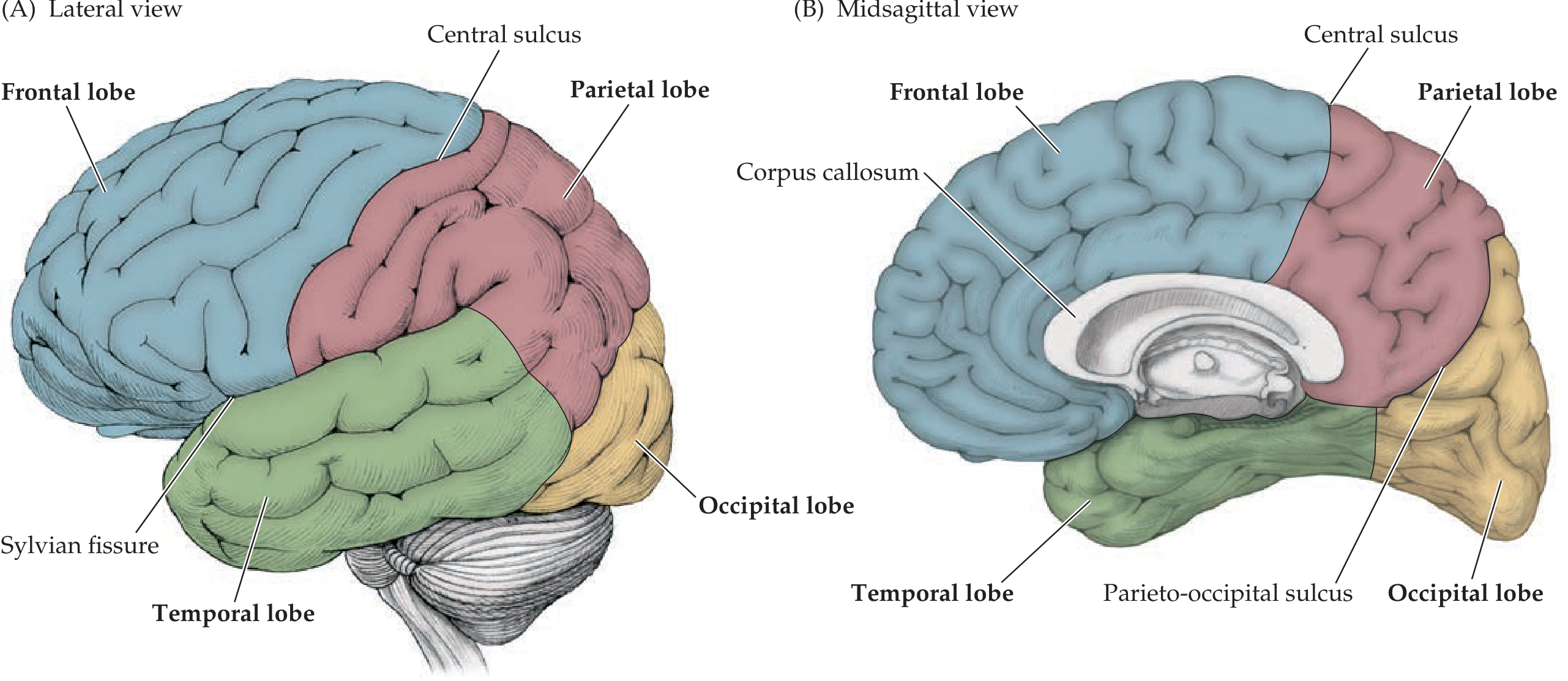
The cerebrum is divided into two cerebral hemispheres by the deep interhemispheric (longitudinal/sagittal) fissure running in the midline. Each hemisphere has three surfaces: superolateral, medial, and inferior (base).
Gyri and Sulci:
The surface of the cerebrum is thrown into folds called gyri (ridges) separated by grooves called sulci. Deeper grooves are termed fissures. This folding dramatically increases the total cortical surface area to approximately 0.25 m² despite fitting within the skull. The total cortex is 2-5 mm thick.
Major Fissures and Sulci:
| Sulcus / Fissure | Location | Significance |
|---|
| Longitudinal (interhemispheric) fissure | Midline | Separates two hemispheres |
| Central sulcus (of Rolando) | Between frontal and parietal lobes | Separates motor from sensory cortex |
| Lateral (Sylvian) fissure | Between frontal/parietal and temporal lobes | One of the deepest sulci |
| Parieto-occipital sulcus | Medial surface | Separates parietal from occipital lobe |
| Calcarine sulcus | Medial occipital lobe | Contains primary visual cortex |
Named Gyri:
- Precentral gyrus - immediately anterior to the central sulcus (primary motor cortex)
- Postcentral gyrus - immediately posterior to central sulcus (primary somatosensory cortex)
- Superior, middle, and inferior frontal gyri - lateral frontal surface
- Superior, middle, and inferior temporal gyri - lateral temporal surface
- Supramarginal gyrus - surrounds the end of the Sylvian fissure
- Angular gyrus - surrounds the end of the superior temporal sulcus
Corpus Callosum:
The two hemispheres are connected by a large, C-shaped band of white matter called the corpus callosum, visible on the medial (midsagittal) view.
Insula:
A fifth lobe of cortex buried deep within the Sylvian fissure, covered by the frontal operculum anteriorly and parietal operculum posteriorly.
2. LOBES OF THE CEREBRUM
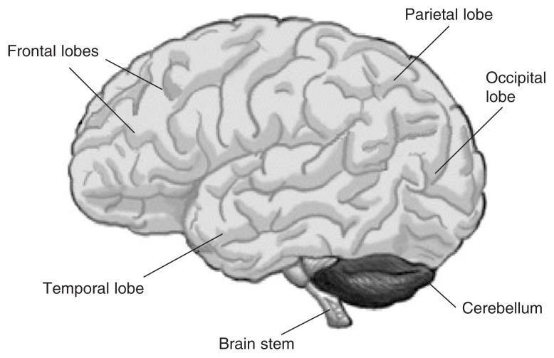
The cerebral cortex is conventionally divided into four main lobes, named after the overlying bones of the skull:
(A) Frontal Lobe
- Boundaries: Anteriorly from the frontal pole; posteriorly bounded by the central sulcus; inferolaterally by the Sylvian fissure.
- Key Gyri: Precentral gyrus, superior/middle/inferior frontal gyri.
- Brodmann Areas (BA): BA 4 (primary motor cortex), BA 6 (premotor/supplementary motor cortex), BA 44/45 (Broca's area in dominant hemisphere), BA 9/10/11/12 (prefrontal association cortex).
(B) Parietal Lobe
- Boundaries: Anteriorly by the central sulcus; antero-inferiorly by the Sylvian fissure; no sharp boundary with occipital and temporal lobes laterally (demarcated by parieto-occipital sulcus on the medial surface).
- Key Gyri: Postcentral gyrus, superior parietal lobule, inferior parietal lobule (supramarginal + angular gyri).
- Brodmann Areas: BA 1, 2, 3 (primary somatosensory cortex), BA 5, 7 (parietal association areas).
(C) Temporal Lobe
- Boundaries: Superiorly bounded by the Sylvian fissure; no distinct posterior boundary with occipital lobe laterally.
- Key Gyri: Superior, middle, and inferior temporal gyri; Heschl's gyri (transverse temporal gyri, buried in superior temporal sulcus = primary auditory cortex).
- Brodmann Areas: BA 41/42 (primary auditory cortex), BA 22 (Wernicke's area in dominant hemisphere), BA 28 (entorhinal cortex), hippocampus and amygdala on medial surface.
(D) Occipital Lobe
- Boundaries: Posterior to the parieto-occipital sulcus and calcarine fissure.
- Key Gyri: Cuneus (above calcarine sulcus), lingual gyrus (below).
- Brodmann Areas: BA 17 (primary visual cortex, striate cortex, in banks of calcarine fissure), BA 18, 19 (visual association cortex).
3. FUNCTIONS
Frontal Lobe
| Region | Function |
|---|
| Primary motor cortex (precentral gyrus, BA 4) | Voluntary movement of contralateral body - homunculus representation |
| Premotor cortex (BA 6) | Motor planning and coordination of complex movements |
| Broca's area (BA 44/45, dominant hemisphere) | Motor speech production (expressive language) |
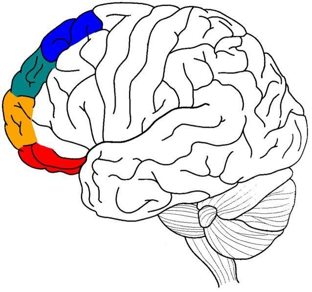
| Prefrontal cortex (BA 9, 10, 11, 46) | Higher cognitive functions - judgment, personality, working memory, attention, planning, social behavior |
| Frontal eye fields (BA 8) | Voluntary saccadic eye movements |
Parietal Lobe
| Region | Function |
|---|
| Primary somatosensory cortex (postcentral gyrus, BA 1, 2, 3) | Conscious perception of touch, pressure, pain, temperature, proprioception from contralateral body |
| Superior parietal lobule (BA 5, 7) | Stereognosis, visuomotor integration, spatial perception |
| Inferior parietal lobule | Body schema, reading, writing, arithmetic (dominant side); visuospatial attention (non-dominant) |
Temporal Lobe
| Region | Function |
|---|
| Primary auditory cortex (Heschl's gyri, BA 41/42) | Conscious auditory perception |
| Wernicke's area (BA 22, dominant hemisphere) | Comprehension of spoken and written language |
| Inferior temporal cortex (BA 20, 21) | Object recognition, face recognition |
| Medial temporal lobe (hippocampus, parahippocampal gyrus) | Declarative memory formation and retrieval |
| Amygdala | Emotional processing, fear conditioning |
Occipital Lobe
| Region | Function |
|---|
| Primary visual cortex (BA 17, striate cortex) | Conscious visual perception; receives input from contralateral visual field |
| Visual association cortex (BA 18, 19) | Visual processing - depth, color, motion, form recognition |
| Dorsal stream ("where" pathway) | Spatial location and visuomotor guidance |
| Ventral stream ("what" pathway) | Object and face identification |
Cortical neuron types: The cortex contains granular (stellate) cells, fusiform cells, and pyramidal cells. Incoming sensory signals terminate in layer IV; output signals arise from layers V and VI (Guyton & Hall, p. 716). The cortex contains over 80 billion neurons with an elaborate laminar organization.
4. APPLIED ANATOMY
Cerebrovascular Disease (Stroke)
- Middle cerebral artery (MCA) territory - most commonly affected; involves lateral frontal, parietal, and temporal lobes → contralateral hemiplegia (arm > leg), contralateral hemisensory loss, homonymous hemianopia. Dominant hemisphere: aphasia (Broca's or Wernicke's). Non-dominant: hemispatial neglect.
- Anterior cerebral artery (ACA) territory - medial frontal and parietal lobes → contralateral weakness/numbness of leg > arm, personality change, urinary incontinence.
- Posterior cerebral artery (PCA) territory - occipital lobe → contralateral homonymous hemianopia (with macular sparing if collateral supply preserved).
Frontal Lobe Lesions
- Broca's aphasia - damage to BA 44/45 (dominant hemisphere) → non-fluent, effortful speech with intact comprehension.
- Prefrontal lesions - personality change, disinhibition, poor judgment (classic: Phineas Gage; frontal lobe tumors such as meningioma).
- Frontal lobe epilepsy - focal seizures, often with adversive head/eye turning (frontal eye fields).
Parietal Lobe Lesions
- Dominant parietal lesion → Gerstmann's syndrome: finger agnosia, agraphia, acalculia, left-right disorientation; also conduction aphasia.
- Non-dominant parietal lesion → hemispatial neglect (ignoring contralateral space), constructional apraxia, dressing apraxia, anosognosia.
- Astereognosis - inability to identify objects by touch despite intact sensation; indicates parietal association area damage.
Temporal Lobe Lesions
- Wernicke's aphasia - dominant superior temporal gyrus lesion → fluent but paraphasic speech, poor comprehension ("word salad").
- Bilateral medial temporal lobe damage (hippocampus) - severe anterograde amnesia (classic: patient H.M.).
- Temporal lobe epilepsy - most common form of focal epilepsy; associated with mesial temporal sclerosis; complex partial seizures with automatisms (lip smacking, fumbling), preceded by aura (olfactory hallucinations - "uncinate fits").
- Bilateral superior temporal lesion - cortical deafness.
Occipital Lobe Lesions
- Unilateral occipital lesion → contralateral homonymous hemianopia (macular vision may be spared due to dual blood supply).
- Bilateral occipital lesions → cortical blindness (pupils react normally to light, distinguishing from optic nerve/chiasm lesions).
- Visual agnosia - lesion in visual association cortex; patient can see but cannot recognize objects.
- Prosopagnosia - inability to recognize faces; bilateral fusiform/inferior occipitotemporal lesions.
Raised Intracranial Pressure (ICP) and Herniation
- Expanding cerebral masses (tumors, hemorrhage, edema) can cause transtentorial (uncal) herniation of the medial temporal lobe over the tentorium cerebelli → compression of CN III (ipsilateral dilated pupil, "blown pupil") and the cerebral peduncle → contralateral hemiplegia (or ipsilateral due to Kernohan's notch).
Cortical Representation - Homunculus
- The motor homunculus (precentral gyrus) and sensory homunculus (postcentral gyrus) provide a topographic, distorted map of the body, with the face and hands having disproportionately large representations reflecting their high density of innervation.
- Clinically: a cortical stroke affecting the leg area (paracentral lobule, medial surface) mimics a spinal cord lesion - important to distinguish by imaging.
Summary Table
| Lobe | Key Function | Lesion Sign |
|---|
| Frontal | Motor, speech production, cognition | Hemiplegia, Broca's aphasia, personality change |
| Parietal | Somatosensory, spatial awareness | Sensory loss, Gerstmann's, neglect |
| Temporal | Auditory, memory, language comprehension | Wernicke's aphasia, amnesia, temporal lobe epilepsy |
| Occipital | Vision | Homonymous hemianopia, cortical blindness |
- Neuroanatomy through Clinical Cases 3rd Ed., pp. 50-56 | Guyton & Hall Medical Physiology, p. 716 | Neuroscience: Exploring the Brain 5th Ed., p. 574