
Why we give episiotomy
Reading File
Episiotomy is a surgical incision made in the perineum during childbirth to enlarge the vaginal opening. It is given to facilitate delivery and prevent uncontrolled perineal tears.
Searching sources...
Finding Sources
Searching Images
episiotomy types midline mediolateral perineum diagram
Reading File
Reading File
Reading File
Loading Image
Here is a complete, textbook-based answer on episiotomy:
Episiotomy - Definition, Indications, and Types
An episiotomy is a surgical incision in the perineum made to enlarge the vaginal introitus during the second stage of labor, facilitating delivery of the baby's head. It is one of the most commonly performed obstetric procedures.
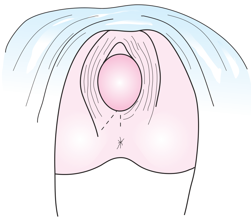
Crowning fetal head distending the perineal tissues. Broken lines depict the location of the incision for a midline (median) or mediolateral episiotomy - Textbook of Family Medicine 9e
Why We Give an Episiotomy (Indications)
Historical / Traditional Indications (now largely disputed)
Historically, episiotomy was done for:
- Substituting an anticipated ragged spontaneous laceration with a more controlled, straight surgical incision
- Shortening the second stage of labor
- Reducing subsequent pelvic floor relaxation and trauma to pelvic musculature
- Protecting the newborn from intracranial hemorrhage
However, evidence since 1980 has not substantiated these benefits. Routine episiotomy has been shown to not prevent perineal damage, pelvic floor relaxation, urinary/fecal incontinence, pelvic organ prolapse, or sexual dysfunction. In fact, in primiparous women, episiotomy is associated with increased 3rd and 4th degree lacerations.
Current Accepted / Selective Indications
Episiotomy is now restricted to specific situations:
| Indication | Rationale |
|---|---|
| Fetal distress / hypoxia | To expedite delivery urgently |
| Shoulder dystocia | To create more room for maneuvers |
| Breech delivery | To facilitate delivery of the after-coming head |
| Occiput-posterior presentation | Larger diameter presents, more room needed |
| Imminent uncontrolled perineal tear | When perineal skin blanches (turns white) - indicating severe stretch with reduced blood flow |
| Instrumental delivery (forceps/vacuum) | To allow space for instruments |
| Rigid or scarred perineum | When spontaneous stretching is inadequate |
| Large baby / macrosomia | To hasten birth and prevent tearing |
| Preterm infant | To reduce trauma to the fragile fetal head |
The primary purpose of episiotomy is to expedite delivery of a baby at risk for hypoxia during the expulsive stage.
Timing
- Early episiotomy: Made before the head crowns (head visible with contractions but recedes between them)
- Timely episiotomy: Made after the head has crowned, when there is maximum tension on the perineal skin - performed when the fetal head distends the perineum to a visible diameter of 3 to 4 cm during a contraction
Types of Episiotomy
| Type | Description | Advantages | Disadvantages |
|---|---|---|---|
| Midline (Median) | Straight incision from posterior vaginal fourchette toward the anus | Easy to repair, heals well, least blood loss, least pain | Risk of extension to 3rd/4th degree laceration (into anal sphincter/rectum) |
| Mediolateral | Oblique incision directed toward the lateral margin of the anal sphincter | Seldom extends into anal sphincter, gains more room | More blood loss, harder to repair, more painful healing, dyspareunia |
| Lateral | Lateral incision from lower third of the vulva | Gains most room | Heaviest bleeding, greatest postpartum complaints, potential anal incontinence - rarely used |
Perineal Laceration Degrees (for context)
| Degree | Extent |
|---|---|
| 1st | Superficial skin/mucosa only |
| 2nd | Mucosa + deeper vaginal/perineal tissues |
| 3rd | Involves anal sphincter |
| 4th | Involves rectal mucosa, transects anal sphincter |
Current Recommendation
Routine episiotomy is no longer recommended. It is an unproven, controversial procedure best restricted to specific fetal and maternal indications listed above. The decision should be individualized to each patient and clinical scenario.
- Textbook of Family Medicine 9e, p. 514
- Roberts and Hedges' Clinical Procedures in Emergency Medicine
- General Anatomy and Musculoskeletal System, THIEME Atlas of Anatomy
Introitus
The introitus (vaginal introitus) is the external opening of the vagina - the entrance to the vaginal canal, located in the vestibule of the vulva. It is partially covered by the hymen in virgins.
Searching sources...
Finding Sources
Finding Sources
Reading File
Finding Sources
Reading File
Finding Sources
Reading File
Reading File
Loading Image
Loading Image
Introitus (Vaginal Introitus)
The introitus (Latin: "entrance") is the external opening of the vagina - the visible entry point into the vaginal canal. It sits within the vestibule, posteriorly to the urethral orifice, and is ringed by the hymen (or its remnants).
Anatomical Location
The introitus is part of the vulva, which is the collective term for all external female genitalia. The vulva contains:
- Mons pubis
- Labia majora and labia minora
- Clitoris
- Vestibule (the space between the labia minora)
- External urethral orifice
- Vaginal introitus (vaginal opening)
- Hymen
- Fourchette (posterior junction of labia minora)
- Perineal body
- Bartholin's glands and Skene's glands
The introitus lies posterior to the external urethral orifice within the vestibule. From this opening, the vagina courses posterosuperiorly through the perineal membrane and into the pelvic cavity.
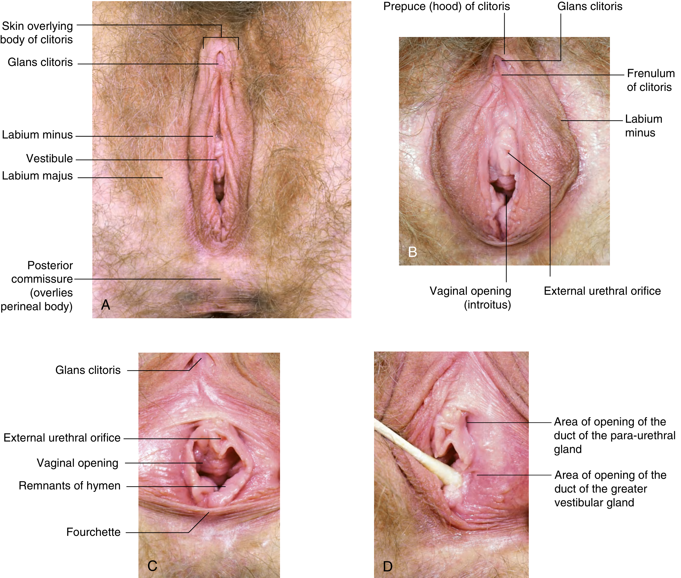
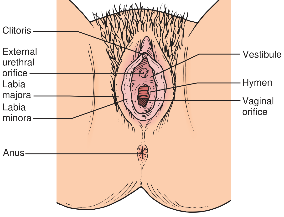
The Hymen
The introitus is ringed by the hymen - a thin mucosal fold that originally closes the vaginal orifice and is usually ruptured during first sexual intercourse. After rupture, remnants (called carunculae myrtiformes) remain around the introitus.
The Vestibule
The vulvar vestibule is the non-keratinized mucosal space between the labia minora that is immediately adjacent to the vaginal introitus. Two important glands open into the vestibule:
| Gland | Location of opening |
|---|---|
| Bartholin's glands (greater vestibular glands) | Skin crease between hymen and labia minora - posterolaterally |
| Skene's glands (para-urethral glands) | Skin crease between urethra and labia minora |
The Vagina (from the introitus upward)
The vagina extends from the introitus at the level of the hymen to the cervix. It is a distensible fibromuscular tube. Its walls:
- Anterior - related to base of bladder and urethra (urethra is embedded in anterior vaginal wall)
- Posterior - related to the rectum
- Superior end - expands into the vaginal vault; the cervix attaches here, creating the fornices (anterior, posterior, and two lateral)
Clinical Relevance
- In episiotomy, the incision enlarges the introitus to allow passage of the fetal head
- The introitus is the point at which the fetal head becomes visible (crowning) during delivery
- Vulvodynia / vestibulodynia - chronic pain at the vestibule/introitus that causes difficulty with intercourse
- Vaginal atresia - absent or stenotic introitus due to failure of vertical fusion
- Gray's Anatomy for Students; Campbell Walsh Wein Urology; Smith and Tanagho's General Urology 19e
This is a shared conversation. Sign in to Orris to start your own chat.