
Answer the questions
python3 -c " import pdfplumber with pdfplumber.open('/tmp/workspace/attachments/ebc2fac4-5ab6-4c04-9ba9-ecdf1a13ff99/Case2 jul27.pdf') as pdf: for i, page in enumerate(pdf.pages): print(f'=== PAGE {i+1} ===') print(page.extract_text()) "
python3 -c " from docx import Document doc = Document('/tmp/workspace/attachments/ebc2fac4-5ab6-4c04-9ba9-ecdf1a13ff99/General Data,cheif complaint,History of present illness..docx') for p in doc.paragraphs: if p.text.strip(): print(f'[{p.style.name}] {p.text}') "
thyroglossal duct cyst anterior neck mass child midline
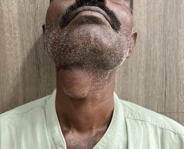
A clinical photograph of an adult male depicting a prominent midline neck mass, consistent with a thyroglossal duct cyst. The patient's neck is slightly extended, revealing a well-circumscribed, ovoid, and globular swelling located in the anterior midline of the neck, specifically at the level of the hyoid bone and inferior to the submental region. The overlying skin is intact and appears unremarkable, without signs of erythema, inflammation, ulceration, or fistulous tracts. This visual presentation is characteristic of a congenital thyroglossal duct anomaly, which typically manifests as a painless, mobile midline mass that often moves superiorly with tongue protrusion or swallowing. The image serves as a clinical example for Otolaryngology and General Surgery education, emphasizing the anatomical localization and physical characteristics necessary for the differential diagnosis of midline neck swellings, such as dermoid cysts or ectopic thyroid tissue.
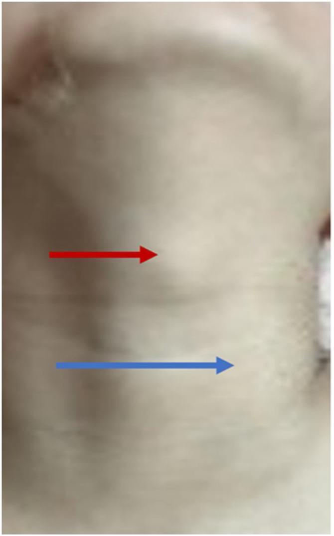
This clinical photograph displays the anterior and lateral neck of a 7-year-old patient, highlighting two distinct areas of subcutaneous swelling. A red arrow indicates a midline mass located in the infrahyoid region, specifically between the thyroid and cricoid cartilages; clinically, this represents a thyroglossal duct cyst. A blue arrow points to a second, more lateral protrusion situated anterior to the left carotid artery, which represents ectopic thymic tissue with cyst formation (thymic ectasia). The overlying skin appears normal, with no evidence of erythema, inflammation, or ulceration. This image is an educational example of concurrent congenital neck masses, illustrating the clinical presentation of a common midline lesion (thyroglossal cyst) alongside a rarer lateral developmental anomaly (ectopic thymus). It is intended for pediatric surgery and otolaryngology training to aid in the differential diagnosis of pediatric neck masses.
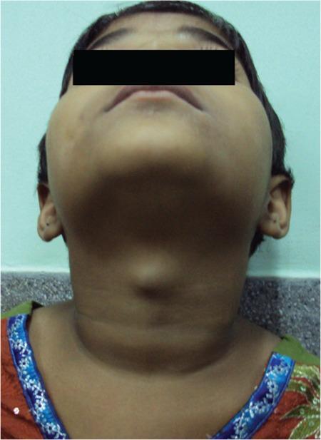
Clinical photograph of a pediatric patient's neck, viewed from an inferior-anterior perspective (looking up toward the chin). The image demonstrates a prominent, well-circumscribed, midline cystic swelling in the anterior neck, located superior to the thyroid cartilage. The overlying skin appears normal with no visible erythema, inflammation, or ulceration. The morphology is characteristic of a thyroglossal duct cyst, a common congenital abnormality occurring along the descent pathway of the thyroid gland. In this clinical context, such a mass often contains ectopic functional thyroid tissue and typically moves upward upon protrusion of the tongue or swallowing. The visual findings illustrate a classic presentation of a midline neck mass in childhood, serving as an educational example for differential diagnosis in pediatric otolaryngology and endocrinology, specifically regarding ectopic thyroid tissue and thyroglossal duct remnants.
Case 2: Anterior Neck Mass - AB, 6-year-old male
Question 1: Primary Working Impression
| Feature | This Patient | Classic TDC |
|---|---|---|
| Age | 6 years old | Most common in childhood |
| Location | Anterior midline neck | Midline, anywhere from foramen cecum to thyroid |
| Movement with deglutition | Yes | Yes |
| Movement with tongue protrusion | Yes | Yes (pathognomonic) |
| Non-tender, well-circumscribed | Yes | Yes (when uninfected) |
- Dermoid cyst - also midline, but does NOT elevate with tongue protrusion; contains keratin (cheesy content) rather than mucus
- Ectopic thyroid - rare; usually the only thyroid tissue present; ruled out with imaging
- Lymphadenopathy - typically lateral, not midline; this patient has no cervical LAD
- Branchial cleft cyst - characteristically lateral neck (anterior to SCM), not midline
Question 2: Diagnostic Tests to Request
1. Neck Ultrasound (FIRST-LINE - most important)
- Confirms the presence of a normal orthotopic thyroid gland in the pretracheal position - this rules out the possibility that the cyst is the patient's only functioning thyroid tissue (failure of caudal thyroid migration)
- Distinguishes TDC from dermoid cyst - the SIST scoring system uses ultrasound features: presence of septa, irregular walls, and solid components are more indicative of TDC than dermoid cyst
- Characterizes the cyst - size, location, cystic vs. solid components, relationship to hyoid bone
2. Thyroid Function Tests (TFTs)
- TSH, Free T3, Free T4
- Rule out hypothyroidism, especially if ectopic thyroid is suspected
- Baseline thyroid function assessment before any surgical intervention
3. Nuclear Radionuclide Imaging (Tc-99m scan) - if indicated
- Used when orthotopic thyroid is not clearly visualized on ultrasound
- Confirms whether the cyst is the only functioning thyroid tissue before surgery
- Prevents inadvertent removal of the patient's sole thyroid gland
4. Fine Needle Aspiration Cytology (FNAC) - optional/selective
- May be done if malignancy is suspected (rare in this age group, but TDC can rarely undergo malignant transformation)
- In this case (6-year-old, no red flags), not routinely required
Question 3: Treatment
Step 1 - Acute Phase (if infected)
- Oral or IV antibiotics (appropriate coverage for skin/oral flora)
- Incision and drainage if abscess formation occurs
- Allow complete resolution of infection before definitive surgery
Step 2 - Definitive Surgical Treatment: The Sistrunk Procedure
- Complete excision of the cyst
- Excision of the entire tract in continuity with the cyst
- Resection of the central (middle) third of the hyoid bone - because the tract passes through or around it
- Resection of the tract superior to the hyoid, including a cuff of tongue base musculature (genioglossus), all the way to the foramen cecum
Intraoperative tip
- Mucus = thyroglossal duct cyst (proceed with full Sistrunk)
- Keratin/cheesy material = dermoid cyst (simpler excision sufficient)
Post-operative considerations
- Thyroid function monitoring if significant thyroid tissue was associated with the cyst
- Histopathologic examination of the excised specimen (rare malignancy ~1% - usually papillary thyroid carcinoma arising in TDC)
- If malignancy is found in the TDC specimen: the identification of malignancy within a TDC is associated with a 6.6% risk of microcarcinoma in the normal-appearing thyroid gland - Cummings Otolaryngology, block 44
Summary Table
| Question | Answer |
|---|---|
| Working Impression | Thyroglossal Duct Cyst |
| Key diagnostic sign | Moves with tongue protrusion + deglutition |
| Primary investigation | Neck ultrasound |
| Additional test if orthotopic thyroid unclear | Tc-99m radionuclide scan |
| Definitive treatment | Sistrunk procedure |
| Recurrence rate (simple excision only) | 40-50% |
| Recurrence rate (Sistrunk) | <5% |
Why do you need to do an ultrasound again?
1. Confirm the orthotopic thyroid gland exists (most critical)
2. Distinguish TDC from a dermoid cyst
| Feature on USS | TDC | Dermoid Cyst |
|---|---|---|
| Internal septa | Present | Absent |
| Wall regularity | Irregular | Smooth |
| Solid components | May be present | Absent |
| Contents | Anechoic/mucoid | Echogenic (keratin) |
3. Characterize the lesion pre-operatively
- Exact size and depth
- Relationship to the hyoid bone
- Single vs. multiloculated
- Any signs of infection or inflammation
- Helps plan the extent of surgery and obtain proper informed consent