
Buffer systems for acid base regulations 20 mark answer
Reading File
Now let me search the medical library for buffer systems content.
Finding Sources
Finding Sources
Excellent results. Now let me read the key sections from Guyton and Medical Physiology in parallel.
Reading File
Reading File
Finding Sources
Reading File
Reading File
Reading File
Reading File
Searching Images
buffer systems acid base regulation bicarbonate phosphate hemoglobin
Reading File
Reading File
Reading File
Reading File
I now have comprehensive content from the textbooks. Let me fetch the bicarbonate buffer system diagram for visual illustration.
Loading Image
Finding Sources
I now have all the content needed to write a comprehensive 20-mark answer. Let me compile it.
Buffer Systems for Acid-Base Regulation
Introduction
The body produces approximately 22,000 mEq of acid per day through metabolism (primarily as CO2), yet the arterial blood pH is maintained within the narrow range of 7.36-7.44. If all metabolic acid were dissolved at once in unbuffered body fluids, the pH would fall below 1. This constancy is achieved by three lines of defense acting in sequence: (1) chemical buffer systems (seconds), (2) the respiratory system (minutes), and (3) the renal system (hours to days). Buffer systems form the first and most immediate line. - Guyton and Hall Textbook of Medical Physiology
A buffer is any substance that can reversibly bind H+:
Buffer + H⁺ ⇌ H-Buffer
When [H+] rises, the reaction shifts right (H+ is bound); when [H+] falls, H-Buffer dissociates to release H+. Buffers do not eliminate H+ from the body - they merely "hold" it until the lungs and kidneys can restore balance.
The Four Major Buffer Systems
| Buffer System | Location | pKa | Relative Contribution |
|---|---|---|---|
| Bicarbonate / Carbonic Acid | ECF (plasma, interstitial) | 6.1 | ~53% of total |
| Hemoglobin | RBCs | ~6.8 | ~35% of total |
| Phosphate | ICF, renal tubular fluid | 6.8 | ~5% |
| Plasma proteins | Plasma, ICF | ~6.7 | ~7% |
1. Bicarbonate Buffer System (Most Important ECF Buffer)
This is the quantitatively most important buffer in extracellular fluid. It consists of:
- Weak acid: H₂CO₃ (carbonic acid)
- Conjugate base: HCO₃⁻ (bicarbonate)
The complete reaction sequence is:
CO₂ + H₂O  ⇌  H₂CO₃  ⇌  H⁺ + HCO₃⁻
         carbonic anhydrase
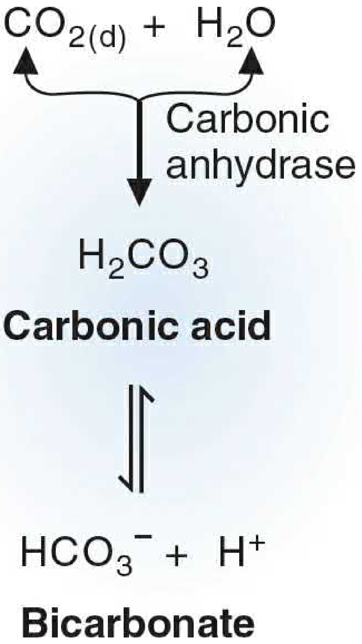
The Henderson-Hasselbalch Equation:
pH = pKa + log [HCO₃⁻] / [H₂CO₃] pH = 6.1 + log [HCO₃⁻] / (0.03 × PaCO₂)
At normal arterial blood: pH 7.40, HCO₃⁻ = 24 mEq/L, PaCO₂ = 40 mmHg
- The ratio [HCO₃⁻]/[H₂CO₃] = 20:1
Buffering strong acid (e.g., HCl):
↑H⁺ + HCO₃⁻ → H₂CO₃ → CO₂ + H₂O
The excess CO₂ stimulates respiration, which blows it off. The strong acid HCl is thus replaced by the weak acid H₂CO₃, which is rapidly converted to CO₂ and exhaled.
Buffering strong base (e.g., NaOH):
OH⁻ + H₂CO₃ → HCO₃⁻ + H₂O
Why is it so powerful despite a pKa of 6.1? Ordinarily, a buffer works best when the pH equals its pKa. The blood pH (7.4) is nearly 1.3 units away from the pKa of 6.1, which should make it ineffective. However, the bicarbonate system is an open system - CO₂ is continuously removed by ventilation and HCO₃⁻ is regulated by the kidneys. This "openness" gives it far greater effective buffering power than any closed system could achieve. - Guyton and Hall
Normal values:
- Plasma HCO₃⁻: 22-26 mEq/L
- Dissolved CO₂ (= 0.03 × PaCO₂): ~1.2 mEq/L
- 20:1 ratio maintained at pH 7.4
2. Phosphate Buffer System
The phosphate buffer consists of two species:
- Weak acid: H₂PO₄⁻ (dihydrogen phosphate)
- Conjugate base: HPO₄²⁻ (monohydrogen phosphate)
Buffering a strong acid:
HCl + Na₂HPO₄ → NaH₂PO₄ + NaCl
The strong acid HCl is replaced by the weak acid NaH₂PO₄, minimising the pH drop.
Buffering a strong base:
NaOH + NaH₂PO₄ → Na₂HPO₄ + H₂O
Characteristics:
- pKa = 6.8, which is close to the pH of intracellular fluid (~7.1) and renal tubular fluid (~5-7)
- Concentration in plasma is low (~1 mEq/L), so it is NOT an important plasma buffer
- Critical in two settings: (1) intracellular fluid where phosphate concentration is much higher, and (2) renal tubular fluid where it buffers secreted H⁺ and allows titratable acid excretion
- Inorganic phosphate + organic phosphates (ATP, glucose-6-phosphate) both contribute intracellularly - Basic Medical Biochemistry, Lippincott
3. Protein Buffer System (Largest Intracellular Buffer)
Proteins are the most abundant buffers in the body due to their high concentration in all cells. They contribute approximately 60-70% of total body buffering capacity (when intracellular and plasma proteins are combined).
Mechanism:
Proteins contain multiple ionisable amino acid side chains, chiefly:
- Histidine residues (imidazole group, pKa ~6.0-7.0) - most important
- Carboxyl groups (-COOH ⇌ -COO⁻ + H⁺)
- Amino groups (-NH₂ + H⁺ ⇌ -NH₃⁺)
In plasma, albumin is the dominant protein buffer, providing buffering through its histidine and other amino acid residues. Albumin contributes significantly because interstitial fluid has too little protein to buffer effectively.
Intracellular proteins buffer approximately 60-70% of acid-base disturbances, but they act more slowly (hours) because H⁺ and HCO₃⁻ equilibrate slowly across cell membranes, except in red blood cells. - Guyton and Hall
4. Hemoglobin Buffer System (Dominant RBC Buffer)
Hemoglobin (Hb) is the most important non-bicarbonate intravascular buffer and works intimately with the bicarbonate system in RBCs.
Reaction:
H⁺ + Hb ⇌ HHb (deoxyhemoglobin)
The Bohr effect explains the physiological integration: deoxyhemoglobin (in tissues) has a higher pKa and is a weaker acid, so it accepts H⁺ more readily. This is critical because:
In peripheral tissues (where CO₂ is produced):
- CO₂ enters RBCs → carbonic anhydrase → H₂CO₃ → H⁺ + HCO₃⁻
- H⁺ is immediately buffered by Hb → HHb
- HCO₃⁻ exits RBC into plasma (in exchange for Cl⁻ via the chloride shift)
- Deoxygenated Hb accepts H⁺ more readily than oxyHb
In the lungs:
- Hb picks up O₂ → oxyhemoglobin (stronger acid, lower pKa)
- OxyHb releases H⁺
- H⁺ + HCO₃⁻ → H₂CO₃ → CO₂ (exhaled)
- HCO₃⁻ re-enters RBCs (chloride shift reverses)
This linkage between O₂/CO₂ transport and acid-base buffering is called the Haldane effect. Hemoglobin buffers approximately 35% of total body acid-base load. - Basic Medical Biochemistry, Lippincott
The Isohydric Principle
All buffer systems in the body fluids are in equilibrium with the same [H⁺]. This is expressed as:
H⁺ = K₁ × [HA₁]/[A₁⁻] = K₂ × [HA₂]/[A₂⁻] = K₃ × [HA₃]/[A₃⁻]
Implication: When any one buffer system changes, all buffer systems change simultaneously because they all share a common H⁺. This means measuring just one buffer pair (the bicarbonate system) gives information about the entire buffer status of the body. - Guyton and Hall
The Respiratory System as the "Second Line"
Although technically not a buffer, the respiratory system amplifies buffering by acting as an open arm of the bicarbonate system:
- Increased PaCO₂ → stimulates peripheral and central chemoreceptors → increases ventilation → CO₂ blown off → pH rises
- Decreased PaCO₂ → reduced ventilation → CO₂ retained → pH falls
- Doubling alveolar ventilation raises pH by ~0.23 units
- Reducing ventilation to 1/4 normal lowers pH by ~0.45 units
The respiratory system responds within 1-3 minutes and can restore up to 50-75% of a pH disturbance. - Guyton and Hall
The Kidneys: Third and Most Powerful Line
The kidneys respond over hours to days but provide the only mechanism for actually eliminating non-volatile acid from the body. They do so by:
- Reabsorbing filtered HCO₃⁻ (mostly proximal tubule, carbonic anhydrase-dependent)
- Generating new HCO₃⁻ (via ammoniagenesis and titratable acid excretion)
- Excreting H⁺ into tubular fluid (buffered by phosphate and ammonia)
Summary Table: Comparison of Buffer Systems
| Feature | Bicarbonate | Phosphate | Protein | Hemoglobin |
|---|---|---|---|---|
| Location | ECF (plasma) | ICF, renal tubule | ICF, plasma | RBCs |
| pKa | 6.1 | 6.8 | ~6.7 | ~6.8 |
| Speed | Immediate | Immediate | Slow (hrs) | Immediate |
| Regulated by | Lungs + kidneys | Kidneys | - | O₂ levels |
| Main role | ECF regulation | ICF + renal excretion | ICF buffering | CO₂ transport + buffering |
| % contribution | ~53% | ~5% | ~7% plasma | ~35% |
Clinical Significance
- In metabolic acidosis (e.g., diabetic ketoacidosis): HCO₃⁻ is consumed buffering ketoacid H⁺; the fall in HCO₃⁻ stimulates Kussmaul breathing (deep, rapid) to compensate by blowing off CO₂.
- In respiratory acidosis (CO₂ retention): Hb and protein buffers act immediately; kidneys increase HCO₃⁻ reabsorption over days.
- In respiratory alkalosis (hyperventilation): CO₂ falls, pH rises; re-breathing into a paper bag increases PaCO₂ and corrects the alkalosis.
- Acid-base abnormalities beyond pH 6.8-7.8 are incompatible with sustaining cardiac, hepatic, and neural function.
Sources: Guyton and Hall Textbook of Medical Physiology (Ch. 31); Basic Medical Biochemistry: A Clinical Approach, Lippincott 6th ed. (Ch. 4)
This is a shared conversation. Sign in to Orris to start your own chat.