
15 marker Physiology university exam ABVMU Describe the connections and functions of Cerebellum
Reading File
Now let me search the medical library for cerebellum connections and functions in parallel.
Finding Sources
Finding Sources
Excellent - found great sources. Let me read the key physiology textbooks in parallel - Costanzo Physiology and Ganong's (both standard for university physiology exams).
Reading File
Reading File
Good start. Let me now read the deeper cerebellum content from both Ganong's and Costanzo, and also check Neuroscience: Exploring the Brain which had a dedicated cerebellum chapter.
Reading File
Reading File
Reading File
I have rich content from Costanzo and Ganong's. Let me now get more detail on afferent/efferent connections and the functional divisions from Costanzo, and also fetch the key cerebellar cortex diagram.
Reading File
Finding Sources
Excellent! Found Adams & Victor with a table on cerebellar peduncles. Let me read that and also get the diagram image.
Reading File
Loading Image
I now have comprehensive material. Let me also fetch the cerebellar projections diagram from Adams & Victor.
Loading Image
Reading File
I now have comprehensive, authoritative material from multiple physiology/neurology textbooks. I have enough to write a complete, detailed 15-marker answer. Let me compile everything now.
Cerebellum: Connections and Functions
Standard answer for ABVMU Physiology (15 marks)
1. Introduction
The cerebellum ("little brain") is a foliated structure located in the posterior fossa, dorsal to the pons and medulla, and below the occipital lobe. Although it weighs only ~10% of the cerebral cortex, its surface area is about 75% of the cortex due to extensive folding. It acts as a comparator and coordinator of motor activity, receiving both motor commands and sensory feedback to fine-tune movement without initiating voluntary movement itself.
2. Gross Anatomy and Divisions
The cerebellum is divided anatomically by two transverse fissures:
- Posterolateral fissure separates the flocculonodular lobe from the rest
- Primary fissure divides the body into anterior and posterior lobes
Functionally, three zones are recognized:
| Functional Division | Anatomical Region | Dominant Input | Function |
|---|---|---|---|
| Vestibulocerebellum | Flocculonodular lobe | Vestibular organs | Balance and eye movements |
| Spinocerebellum | Vermis + intermediate hemisphere | Spinal cord (proprioception) | Synergy/coordination of ongoing movement |
| Pontocerebellum (Cerebrocerebellum) | Lateral hemispheres | Cerebral cortex via pontine nuclei | Planning and initiation of voluntary movement |
3. Internal Structure
3.1 Layers of the Cerebellar Cortex (3 layers)
| Layer | Position | Cell Types |
|---|---|---|
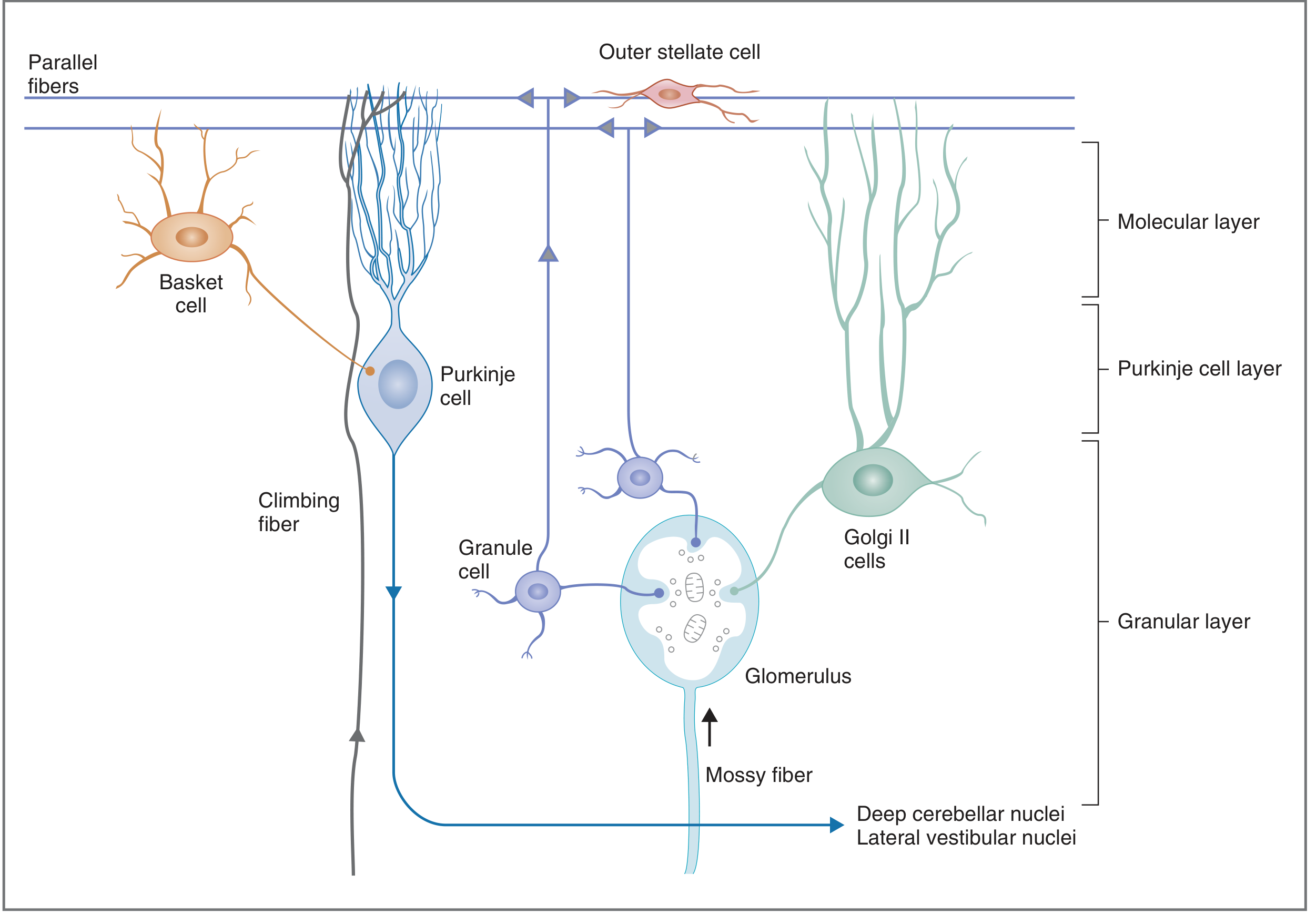
| Molecular layer | Outermost | Basket cells, stellate cells, Purkinje dendrites, parallel fibers |
| Purkinje cell layer | Middle | Purkinje cells (one cell thick) |
| Granular layer | Innermost | Granule cells, Golgi II cells, glomeruli |
3.2 Deep Cerebellar Nuclei (4 nuclei - medial to lateral)
- Fastigial nucleus - receives from vermis; projects to vestibular and reticular nuclei
- Globose nucleus - receives from intermediate zone
- Emboliform nucleus - receives from intermediate zone (globose + emboliform = interpositus nucleus)
- Dentate nucleus - largest; receives from lateral hemisphere (pontocerebellum); projects to thalamus and red nucleus
Mnemonic: "Don't Eat Greasy Food" - Dentate, Emboliform, Globose, Fastigial (lateral to medial)
4. Connections of the Cerebellum
The cerebellum connects to the brainstem via three pairs of cerebellar peduncles:
4.1 Superior Cerebellar Peduncle (Brachium Conjunctivum)
| Direction | Tracts |
|---|---|
| Afferent | Ventral spinocerebellar tract; tectocerebellar tract (via mossy fibers) |
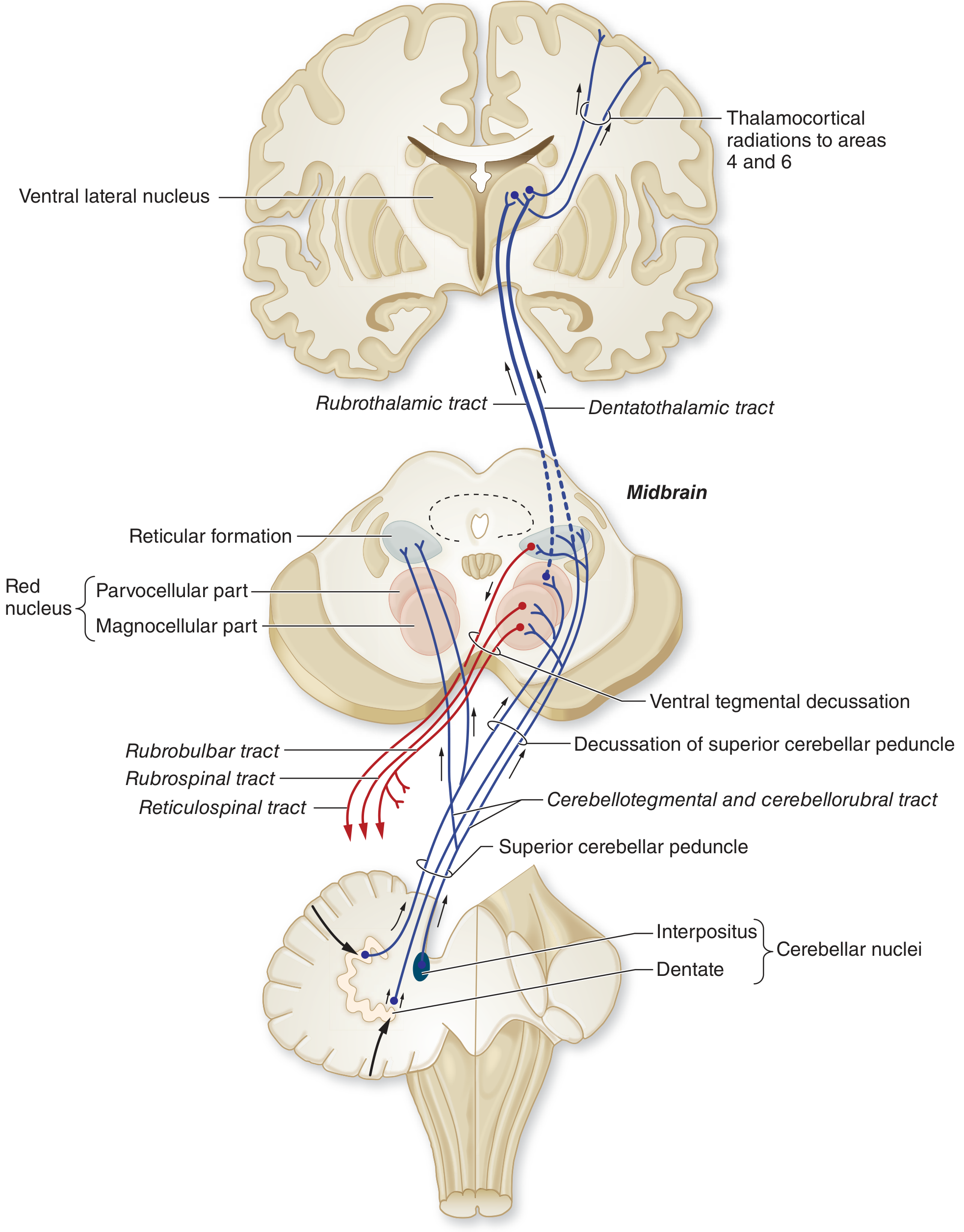
| Efferent (MAIN OUTPUT) | Cerebellothalamic, cerebellorubral, cerebelloreticular, and cerebellovestibular fibers |
- The dentate and interpositus nuclei send fibers through this peduncle, which decussate at the level of the inferior colliculus in the midbrain tegmentum, then ascend to the ventrolateral nucleus of the thalamus
- Thalamus projects to motor cortex (areas 4 and 6) - completing the cerebello-thalamo-cortical loop
- Some fibers synapse in the red nucleus (cerebellorubral fibers) before continuing to thalamus
4.2 Middle Cerebellar Peduncle (Brachium Pontis)
| Direction | Tracts |
|---|---|
| Afferent only | Pontocerebellar fibers from contralateral pontine nuclei (carrying cortical motor commands) |
| Efferent | None |
- This is the largest peduncle and carries only afferent fibers (corticopontocerebellar pathway)
4.3 Inferior Cerebellar Peduncle (Restiform Body)
| Direction | Tracts |
|---|---|
| Afferent | Dorsal spinocerebellar tract; cuneocerebellar tract; vestibulocerebellar fibers; olivocerebellar fibers from contralateral inferior olive (via climbing fibers) |
| Efferent | Cerebellovestibular fibers (from fastigial nucleus to vestibular nuclei) |
4.4 Summary Table of Cerebellar Peduncles (from Adams & Victor's Neurology)
| Peduncle | Afferent (into cerebellum) | Efferent (from cerebellum) |
|---|---|---|
| Superior | Ventral spinocerebellar, tectocerebellar | Cerebellothalamic, cerebellorubral, cerebelloreticular, cerebellovestibular |
| Middle | Pontocerebellar (from contralateral pons) | None |
| Inferior | Dorsal + cuneospinocerebellar, contralateral inferior olive | Some to vestibular nuclei |
5. Afferent Fiber Systems (Input to the Cerebellar Cortex)
Two excitatory fiber systems carry input to the cerebellar cortex:
5.1 Mossy Fiber System
- Sources: Vestibular nuclei, spinal cord (spinocerebellar tracts), pontine nuclei (cortical relay)
- Pathway: Mossy fibers → synapse on granule cells in glomeruli → granule cell axons ascend to molecular layer → bifurcate as parallel fibers → excite Purkinje cell dendrites
- Each Purkinje cell may receive input from up to 250,000 parallel fibers
- Produce simple spikes in Purkinje cells
5.2 Climbing Fiber System
- Source: Inferior olivary nucleus of the contralateral medulla
- Pathway: Climbing fibers make multiple direct synaptic contacts along the dendrites of Purkinje cells
- Each Purkinje cell receives input from only one climbing fiber, but that one fiber makes many synapses
- Produce powerful complex spikes in Purkinje cells
- Role in cerebellar learning and error correction (conditioning Purkinje cells to modulate their response to mossy fiber input)
6. Output of the Cerebellar Cortex
- The only output of the cerebellar cortex is via Purkinje cell axons
- Purkinje cell output is always inhibitory (neurotransmitter: GABA)
- They project to:
- Deep cerebellar nuclei (topographically organized)
- Lateral vestibular nuclei (direct)
- Deep cerebellar nuclei neurons are tonically active and excitatory; Purkinje cells modulate this tonic output
Vermis → Fastigial | Intermediate zone → Globose + Emboliform (Interpositus) | Lateral hemisphere → Dentate
7. Interneurons of the Cerebellar Cortex
All interneurons except granule cells are inhibitory:
| Cell | Action |
|---|---|
| Granule cell | Excitatory - excites Purkinje cells via parallel fibers |
| Basket cell | Inhibitory - inhibits Purkinje cells (via parallel fibers) |
| Stellate cell | Inhibitory - inhibits Purkinje cells |
| Golgi II cell | Inhibitory - inhibits granule cells (negative feedback) |
8. Cerebellar Circuits (Diagrams)
Cerebellar cortex structure (Costanzo Physiology):
Efferent projections from cerebellum to thalamus and cortex (Adams & Victor):
9. Functions of the Cerebellum
The cerebellum does NOT initiate movement - it acts as a comparator, comparing the intended movement (from cortex) with actual movement (from proprioceptive feedback) and correcting errors in real time.
9.1 Coordination of Movement (Synergy)
- Controls the rate, range, force, and direction of movements
- Ensures smooth, accurate execution of voluntary movements
9.2 Maintenance of Posture and Equilibrium
- Vestibulocerebellum uses vestibular input to maintain balance and upright posture
- Fastigial nucleus relays to vestibular nuclei and reticulospinal pathways
9.3 Control of Eye Movements
- Flocculonodular lobe controls smooth pursuit eye movements
- Helps coordinate head-and-eye movements together
9.4 Planning and Initiation of Voluntary Movement
- Pontocerebellum (lateral hemispheres) receives cortical input via pontine nuclei
- Projects back to cortex (via dentate → thalamus → cortex) to participate in motor planning
9.5 Motor Learning
- Climbing fibers from the inferior olive act as a "teaching signal"
- Long-term depression (LTD) at parallel fiber-Purkinje cell synapses underlies adaptation and motor learning (e.g., learning to throw accurately)
9.6 Regulation of Muscle Tone
- Cerebellum facilitates motor neurons and maintains appropriate muscle tone through connections with vestibular and reticular nuclei
10. Cerebellar Feedback Loops
Cortico-ponto-cerebello-thalamo-cortical Loop
- Motor cortex → corticospinal collaterals → pontine nuclei → middle cerebellar peduncle → cerebellar cortex → deep nuclei (dentate) → superior cerebellar peduncle → decussates → ventrolateral thalamus → motor cortex
- Since the peduncle decussates AND corticospinal tract decussates, the net result is: cerebellar lesion causes ipsilateral signs (double-crossing)
Guillain-Mollaret Triangle
- Dentate nucleus → red nucleus → central tegmental fasciculus → inferior olive → back to cerebellum
- Clinical relevance: lesions cause oculopalatal tremor and hypertrophy of the inferior olive
11. Effects of Cerebellar Lesions (Clinical Correlations)
Cerebellar lesions produce ataxia - incoordination due to errors in rate, range, force, and direction of movement:
| Sign | Description |
|---|---|
| Ataxia | Incoordination of movement (ipsilateral) |
| Dysmetria | Overshoot or undershoot of target (past-pointing) |
| Dysdiadochokinesis | Inability to perform rapid alternating movements |
| Intention tremor | Tremor increasing as limb approaches target (unlike resting tremor of Parkinson's) |
| Rebound phenomenon | Inability to stop a movement when resistance is suddenly removed |
| Nystagmus | Disorder of eye movement coordination |
| Scanning (cerebellar) dysarthria | Slow, slurred, ataxic speech |
| Hypotonia | Reduced muscle tone |
Summary
The cerebellum serves as the brain's motor coordinator. It receives proprioceptive, vestibular, and cortical motor information via three peduncles and two fiber systems (mossy and climbing fibers). Its output, via inhibitory Purkinje cells → deep cerebellar nuclei → superior cerebellar peduncle, modulates the motor cortex and brainstem nuclei. The three functional divisions - vestibulo-, spino-, and pontocerebellum - handle balance, coordination, and motor planning respectively. Damage produces the characteristic ipsilateral cerebellar syndrome of ataxia, dysmetria, and intention tremor.
Sources: Costanzo Physiology 7e; Ganong's Review of Medical Physiology 26e; Adams & Victor's Principles of Neurology 12e
This is a shared conversation. Sign in to Orris to start your own chat.