
Explain diabetic retinopathy Staging Clinical features Investigation Treatment Complications
"diabetic retinopathy"[MeSH Terms] AND treatment
Use ak khurana
- Kanski's Clinical Ophthalmology: A Systematic Approach (10th ed.)
- The Wills Eye Manual
Ok continue
Definition & Overview
Pathogenesis
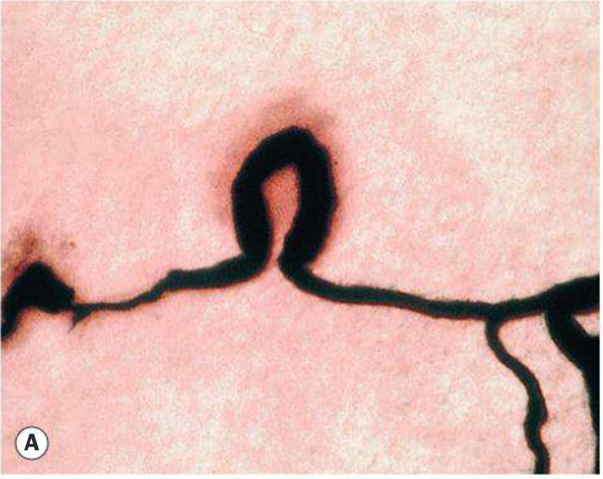
- Pericyte loss - pericytes surround retinal capillaries and provide structural support; their selective loss under hyperglycaemia is the hallmark early change
- Basement membrane thickening - increases capillary permeability and rigidity
- Endothelial cell damage - loss of the blood-retinal barrier
- Capillary closure / non-perfusion - leads to retinal ischaemia
- VEGF upregulation - ischaemia drives Vascular Endothelial Growth Factor (VEGF) release, promoting pathological new vessel formation
Risk Factors
| Factor | Details |
|---|---|
| Duration of diabetes | Most important; DR rarely appears in first 5 years of type 1 |
| Poor glycaemic control | HbA1c is a key modifiable risk |
| Hypertension | Independent risk factor; control reduces progression |
| Pregnancy | DR may worsen rapidly; review every trimester or monthly in PDR |
| Hyperlipidaemia | Promotes hard exudate formation |
| Anaemia | Worsens retinal hypoxia |
| Renal disease | Associated with accelerated DR |
| Cataract surgery | Can accelerate macular oedema |
Staging / Classification
ETDRS Classification with Management Guidelines
| Stage | Features | Review Interval |
|---|---|---|
| No DR | Normal fundus | 12 months |
| Very mild NPDR | Microaneurysms only | 12 months |
| Mild NPDR | Microaneurysms, dot/blot haemorrhages, exudates, cotton-wool spots - up to but not reaching moderate NPDR | 6-12 months |
| Moderate NPDR | >mild NPDR but <severe NPDR; may have CWS, venous beading | 3-6 months |
| Severe NPDR (Pre-PDR) | Any one of the 4-2-1 rule: >20 haemorrhages in all 4 quadrants, venous beading in ≥2 quadrants, IRMA in ≥1 quadrant | 3 months |
| PDR | New vessels on disc (NVD) or elsewhere (NVE), ± vitreous/preretinal haemorrhage | Treat; review 4-6 weeks |
| High-risk PDR | NVD >1/3-disc area, any NVD + VH, NVE >1/2-disc area + VH, or any NVI/NVA | Treat urgently |
| Advanced diabetic eye disease | Tractional retinal detachment, persistent VH, neovascular glaucoma | Vitrectomy ± medical Rx |
Descriptive Categories (Widely Used in Practice)
- Background DR (BDR): Microaneurysms, dot/blot haemorrhages, hard exudates - earliest signs
- Diabetic maculopathy: Significant macular changes - oedema (most common cause of visual loss in type 2) or ischaemia
- Pre-proliferative DR (PPDR): CWS, venous beading, IRMA, moderate-severe haemorrhages - indicates progressive ischaemia
- Proliferative DR (PDR): NVD and/or NVE; driven by VEGF
- Advanced diabetic eye disease: Tractional RD, persistent VH, neovascular glaucoma
Clinical Features (Signs)
1. Microaneurysms
- Earliest ophthalmoscopic sign of DR
- Saccular outpouchings of capillary walls, from focal weakness after pericyte loss
- Appear as tiny red dots, typically at the posterior pole
- On FA: hyperfluorescent dots in early frames; late leakage
- On OCT: located in middle retinal layers
2. Retinal Haemorrhages
- Dot and blot haemorrhages: Arise from the venous end of capillaries; located in compact middle retinal layers; round/oval red spots
- Flame-shaped haemorrhages: From superficial precapillary arterioles in the nerve fibre layer; shaped by the NFL architecture
- Dark round ("blot") haemorrhages: Represent haemorrhagic retinal infarcts; located in middle layers; their extent is a significant marker for progression to PDR
3. Hard Exudates
- Caused by chronic localised leakage from microaneurysms and abnormal capillaries
- Intraretinal deposits of lipids and lipoproteins - bright yellow-white waxy lesions
- Located at the outer plexiform layer
- Arranged in circinate (ring) patterns around leaking microaneurysms
- When at the macula: threaten central vision
4. Cotton-Wool Spots (CWS)
- Superficial white fluffy patches from localised ischaemia of the NFL - accumulation of axoplasmic debris (cytoid bodies)
- Marker of increasing retinal ischaemia - signal progression toward PPDR
- Transient; resolve in weeks to months
5. Venous Changes (PPDR)
- Venous beading: Sausage-like segmental dilatation of veins - indicates severe ischaemia
- Venous loops and reduplication
- Venous dilatation and tortuosity
6. Intraretinal Microvascular Abnormalities (IRMA)
- Dilated capillary channels within the retina that bypass areas of capillary non-perfusion
- Flat on FA (no leakage beyond retina - distinguishes from NVE)
- Marker of severe NPDR / impending PDR
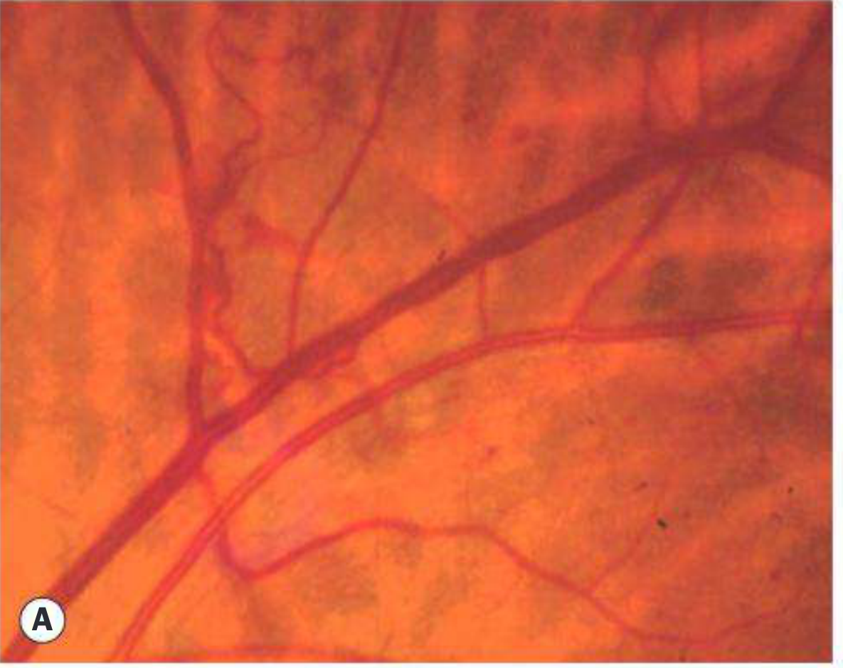
7. New Vessels (PDR)
- NVD (new vessels on the disc): On or within 1 disc diameter of optic disc; arise from optic disc surface or adjacent retina
- NVE (new vessels elsewhere): Arise from retinal surface; may be flat initially then elevated; associated with fibrous tissue proliferation
8. Rubeosis Iridis (NVI)
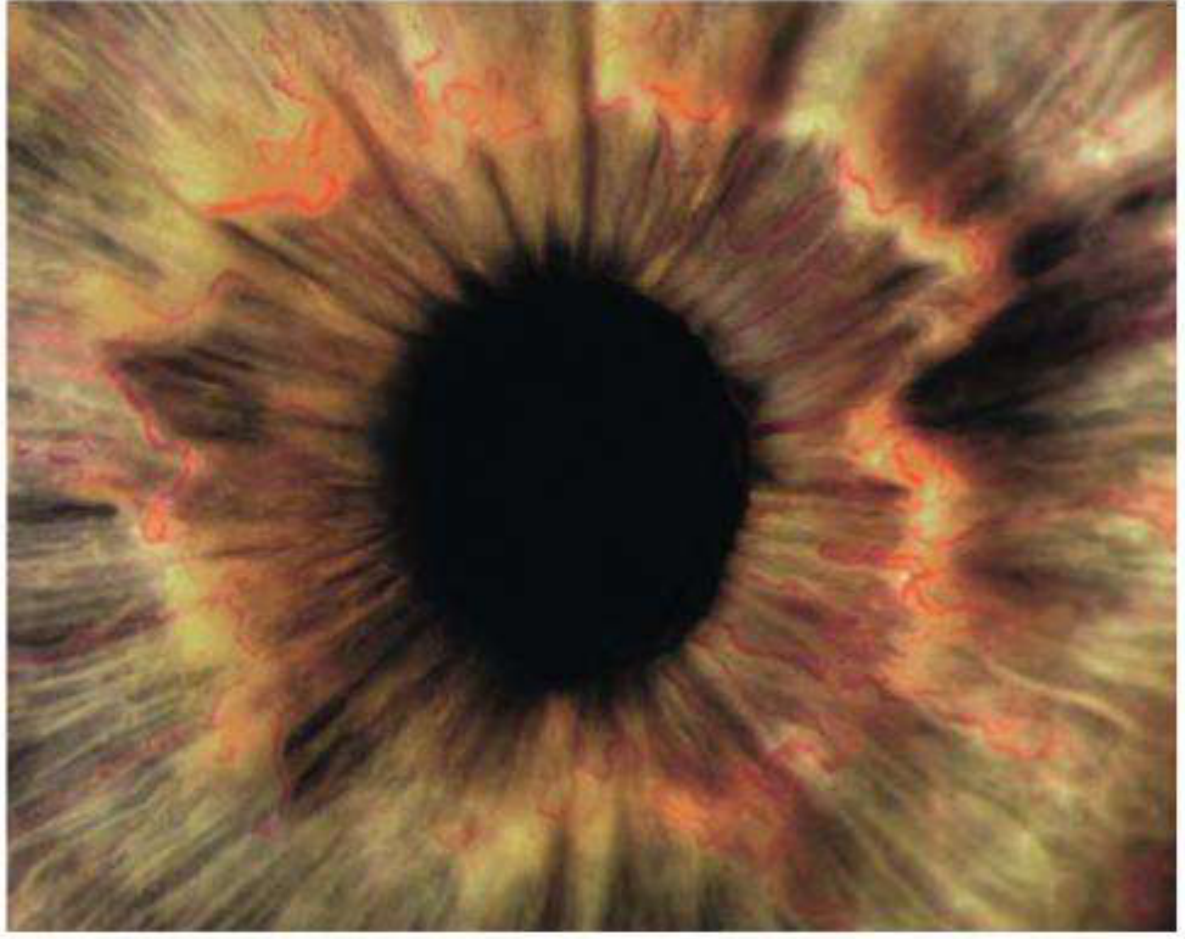
- New vessels on the iris surface; arise at the pupil margin and spread to the angle
- Sign of severe ischaemia and high risk of neovascular glaucoma
9. Diabetic Maculopathy
- Focal: Localised area of retinal thickening with hard exudates associated with microaneurysm clusters
- Diffuse: Generalised macular thickening from extensive leakage
- Ischaemic: Foveal avascular zone (FAZ) enlargement; poor prognosis; no treatment modifies ischaemia
- Retinal thickening within 500 μm of fovea
- Hard exudates within 500 μm of fovea with adjacent thickening
- Retinal thickening ≥1 disc area, any part within 1 disc diameter of fovea
Investigations
1. Fundus Examination
- Dilated fundus examination using 90D or 60D lens at slit lamp - gold standard for grading
- Indirect ophthalmoscopy for peripheral retinal examination
2. Fluorescein Angiography (FA)
- Differentiates dot haemorrhages from microaneurysms (MA leaks, haemorrhage does not)
- Identifies areas of capillary non-perfusion (ischaemia)
- Detects foveal ischaemia (FAZ enlargement)
- Identifies NVD/NVE (early hyperfluorescence with late leakage)
- Guides focal/grid laser therapy
- Distinguishes IRMA (no leakage) from NVE (leakage)
3. Optical Coherence Tomography (OCT)
- Investigation of choice for DME - quantifies retinal thickness, detects subretinal fluid, cystoid changes
- Identifies vitreomacular traction
- Guides anti-VEGF therapy and monitors response
- OCT Angiography (OCTA): Non-invasive assessment of the FAZ and capillary non-perfusion; detects significant central macular ischaemia
4. Systemic Investigations
- Fasting blood glucose + HbA1c - glycaemic control assessment
- Blood pressure
- Lipid panel (elevated lipids worsen hard exudate formation)
- Renal function (urea, creatinine)
- BMI, urinalysis
5. Additional Ocular Investigations
- Gonioscopy - before dilation, to check for NVI/NVA in suspected PDR
- B-scan ultrasound - when vitreous haemorrhage obscures the fundus view; rules out tractional retinal detachment
Treatment
A. Medical / Systemic Control (Cornerstone)
- Tight glycaemic control (target HbA1c per UKPDS/DCCT trials) - reduces incidence and slows progression
- Blood pressure control - target <130/80 mmHg; reduces progression
- Lipid control - statins/fenofibrate reduce hard exudate deposits
- Treatment of anaemia, renal disease
B. Diabetic Macular Oedema (DME) Treatment
1. Anti-VEGF Therapy (First Line for Centre-Involving DME)
- Ranibizumab (Lucentis) - 0.3 mg intravitreal
- Aflibercept (Eylea) - 2 mg intravitreal; superior for eyes with VA ≤6/15
- Faricimab (Vabysmo) - newest agent; dual mechanism: neutralises angiopoietin-2 AND VEGF-A (YOSEMITE and RHINE studies); as effective as aflibercept and ranibizumab; 4 initial monthly injections then treat-and-extend up to 16-week intervals
- Bevacizumab (Avastin) - off-label but widely used; comparable to ranibizumab for VA >6/15
2. Intravitreal Corticosteroids (Second Line)
- Dexamethasone implant (Ozurdex) - biodegradable 0.7 mg implant
- Fluocinolone acetonide implant (Iluvien) - long-acting (3 years)
- Off-label: triamcinolone acetonide 1-4 mg
- Side effects: cataract formation, raised IOP
3. Laser Photocoagulation (Adjunctive)
- No longer first-line; reserved for:
- Off-centre thickening with exudates threatening the fovea
- Extrafoveal microaneurysms causing significant oedema
- When anti-VEGF/steroids are contraindicated (e.g. pregnancy)
- Focal laser: Burns applied to leaking microaneurysms 500-3000 μm from foveola; 50-100 μm spot, 0.02-0.1 sec, mild grey reaction
- Modified grid laser: Focal treatment of leaking foci
- Subthreshold macular laser (micropulse): Short pulses (100-300 μs on, 1700-1900 μs off); minimises collateral damage while stimulating RPE; no visible burn
C. Proliferative DR (PDR) Treatment
1. Panretinal Photocoagulation (PRP) - Mainstay
- Indicated for high-risk PDR characteristics:
- NVD >1/3-disc area
- Any NVD with preretinal haemorrhage or VH
- NVE >1/2-disc area with preretinal haemorrhage or VH
- Any NVI or NVA
- Mechanism: destroys ischaemic retina → reduces VEGF production → regression of new vessels
- Technique: 1200-1600 burns, 500 μm spot, 0.05-0.1 sec, ~2 mm apart, from posterior retina to periphery
- Complications: loss of peripheral/night vision, macular oedema, decreased colour vision, exacerbation of pre-existing DME
Clinical tip: PRP remains the mainstay of PDR treatment in most healthcare systems.
2. Anti-VEGF Therapy for PDR
- Alternative to PRP; preferred initial therapy when:
- DME is co-present
- View to peripheral retina is limited by VH
- Agents: ranibizumab, aflibercept, bevacizumab
- Caution: patients lost to follow-up show worse anatomic and visual outcomes than PRP-treated patients
- Recent 2025 meta-analysis (PMID 39128789) confirms anti-VEGF and PRP have comparable visual outcomes in PDR, with anti-VEGF having lower risk of visual field loss
3. Vitrectomy (Pars Plana Vitrectomy - PPV)
- Dense, non-clearing or recurrent vitreous haemorrhage causing significant vision loss
- Tractional retinal detachment involving or progressing within the macula
- Macular epiretinal membranes or vitreomacular traction causing significant symptoms
- Dense premacular haemorrhage
- Chronic DME unresponsive to other treatment
- Severe neovascularisation and fibrous proliferation unresponsive to laser or anti-VEGF
Note: Young type 1 diabetics have more aggressive PDR and may benefit from earlier vitrectomy or anti-VEGF therapy. B-scan US is required if dense VH obscures fundus view.
Complications
Ocular Complications
| Complication | Mechanism |
|---|---|
| Vitreous haemorrhage | Bleeding from fragile new vessels (NVD/NVE); may be dense, causing sudden visual loss |
| Tractional retinal detachment | Fibrovascular proliferation contracts over time, pulling retina from RPE |
| Rhegmatogenous RD | Vitreous traction creates retinal tear → combined tractional-rhegmatogenous RD |
| Neovascular glaucoma (NVG) | NVI extends to angle → fibrovascular membrane closes trabecular meshwork → raised IOP → painful blind eye |
| Diabetic macular oedema | Breakdown of blood-retinal barrier → fluid in macula → central vision loss (main cause of VA loss in type 2) |
| Macular ischaemia | FAZ enlargement from capillary non-perfusion → poor vision, no effective treatment |
| Epiretinal membrane | Fibrous proliferation on retinal surface from organised haemorrhage or traction |
| Accelerated cataract | Sorbitol accumulation in lens; posterior subcapsular cataracts |
| Rubeosis iridis / NVI | Precursor to NVG; sign of very advanced ischaemia |
| Diabetic papillopathy | Optic disc swelling (usually benign, self-limiting); more common in type 1 |
| Ocular motor nerve palsies | CN III, IV, VI - from ischaemia of vasa nervorum; CN III palsy is pupil-sparing |
| Reduced corneal sensitivity | Diabetic neuropathy |
Systemic Associations
- Diabetic nephropathy - DR and nephropathy share the same microangiopathic process
- Cardiovascular disease - patients with PDR have significantly elevated cardiovascular risk
- Diabetic neuropathy
Screening Protocol
| Retinopathy Status | Review Interval |
|---|---|
| No DR / Very mild NPDR | Annual |
| Mild NPDR | Every 6-9 months |
| Moderate NPDR | Every 3-6 months |
| Severe NPDR / PPDR | Every 3 months |
| PDR | Treat; review every 4-6 weeks |
| Pregnancy + Mild-Moderate NPDR | Every trimester |
| Pregnancy + High-risk NPDR or PDR | Monthly; treat with PRP |
"The risk of severe visual loss can be reduced to less than 5% with effective screening and implementation of ETDRS trial conclusions and anti-VEGF clinical trials." - Kanski's Clinical Ophthalmology, 10th Ed.
?
- Asking about something specific from the diabetic retinopathy explanation above?
- Confused about a particular section (staging, treatment, etc.)?
- Wanting more detail on a specific topic?
- Something else entirely?
Create a visual summary/infographic of DR stages and treatment
Medical infographic titled "Diabetic Retinopathy: Stages & Treatment" with a clean clinical style on a white background. Layout: vertical flowchart with 6 color-coded stages connected by downward arrows showing progression. Stage 1 - GREEN box: "No DR / Very Mild NPDR" - Microaneurysms only - Review: 12 months - Treatment: Systemic control (glucose, BP, lipids) Stage 2 - LIGHT YELLOW box: "Mild NPDR" - Microaneurysms, dot/blot haemorrhages, hard exudates - Review: 6-12 months - Treatment: Systemic control Stage 3 - YELLOW box: "Moderate NPDR" - Above + cotton-wool spots, venous beading - Review: 3-6 months - Treatment: Systemic control; treat DME if present Stage 4 - ORANGE box: "Severe NPDR (Pre-PDR) - 4-2-1 Rule" - >20 haemorrhages in all 4 quadrants OR venous beading in 2+ quadrants OR IRMA in 1+ quadrant - Review: 3 months - Treatment: Prepare for PRP; anti-VEGF if DME Stage 5 - RED box: "Proliferative DR (PDR)" - NVD, NVE, vitreous/preretinal haemorrhage - Review: 4-6 weeks - Treatment: Panretinal Photocoagulation (PRP) OR Anti-VEGF (ranibizumab, aflibercept, faricimab) Stage 6 - DARK RED box: "Advanced Diabetic Eye Disease" - Tractional RD, persistent VH, neovascular glaucoma - Treatment: Vitrectomy (PPV) On the right side: a separate teal-colored panel titled "Diabetic Macular Oedema (DME) - Any Stage" listing: 1st line: Anti-VEGF (aflibercept, ranibizumab, faricimab) | 2nd line: Intravitreal steroids (dexamethasone implant) | 3rd line: Focal/grid laser At the bottom: a blue bar titled "Systemic Control (All Stages): HbA1c control | BP <130/80 | Lipid management" Clean sans-serif font, professional medical infographic style, clear hierarchy, no clutter.
404 Not Found. {'message': '{\n "error": {\n "code": 404,\n "message": "Publisher model `projects/engaged-beaker-474810-u2/locations/global/publishers/google/models/gemini-3-pro-image-preview` was not found or your project does not have access to it. Ensure you are using a valid model name and that the model is available in the specified region. For more information, see: https://docs.cloud.google.com/gemini-enterprise-agent-platform/resources/locations.",\n "status": "NOT_FOUND"\n }\n}\n', 'status': 'Not Found'}
We encountered an error while processing your request. Please try again or reach out to us for help.