
Amoxicillin, clarithromycin, metronidazole, levoflooxacin, ppi Give each drug separate class , short moa, dose, adr, frequency
Drug Reference Table: Amoxicillin | Clarithromycin | Metronidazole | Levofloxacin | PPI
1. Amoxicillin
| Parameter | Details |
|---|---|
| Class | Aminopenicillin (Beta-lactam antibiotic) |
| MOA | Inhibits bacterial cell wall synthesis by binding penicillin-binding proteins (PBPs), blocking peptidoglycan cross-linking → bactericidal |
| Dose | 500 mg TDS or 875 mg BD (oral); up to 80-90 mg/kg/day in children |
| Frequency | 3x/day (TDS) for 500 mg; 2x/day (BD) for 875 mg formulation |
| ADRs | Diarrhea, nausea, skin rash, urticaria, anaphylaxis (rare), pseudomembranous colitis, maculopapular rash (esp. in EBV/mono) |
2. Clarithromycin
| Parameter | Details |
|---|---|
| Class | Macrolide antibiotic |
| MOA | Binds the 50S ribosomal subunit (23S rRNA, domain V) → inhibits peptide chain elongation and aminoacyl translocation → bacteriostatic (bactericidal at high doses) |
| Dose | 250-500 mg BD (oral); for H. pylori: 500 mg BD |
| Frequency | Twice daily (BD) |
| ADRs | GI upset (nausea, abdominal pain, diarrhea), metallic taste, QT prolongation, hepatotoxicity (rare), CYP3A4 drug interactions (strong inhibitor) |
3. Metronidazole
| Parameter | Details |
|---|---|
| Class | Nitroimidazole antibiotic / antiprotozoal |
| MOA | Nitro group is reduced by anaerobic bacterial enzymes → forms reactive intermediates → causes DNA strand breakage and disrupts DNA replication → bactericidal against anaerobes and certain protozoa |
| Dose | 400-500 mg TDS (oral); 500 mg IV TDS for severe infections; H. pylori: 400-500 mg BD/TDS |
| Frequency | 3x/day (TDS) standard; BD for some regimens |
| ADRs | Metallic/bitter taste, nausea, headache, peripheral neuropathy (prolonged use), disulfiram-like reaction with alcohol, CNS effects (seizures, encephalopathy - rare), dark/reddish-brown urine |
4. Levofloxacin
| Parameter | Details |
|---|---|
| Class | Fluoroquinolone antibiotic (3rd generation) |
| MOA | Inhibits bacterial DNA gyrase (topoisomerase II) and topoisomerase IV → prevents DNA supercoiling/relaxation and strand separation → blocks DNA replication and transcription → bactericidal |
| Dose | 500-750 mg OD (oral or IV); H. pylori: 500 mg OD; CAP: 500 mg OD x 7-14 days; UTI: 250-500 mg OD |
| Frequency | Once daily (OD) |
| ADRs | Nausea, diarrhea, headache, dizziness, QT prolongation, tendinopathy/tendon rupture (esp. Achilles, in elderly/steroid users), photosensitivity, peripheral neuropathy, seizures (rare), hypoglycemia/hyperglycemia in diabetics |
5. PPI (Proton Pump Inhibitors) - e.g., Omeprazole, Pantoprazole, Rabeprazole
| Parameter | Details |
|---|---|
| Class | Proton pump inhibitor (antisecretory agent) |
| MOA | Irreversibly inhibits H⁺/K⁺-ATPase (the proton pump) on the luminal surface of gastric parietal cells → blocks the final step of acid secretion → profound, long-lasting suppression of both basal and stimulated gastric acid |
| Dose | Omeprazole/Pantoprazole: 20-40 mg OD; Rabeprazole: 20 mg OD; Esomeprazole: 20-40 mg OD |
| Frequency | Once daily (OD), ideally 30-60 minutes before a meal (breakfast) for maximum efficacy |
| ADRs | Headache, diarrhea, nausea, abdominal pain; long-term: hypomagnesemia, vitamin B12 deficiency, C. difficile infection risk, bone fractures (osteoporosis risk), iron malabsorption, fundic gland polyps, rebound acid hypersecretion on abrupt withdrawal |
Diagnostic test of peptic ulcer
peptic ulcer endoscopy diagnosis
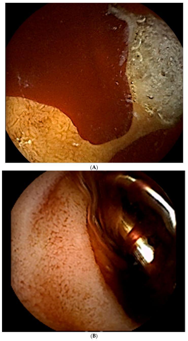
This composite educational image features two frames (A and B) from an upper gastrointestinal endoscopy, illustrating the diagnosis and treatment of acute peptic ulcer bleeding in the duodenal bulb. Frame A demonstrates clinical findings of hematemesis and melena, showing two cratered, oozing ulcers located in the duodenum. Significant active hemorrhage is visible as bright red blood pooling over the mucosal surface. The largest ulcer measures approximately 10 mm in diameter with a visible base. Frame B shows the immediate post-intervention state following the administration of 1:10,000 epinephrine (4cc) and the deployment of metallic hemostatic clips. A hemostatic clip is clearly visible, mechanically compressing the ulcerated site to achieve primary hemostasis. The surrounding mucosa appears altered in color, showing a reddish-orange hue with reduced active bleeding compared to the initial diagnostic view. This sequence serves as a clinical teaching tool for endoscopic management of gastroduodenal artery-related bleeding and the utilization of endoscopic markers to guide subsequent angiographic interventions.
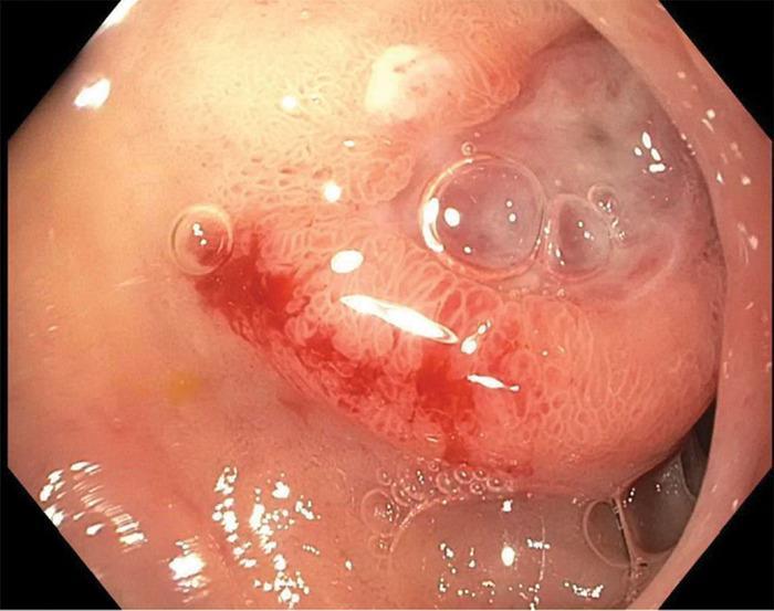
This endoscopic clinical photograph from an upper gastrointestinal endoscopy (EGD) displays the lumen of the distal duodenal bulb. Centered in the frame is a single, approximately 1-cm ulcerated lesion. The ulcer is characterized by a clean base and a slightly protuberant, elevated morphology. Distinctive features include irregular, 'heaped-up' mucosal edges surrounding the ulcer crater, and a cluster of translucent air bubbles typical of the insufflation process. Clinically significant signs of hemorrhage are visible, specifically active oozing of bright red blood (Forrest classification 1B) from the ulcer's margin. The surrounding mucosa shows slight erythema and texture changes indicative of gastric heterotopia or secondary inflammation. This image is an educational example of a peptic ulcer with features suspicious for an underlying mass lesion, requiring differential diagnosis between benign peptic ulcer disease and malignant or stromal tumors such as a Gastrointestinal Stromal Tumor (GIST).
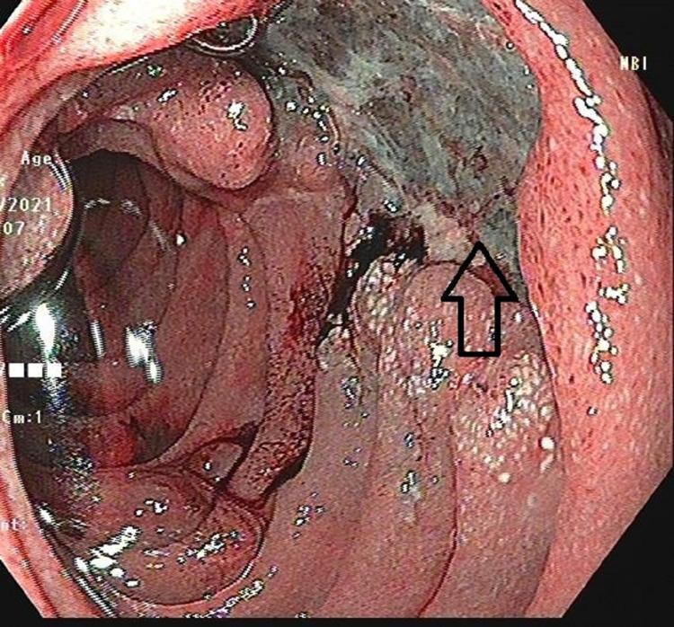
This clinical photograph displays an upper gastrointestinal endoscopy (UGIE) view of the second part of the duodenum. The primary finding is a large, irregularly shaped ulcer approximately 2x2 cm in size, situated on the medial wall. The ulcer features prominent nodular margins and a base covered with dark, necrotic-appearing material and focal areas of hemorrhage. An arrow indicates the close anatomical proximity of the lesion to the duodenal papilla. The surrounding duodenal mucosa is erythematous and inflamed, with thickened, irregular folds and a granular texture suggestive of significant tissue reaction. This endoscopic view is critical for the differential diagnosis of duodenal pathologies, including malignancy (duodenal adenocarcinoma), complex peptic ulcer disease, or atypical presentations of granulomatous diseases like tuberculosis or groove pancreatitis. The image illustrates key markers for malignancy or chronic inflammation, such as nodularity and mucosal distortion.
Diagnostic Tests for Peptic Ulcer Disease
1. Endoscopy (OGD - Oesophago-Gastro-Duodenoscopy)
- What it does: Direct visual inspection of the gastric and duodenal mucosa; allows biopsy, H. pylori testing, and therapeutic intervention
- Sensitivity/Specificity: High - most sensitive and specific test
- Key advantages:
- Visualizes ulcer size, location, depth, and bleeding stigmata
- Biopsy from ulcer edge to exclude malignancy (mandatory for all gastric ulcers)
- Allows biopsy-based H. pylori testing (rapid urease test, histology, culture)
- Can be therapeutic (injection, clipping, cautery for bleeding ulcers)
- Indications (mandatory):
- Age >55 with new-onset dyspepsia
- Any "alarm/red flag" symptoms (weight loss, dysphagia, hematemesis, melena, anemia, palpable mass)
- Gastric ulcer - always biopsy to rule out cancer
- Non-healing or recurrent ulcers
- Limitation: Invasive, needs sedation; PPIs reduce sensitivity slightly
2. Tests for H. pylori (the primary cause of PUD)
A. Non-Invasive Tests
| Test | Principle | Sensitivity | Specificity | Notes |
|---|---|---|---|---|
| Urea Breath Test (UBT) | Patient ingests C-13 or C-14 labelled urea; H. pylori urease splits it → labelled CO₂ detected in exhaled breath | ~95% | ~96% | Best non-invasive test; used for diagnosis AND post-eradication confirmation; stop PPIs 2 weeks before |
| Stool Antigen Test (SAT) | Detects H. pylori antigens in stool using monoclonal antibodies | ~94% | ~97% | Good for diagnosis and confirmation of eradication; monoclonal > polyclonal |
| Serology (IgG antibody) | Detects serum IgG against H. pylori | ~85% | ~79% | Cannot distinguish active vs. past infection; cannot confirm eradication; cheap but least specific |
B. Invasive Tests (Require Endoscopy + Biopsy)
| Test | Principle | Sensitivity | Specificity | Notes |
|---|---|---|---|---|
| Rapid Urease Test (CLO test) | Biopsy placed in urea + pH indicator; urease from H. pylori changes colour | ~90-95% | ~95-100% | Quick result (1-24 hrs); first-line biopsy-based test |
| Histology | Microscopic examination of biopsy with special stains (Giemsa, Warthin-Starry) | ~93-99% | ~95-99% | Gold standard for H. pylori detection; also shows gastritis/dysplasia |
| Culture | Grows H. pylori from biopsy | ~70-80% | ~100% | Allows antibiotic sensitivity testing; slow (5-7 days); used when resistance suspected |
| PCR | Detects H. pylori DNA in biopsy | ~>95% | ~>95% | Detects resistance genes; research/specialist use |
3. Barium Meal / Upper GI Series
- What it shows: Barium fills ulcer crater → "niche" sign on X-ray; Hampton's line (radiolucent line across base of gastric ulcer); mucosal folds radiating toward crater
- Use: When endoscopy is not available or patient cannot tolerate it
- Sensitivity: Lower than endoscopy (~80% for gastric ulcer, 70% for duodenal)
- Limitation: Cannot biopsy, cannot detect H. pylori; less sensitive for small ulcers; cannot differentiate benign from malignant
4. CT Scan / CT Enterography
- Not a primary test for PUD
- Useful for detecting complications - perforation (free air under diaphragm/peritoneum), penetration, gastric outlet obstruction
- Can incidentally show gastric/duodenal thickening but sensitivity for ulcer is low
5. Serum Gastrin Level (for Zollinger-Ellison Syndrome)
- Indicated when: multiple/refractory ulcers, ulcers in unusual locations (distal duodenum/jejunum), recurrent ulcers post-H. pylori eradication, diarrhea + PUD
- Fasting serum gastrin >1000 ng/L + basal acid output >15 mmol/hr → diagnostic of gastrinoma (ZES)
- Secretin stimulation test: Secretin IV → paradoxical rise in gastrin >120 pg/mL in ZES (normally gastrin falls)
6. Gastric Acid Analysis (Basal Acid Output / BAO)
- Measures acid secretion via nasogastric tube aspiration
- BAO >15 mEq/hr suggests ZES
- Rarely used in routine PUD; mainly for suspected hypersecretory states
Summary: When to Use Which Test
| Clinical Situation | Preferred Test |
|---|---|
| Standard PUD workup, age <55, no alarm features | Test and treat: Urea Breath Test or Stool Antigen Test |
| Age >55, alarm symptoms, or gastric ulcer | Endoscopy + biopsy (CLO test + histology) |
| Post-eradication confirmation (4-6 weeks after treatment) | UBT or SAT (not serology) |
| Suspected perforation/complication | CT scan / AXR |
| Recurrent/refractory ulcers - exclude ZES | Fasting serum gastrin + secretin stimulation test |
| Suspected antibiotic resistance | Culture + sensitivity |