
Teach me Meniningiomyocele like a neonataologist/ paeditrician in depth
myelomeningocele neonatal management outcomes
Myelomeningocele (Meningomyelocele) - A Neonatologist's Deep Dive
Terminology note: The terms myelomeningocele (MMC) and meningomyelocele are used interchangeably. "Myelomeningocele" is the preferred modern term. It is a subset of open spina bifida and the most severe common form of neural tube defect (NTD) compatible with life.
1. The Anatomy - What Exactly Is It?
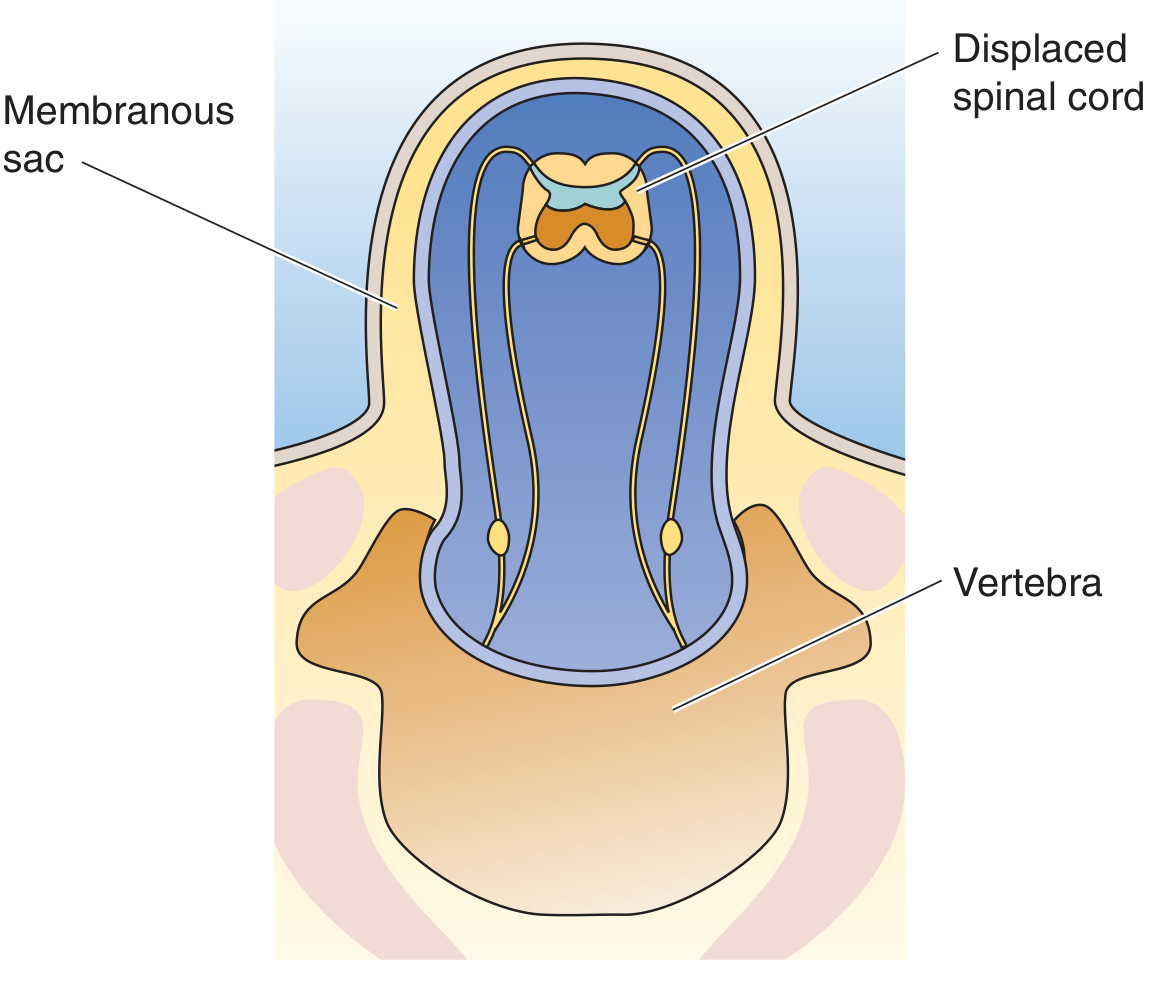
| Type | What herniates | Neural elements? | Neurological deficit |
|---|---|---|---|
| Spina bifida occulta | Nothing - vertebral arch unfused only | No | Usually none |
| Meningocele | Meninges + CSF only | No | Usually none |
| Myelomeningocele | Meninges + spinal cord + nerve roots | Yes | Always present |
| Myeloschisis | Open spinal cord, no sac - exposed neural plate | Yes | Severe |
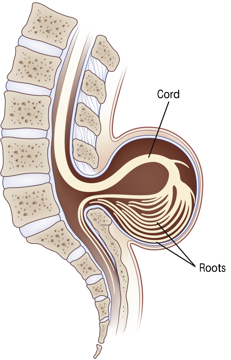
2. Embryology - Why Does It Happen?
- First hit: Primary failure of neural tube closure at ~week 4
- Second hit: Progressive damage from chronic exposure of the open neural placode to neurotoxic amniotic fluid throughout gestation - amniotic fluid contains substances directly toxic to neural tissue, and there is also mechanical trauma
3. Epidemiology
- Incidence: approximately 1 in 3,000 live births (post-folate supplementation era)
- Folate supplementation has reduced MMC prevalence by ~50%, but a plateau has been reached
- 25-40% of MMC pregnancies are terminated following prenatal diagnosis
- Most common in lumbar and sacral regions (>75% of cases)
- Slight female predominance
- Maternal folate/B12 deficiency (most important modifiable factor)
- Valproic acid during early pregnancy (1-2% risk of NTD - one of the highest teratogen risks known)
- Carbamazepine
- Family history of NTDs
- Prior affected pregnancy
- Maternal obesity, diabetes, hyperthermia in early pregnancy
4. Prenatal Diagnosis
- Maternal serum AFP (MSAFP) at 14-16 weeks - elevated in ~85% of open NTDs. Open defects leak AFP into amniotic fluid then into maternal blood.
- Amniotic fluid AFP + acetylcholinesterase (more specific than AFP alone)
- Second-trimester ultrasound (18-20 weeks): Detects >90% of NTDs
| Sign | What you see | Significance |
|---|---|---|
| Lemon sign | Frontal bone scalloping (skull looks like a lemon) | Chiari II pulling on brain |
| Banana sign | Curved cerebellum wrapping around brainstem | Chiari II herniation |
| Splayed vertebrae | U-shaped or V-shaped vertebrae on axial view | Direct sign of spina bifida |
5. Associated Anomalies - What Else to Look For
Chiari II Malformation (THE most important associated anomaly)
- Cerebellar vermis + caudal brainstem (medulla, sometimes pons) herniate through enlarged foramen magnum
- Small posterior fossa
- Kinked medulla
- Polymicrogyria can be seen
- Stridor (inspiratory) - from vocal cord palsy due to cranial nerve X dysfunction
- Apnea - central (brainstem compression) or obstructive
- Swallowing dysfunction - aspiration risk
- Central hypoventilation - life-threatening
- Opisthotonus in severe cases
- These symptoms = brainstem dysfunction = 35% mortality by 5 years even with repair
Hydrocephalus
- Present in 64-85% of MMC cases requiring VP shunt (lumbosacral lesions)
- Mechanism: obstruction of CSF flow at the fourth ventricle due to Chiari II malformation
- Higher lesion level = higher risk of shunt-dependent hydrocephalus
- Mean IQ with VP shunt: ~80 (low normal); lower than those who don't require a shunt
- Monitor for shunt malfunction throughout childhood
Other associations:
- Tethered cord syndrome - progressive neurological deterioration post-repair as cord gets re-tethered
- Latex allergy - critically important. Children with MMC have a dramatically higher prevalence (10-40%) of latex allergy from repeated early latex exposure during multiple procedures. Strict latex-free environment from day 1.
- Craniolacunia (Lückenschädel) - depressed non-ossified areas on inner calvaria
- Syringomyelia
- Orthopedic deformities (hip dislocation, clubfoot, scoliosis, kyphosis)
- Neurogenic bladder and bowel
6. The Lesion Level - Functional Anatomy for Neonatologists
| Lesion Level | Motor Function Preserved | Ambulation Potential | Bladder/Bowel |
|---|---|---|---|
| Thoracic | Arms, trunk partially | Wheelchair-dependent | Neurogenic |
| L1-L2 | Hip flexors (iliopsoas) | Braces + wheelchair | Neurogenic |
| L3-L4 | Knee extensors (quads) | May walk with braces | Neurogenic |
| L4-L5 | Ankle dorsiflexors, knee flexors | Walk with AFOs | Neurogenic (partial) |
| Sacral | Intrinsic foot muscles | Independent walking | Variable |
- Urinary retention with overflow incontinence if untreated
- Risk of upper urinary tract damage (hydronephrosis, reflux, renal failure)
- Clean intermittent catheterization (CIC) must be started early - often within days of birth
- Baseline renal ultrasound and urodynamics in first weeks of life
7. Neonatal Presentation and Initial Assessment
At Delivery - What You See
- Intact sac: Translucent membrane covered sac containing CSF, meninges, and neural tissue - relatively protected
- Ruptured/exposed placode: Raw, red, weeping area - urgent surgical priority (meningitis risk)
- Look for motor posturing of lower limbs (flaccid = bad)
Neurological Examination in the Delivery Room
| What to assess | How | What it tells you |
|---|---|---|
| Spontaneous leg movement | Observe | Level of function |
| Response to pin-prick on lower limbs | Careful dermatomal testing | Sensory level |
| Anal wink reflex | Perianal stimulation | Sacral root integrity |
| Tone of lower limbs | Passive movement | LMN injury pattern |
| Head circumference | OFC measurement | Pre-existing hydrocephalus |
| Fontanelle tension | Palpation | ICP |
| Respiratory pattern | Observe | Brainstem function |
| Cry/voice quality | Listen | Vocal cord palsy |
Immediate Neonatal Orders (First Hour)
- Positioning: Prone or lateral decubitus - never supine over the sac. Place on a "doughnut" (ring pad) to protect the sac.
- Sterile moist saline gauze over the defect (non-adherent) - prevents desiccation and infection
- Latex-free environment immediately - use only latex-free gloves, equipment
- Thermoregulation: High insensible losses from the exposed defect - prone to hypothermia and heat loss. Incubator.
- Fluid management: Increase maintenance fluids to account for insensible losses from the sac
- IV access - anticipate early surgery
- Cranial ultrasound - assess ventricular size, rule out acute hydrocephalus
- Renal ultrasound - baseline upper tract
- Neurosurgical consultation - within hours for surgical planning
- Urology consultation - for bladder management plan
8. Timing and Type of Repair
Postnatal Repair
- Standard of care for decades
- Goal: Close within 24-72 hours of birth (reduces meningitis risk, though exact timing evidence varies)
- Rationale for urgency: open defect = direct route for ascending infection; CSF leak
- Surgical steps: separation of placode from surrounding skin, dural closure, fascial closure, skin closure
- Anesthesia considerations: prone positioning for intubation (doughnut under the sac), risk of brainstem herniation on induction, anticipate significant insensible fluid loss, vocal cord palsy risk
In Utero (Fetal) Repair - The MOMS Trial Revolution
- Reduced need for VP shunt placement before 1 year (40% vs 82%)
- Reversal of hindbrain herniation (Chiari II improvement)
- Better lower extremity motor function: 44.8% vs 23.9% walked independently
- Function ≥2 levels better than anatomic level: 26.4% vs 11.4%
- Better Bayley Mental Development Index scores
- Better Peabody Motor Scale scores
- Higher risk of preterm birth, preterm PROM
- Uterine dehiscence at delivery (11%)
- Maternal hysterotomy scar (affects future pregnancies - cesarean delivery required)
- Two perinatal deaths in each group
- Singleton pregnancy
- MMC between T1 and S1 with hindbrain herniation
- Gestational age 19-25+6 weeks at evaluation
- Normal chromosomes
- No other major fetal anomalies
- No maternal contraindications
9. Postoperative Neonatal Care
- Positioning: Still prone for 5-7 days to allow wound healing; avoid pressure on repair
- Monitor for hydrocephalus: Head circumference daily; serial cranial ultrasounds; fontanelle examination. May develop or worsen post-repair as CSF dynamics change.
- VP shunt decision: Most will need a shunt (64-85% of lumbosacral MMC). Usually placed days to weeks after closure.
- Wound care: Monitor for CSF leak at repair site, infection
- Ventilatory monitoring: Apnea, central hypoventilation from Chiari II - may require prolonged monitoring, some require home apnea monitor
- Feeding: Assess swallow function before oral feeds - risk of aspiration with Chiari II-related bulbar dysfunction. NG feeds initially.
- Bladder management: Initiate CIC as soon as urology clears (typically within days)
- Ophthalmology: strabismus, papilledema
- Audiology: sensorineural hearing loss can be associated
- Neurodevelopmental team engagement
- Social work / family counseling
10. Long-Term Complications and Follow-up (The Lifespan Perspective)
| System | Problem | When |
|---|---|---|
| CNS | Shunt malfunction/infection | Anytime |
| CNS | Tethered cord syndrome (progressive deficits) | Childhood, adolescence |
| CNS | Syringomyelia | Any age |
| Urological | Neurogenic bladder, UTIs, vesicoureteral reflux, CKD | Lifelong |
| Orthopedic | Scoliosis, kyphosis, hip dislocation, clubfoot, pressure ulcers | Childhood onwards |
| Cognitive | Learning disabilities, executive function deficits, attention problems | School age |
| Skin | Pressure ulcers (insensate skin) | Any age |
| Allergy | Latex reactions (anaphylaxis risk) | Lifelong |
| Endocrine | Precocious puberty (associated with hydrocephalus) | Childhood |
| Respiratory | Central sleep apnea | Any age |
11. Prevention - What You Tell Parents and Primary Care
- ALL women of childbearing age: 0.4 mg (400 mcg) daily (CDC recommendation)
- Women with a prior NTD pregnancy or on antiepileptic drugs: 4 mg/day (10x dose), starting 1 month before conception and continuing for first 3 months
- Mechanism: folate is essential for DNA synthesis, methylation reactions, and neural tube closure. Deficiency impairs closure at exactly the critical window (days 18-28)
- Fortification of cereal/grain products in many countries has reduced NTD rates by ~30-50%
- Valproic acid (1-2% NTD risk)
- Carbamazepine
- Trimethoprim (folate antagonist)
- Methotrexate
12. Quick Summary for the Bedside
- Posterior midline sac (lumbosacral most common)
- Flaccid lower limbs or absent voluntary movement
- Absent anal wink
- Macrocephaly / bulging fontanelle (hydrocephalus)
- Possible stridor (Chiari II vocal cord palsy)
- Latex-free protocol - from this moment
- Prone positioning with sac protected (sterile moist gauze + doughnut)
- Thermoregulation in incubator (high insensible losses)
- IV access + extra fluids
- Cranial ultrasound + renal ultrasound
- Urgent Neurosurgery + Urology consults
Above make in interactive pdf with minimum pages to print in bookish two row in one page type
ls /home/daytona/workspace/ 2>/dev/null && python3 -c "import reportlab; print('reportlab ok')" && python3 -c "from reportlab.lib.pagesizes import A4; print('A4 ok')"
mkdir -p /home/daytona/workspace/mmc-pdf && echo "dir ready"
~/mmc-pdf/generate_mmc_pdf.py
#!/usr/bin/env python3
"""
Myelomeningocele (MMC) - Interactive 2-column print-optimised PDF
Neonatologist/Paediatrician Reference
"""
from reportlab.lib.pagesizes import A4
from reportlab.lib import colors
from reportlab.lib.units import mm, cm
from reportlab.lib.styles import getSampleStyleSheet, ParagraphStyle
from reportlab.lib.enums import TA_LEFT, TA_CENTER, TA_JUSTIFY
from reportlab.platypus import (
SimpleDocTemplate, Paragraph, Spacer, Table, TableStyle,
HRFlowable, KeepTogether, PageBreak
)
from reportlab.platypus.flowables import Flowable
from reportlab.lib.colors import HexColor, white, black
import io
# ─── Colour palette ────────────────────────────────────────────────────────────
C_NAVY = HexColor("#1a3a5c") # header / title bg
C_TEAL = HexColor("#0e7c86") # section heading bg
C_ORANGE = HexColor("#e07b39") # accent / alert
C_SKY = HexColor("#daf0f5") # alternate row / note bg
C_LIME = HexColor("#e8f5e9") # green note bg
C_YELLOW = HexColor("#fff8e1") # warning bg
C_RED = HexColor("#b71c1c") # danger text
C_GREY = HexColor("#f5f5f5") # table alt row
C_LGREY = HexColor("#90a4ae") # subtext
C_PURPLE = HexColor("#6a1b9a") # purple accent
C_DGREY = HexColor("#37474f") # body text
PAGE_W, PAGE_H = A4
# ─── Two-column layout constants ───────────────────────────────────────────────
MARGIN_OUTER = 12 * mm
MARGIN_INNER = 8 * mm
COL_GAP = 6 * mm
COL_W = (PAGE_W - 2*MARGIN_OUTER - COL_GAP) / 2 # ≈ 85 mm
# ─── Style registry ────────────────────────────────────────────────────────────
styles = getSampleStyleSheet()
def S(name, **kw):
return ParagraphStyle(name, **kw)
# Base styles
BASE = S("base", fontName="Helvetica", fontSize=7.2, leading=10, textColor=C_DGREY)
BODY = S("body", fontName="Helvetica", fontSize=7.2, leading=10.5, textColor=C_DGREY, alignment=TA_JUSTIFY)
BODY_SML = S("bodySmall", fontName="Helvetica", fontSize=6.5, leading=9, textColor=C_DGREY, alignment=TA_JUSTIFY)
BOLD = S("bold", fontName="Helvetica-Bold", fontSize=7.2, leading=10, textColor=C_DGREY)
ITALIC = S("italic", fontName="Helvetica-Oblique", fontSize=7, leading=9.5, textColor=C_LGREY)
# Headings
H_PAGE = S("hPage", fontName="Helvetica-Bold", fontSize=10, leading=13, textColor=white, alignment=TA_CENTER)
H_SEC = S("hSec", fontName="Helvetica-Bold", fontSize=7.5,leading=10, textColor=white, alignment=TA_LEFT)
H_SUB = S("hSub", fontName="Helvetica-Bold", fontSize=7.2,leading=10, textColor=C_NAVY, alignment=TA_LEFT)
H_MINI = S("hMini", fontName="Helvetica-Bold", fontSize=6.8,leading=9, textColor=C_TEAL, alignment=TA_LEFT)
# Bullets / lists
BULLET = S("bullet", fontName="Helvetica", fontSize=7, leading=10, textColor=C_DGREY,
leftIndent=8, firstLineIndent=-6)
BULLET_RED = S("bulletRed", fontName="Helvetica-Bold", fontSize=7, leading=10, textColor=C_RED,
leftIndent=8, firstLineIndent=-6)
BULLET_TEAL= S("bulletTeal", fontName="Helvetica", fontSize=7, leading=10, textColor=C_TEAL,
leftIndent=8, firstLineIndent=-6)
NOTE = S("note", fontName="Helvetica-Oblique", fontSize=6.5, leading=9, textColor=C_PURPLE)
WARN = S("warn", fontName="Helvetica-Bold", fontSize=7, leading=10, textColor=C_RED, alignment=TA_CENTER)
SOURCE = S("source", fontName="Helvetica-Oblique", fontSize=5.8, leading=8, textColor=C_LGREY)
# ─── Helper Flowables ──────────────────────────────────────────────────────────
class ColorRect(Flowable):
"""Full-width coloured rectangle used as section header bar."""
def __init__(self, text, bg=C_TEAL, text_style=None, h=7*mm, icon=""):
super().__init__()
self.text = icon + (" " if icon else "") + text
self.bg = bg
self.h = h
self.ts = text_style or H_SEC
self._w = 0
def wrap(self, aw, ah):
self._w = aw
return aw, self.h
def draw(self):
c = self.canv
c.setFillColor(self.bg)
c.roundRect(0, 0, self._w, self.h, 2, fill=1, stroke=0)
p = Paragraph(self.text, self.ts)
pw, ph = p.wrap(self._w - 4*mm, self.h)
p.drawOn(c, 2*mm, (self.h - ph) / 2)
class AlertBox(Flowable):
"""Coloured alert / note box."""
def __init__(self, text, bg=C_YELLOW, border=C_ORANGE, text_style=None, label=""):
super().__init__()
self.text = text
self.bg = bg
self.border = border
self.ts = text_style or BOLD
self.label = label
self._w = self._h = 0
def wrap(self, aw, ah):
self._w = aw
p = Paragraph(("<b>" + self.label + "</b> " if self.label else "") + self.text, self.ts)
_, ph = p.wrap(self._w - 6*mm, 9999)
self._h = ph + 4*mm
return aw, self._h
def draw(self):
c = self.canv
c.setFillColor(self.bg)
c.setStrokeColor(self.border)
c.setLineWidth(0.8)
c.roundRect(0, 0, self._w, self._h, 2, fill=1, stroke=1)
p = Paragraph(("<b>" + self.label + "</b> " if self.label else "") + self.text, self.ts)
p.wrap(self._w - 6*mm, 9999)
p.drawOn(c, 3*mm, 2*mm)
def sp(n=2): return Spacer(1, n)
def hr(color=C_TEAL): return HRFlowable(width="100%", thickness=0.5, color=color, spaceAfter=2)
# ─── Table helper ──────────────────────────────────────────────────────────────
def make_table(data, col_widths, header_bg=C_NAVY, alt_bg=C_GREY,
header_text_color=white, font_size=6.5, leading=9):
"""Build a styled table with alternating rows."""
styled_data = []
for ri, row in enumerate(data):
styled_row = []
for ci, cell in enumerate(row):
if ri == 0:
styled_row.append(Paragraph(str(cell),
ParagraphStyle("th", fontName="Helvetica-Bold",
fontSize=font_size, leading=leading, textColor=header_text_color)))
else:
styled_row.append(Paragraph(str(cell),
ParagraphStyle("td", fontName="Helvetica",
fontSize=font_size, leading=leading, textColor=C_DGREY)))
styled_data.append(styled_row)
ts = TableStyle([
("BACKGROUND", (0,0), (-1,0), header_bg),
("TEXTCOLOR", (0,0), (-1,0), header_text_color),
("GRID", (0,0), (-1,-1), 0.4, HexColor("#cfd8dc")),
("ROWBACKGROUNDS", (0,1), (-1,-1), [white, alt_bg]),
("TOPPADDING", (0,0), (-1,-1), 2),
("BOTTOMPADDING",(0,0),(-1,-1), 2),
("LEFTPADDING", (0,0), (-1,-1), 3),
("RIGHTPADDING",(0,0), (-1,-1), 3),
("VALIGN", (0,0), (-1,-1), "MIDDLE"),
])
return Table(styled_data, colWidths=col_widths, style=ts, repeatRows=1)
# ─── Two-column builder ────────────────────────────────────────────────────────
def two_col(left_content, right_content, col_w=COL_W, gap=COL_GAP):
"""Wrap two lists of flowables into a side-by-side table."""
from reportlab.platypus import KeepInFrame
lf = KeepInFrame(col_w, 9999, left_content, mode='shrink')
rf = KeepInFrame(col_w, 9999, right_content, mode='shrink')
tbl = Table([[lf, rf]], colWidths=[col_w, col_w])
tbl.setStyle(TableStyle([
("VALIGN", (0,0), (-1,-1), "TOP"),
("LEFTPADDING", (0,0), (-1,-1), 0),
("RIGHTPADDING",(0,0), (-1,-1), 0),
("TOPPADDING", (0,0), (-1,-1), 0),
("BOTTOMPADDING",(0,0),(-1,-1), 0),
("INNERGRID", (0,0), (-1,-1), 0, white),
("BOX", (0,0), (-1,-1), 0, white),
]))
return tbl
# ─── Page frame / background decoration ───────────────────────────────────────
def page_header(canvas, doc):
canvas.saveState()
# Top navy bar
canvas.setFillColor(C_NAVY)
canvas.rect(0, PAGE_H - 8*mm, PAGE_W, 8*mm, fill=1, stroke=0)
canvas.setFillColor(white)
canvas.setFont("Helvetica-Bold", 7.5)
canvas.drawCentredString(PAGE_W/2, PAGE_H - 5.5*mm,
"MYELOMENINGOCELE (MMC) | Neonatologist & Paediatrician Reference")
# Bottom footer
canvas.setFillColor(C_NAVY)
canvas.rect(0, 0, PAGE_W, 6*mm, fill=1, stroke=0)
canvas.setFillColor(white)
canvas.setFont("Helvetica", 5.5)
canvas.drawString(MARGIN_OUTER, 2*mm,
"Sources: Bradley & Daroff Neurology 8e | Miller's Anesthesia 10e | Creasy & Resnik MFM | The Developing Human | MOMS Trial")
canvas.drawRightString(PAGE_W - MARGIN_OUTER, 2*mm, f"Page {doc.page}")
# Teal left accent stripe
canvas.setFillColor(C_TEAL)
canvas.rect(0, 6*mm, 1.5*mm, PAGE_H - 14*mm, fill=1, stroke=0)
canvas.restoreState()
# ─── CONTENT BLOCKS ────────────────────────────────────────────────────────────
def build_story():
story = []
# ══════════════════════════════════════════════════════
# ▌ PAGE 1 – Cover banner + Anatomy + Embryology + Epidemiology
# ══════════════════════════════════════════════════════
# ── Title banner ──
story.append(ColorRect("MYELOMENINGOCELE (Meningomyelocele)", bg=C_NAVY, h=14*mm,
text_style=ParagraphStyle("tt", fontName="Helvetica-Bold", fontSize=11,
leading=14, textColor=white, alignment=TA_CENTER)))
story.append(sp(1))
story.append(Paragraph(
"A comprehensive neonatologist/paediatrician reference — 2-column print edition",
ParagraphStyle("sub", fontName="Helvetica-Oblique", fontSize=7, leading=9,
textColor=C_TEAL, alignment=TA_CENTER)))
story.append(sp(3))
# ── Two-column: left = Anatomy spectrum, right = Embryology ──
left = [
ColorRect("1 · ANATOMY & SPECTRUM", bg=C_TEAL, h=6*mm),
sp(2),
Paragraph("<b>The Spina Bifida Spectrum</b>", H_SUB),
sp(1),
make_table([
["Type", "What Herniates", "Neural?", "Deficit"],
["Spina bifida\nocculta", "Nothing\n(arch only)", "No", "None"],
["Meningocele", "Meninges+CSF", "No", "Rarely"],
["<b>Myelomening-\nocele</b>", "<b>Cord+roots\n+meninges</b>", "<b>YES</b>", "<b>Always</b>"],
["Myeloschisis", "Open cord,\nno sac", "Yes", "Severe"],
], col_widths=[22*mm, 24*mm, 14*mm, 16*mm]),
sp(3),
Paragraph("<b>Key Anatomical Fact</b>", H_SUB),
sp(1),
AlertBox(
"The neural PLACODE (dysplastic spinal cord) lies exposed in or at the surface of the sac — directly bathed in amniotic fluid. This is why closure (prenatal or postnatal) is urgent.",
bg=C_SKY, border=C_TEAL,
text_style=ParagraphStyle("ab", fontName="Helvetica", fontSize=6.8, leading=10, textColor=C_NAVY)),
sp(3),
Paragraph("<b>Location of Defect</b>", H_SUB),
sp(1),
Paragraph("● <b>>75%</b> lumbosacral region", BULLET),
Paragraph("● Thoracic lesions — minority but worst deficit", BULLET),
Paragraph("● Cervical lesions — rare", BULLET),
sp(2),
Paragraph("<b>Myelomeningocele vs Meningocele</b>", H_MINI),
sp(1),
Paragraph("If the sac contains nerve roots or spinal cord → <b>myelomeningocele</b>. If neural elements absent → <b>meningocele</b>. The distinction is clinical + MRI.", BODY_SML),
]
right = [
ColorRect("2 · EMBRYOLOGY", bg=C_TEAL, h=6*mm),
sp(2),
Paragraph("<b>Timeline</b>", H_SUB),
sp(1),
make_table([
["Day", "Event"],
["Day 18", "Neural plate forms"],
["Day 22-23", "Neural folds begin fusing (cranial)"],
["Day 26", "Rostral neuropore closes"],
["<b>Day 26-28</b>", "<b>Caudal neuropore closes → FAILURE here = MMC</b>"],
], col_widths=[18*mm, 58*mm]),
sp(3),
Paragraph("<b>The Two-Hit Hypothesis</b>", H_SUB),
sp(1),
AlertBox(
"<b>Hit 1:</b> Primary failure of neural tube closure at ~week 4 (genetic + environmental)\n\n"
"<b>Hit 2:</b> Progressive secondary damage from chronic exposure of the open placode to neurotoxic amniotic fluid + mechanical trauma throughout gestation",
bg=C_YELLOW, border=C_ORANGE,
text_style=ParagraphStyle("twohit", fontName="Helvetica", fontSize=6.8, leading=10, textColor=C_DGREY)),
sp(2),
Paragraph("<i>This two-hit model is the entire scientific rationale for in-utero surgery — closing early stops Hit 2.</i>", NOTE),
sp(3),
Paragraph("<b>3 · EPIDEMIOLOGY</b>", H_SUB),
sp(1),
make_table([
["Parameter", "Data"],
["Incidence", "~1 in 3,000 live births (post-folate era)"],
["Folate effect", "~50% reduction in MMC; plateau reached"],
["Terminations", "25–40% of MMC pregnancies terminated after prenatal Dx"],
["Sex", "Slight female predominance"],
["Most common\nlevel", "Lumbosacral (>75%)"],
], col_widths=[24*mm, 52*mm]),
sp(2),
Paragraph("<b>Risk Factors</b>", H_MINI),
sp(1),
Paragraph("● Maternal folate/B12 deficiency (most important modifiable)", BULLET),
Paragraph("● <b>Valproic acid</b> in 1st trimester → 1–2% NTD risk", BULLET_RED),
Paragraph("● Carbamazepine, trimethoprim (folate antagonist)", BULLET),
Paragraph("● Prior affected pregnancy (10× background risk)", BULLET),
Paragraph("● Maternal obesity, diabetes, early hyperthermia", BULLET),
Paragraph("● Family history of NTDs", BULLET),
]
story.append(two_col(left, right))
story.append(sp(4))
story.append(hr())
# ══════════════════════════════════════════════════════
# Two-column: Prenatal Diagnosis | Associated Anomalies
# ══════════════════════════════════════════════════════
left2 = [
ColorRect("4 · PRENATAL DIAGNOSIS", bg=C_TEAL, h=6*mm),
sp(2),
Paragraph("<b>Screening Timeline</b>", H_SUB),
sp(1),
make_table([
["Test", "When", "Sensitivity"],
["MSAFP", "14–16 wks", "~85%"],
["Amnio AFP +\nAChE", "As indicated", "Higher specificity"],
["<b>2nd trimester USS</b>", "<b>18–20 wks</b>", "<b>>90%</b>"],
["Fetal MRI", "19–25 wks", "Best detail"],
], col_widths=[24*mm, 22*mm, 30*mm]),
sp(3),
Paragraph("<b>Ultrasound Signs — The Classic Triad</b>", H_SUB),
sp(1),
make_table([
["Sign", "What You See", "Mechanism"],
["🍋 Lemon sign", "Frontal bone scalloping", "Chiari II pulling brain"],
["🍌 Banana sign", "Curved cerebellum", "Hindbrain herniation"],
["Splayed vertebrae", "U/V-shaped on axial", "Direct spinal defect"],
], col_widths=[26*mm, 28*mm, 22*mm]),
sp(3),
Paragraph("<b>Fetal MRI advantages</b>", H_MINI),
sp(1),
Paragraph("● Precise lesion level characterisation", BULLET),
Paragraph("● Degree of hindbrain herniation", BULLET),
Paragraph("● Cortical anomalies (polymicrogyria)", BULLET),
Paragraph("● Ventricular size assessment", BULLET),
sp(2),
AlertBox("AFP is elevated because open neural defects allow CSF-AFP to leak into amniotic fluid → absorbed into maternal circulation. <b>Closed defects do NOT elevate AFP.</b>",
bg=C_SKY, border=C_TEAL,
text_style=ParagraphStyle("afp", fontName="Helvetica", fontSize=6.5, leading=9, textColor=C_DGREY)),
]
right2 = [
ColorRect("5 · ASSOCIATED ANOMALIES", bg=C_ORANGE, h=6*mm,
text_style=ParagraphStyle("h5", fontName="Helvetica-Bold", fontSize=7.5, leading=10, textColor=white)),
sp(2),
AlertBox("THE TRIAD: Chiari II + Hydrocephalus + Neurogenic Bladder\nAll three need active management from Day 1.",
bg=HexColor("#fff3e0"), border=C_ORANGE,
text_style=ParagraphStyle("triad", fontName="Helvetica-Bold", fontSize=7.2, leading=10.5, textColor=C_NAVY)),
sp(3),
Paragraph("<b>A. Chiari II Malformation</b> — present in virtually 100%", H_SUB),
sp(1),
Paragraph("Downward traction of tethered cord pulls hindbrain through foramen magnum:", BODY_SML),
sp(1),
Paragraph("● Cerebellar vermis + caudal brainstem herniate through enlarged foramen magnum", BULLET),
Paragraph("● Small posterior fossa, kinked medulla, polymicrogyria", BULLET),
sp(1),
Paragraph("<b>Neonatal/Infant Signs of Chiari II:</b>", H_MINI),
sp(1),
make_table([
["Sign", "Mechanism", "Severity"],
["Inspiratory stridor", "Vocal cord palsy (CN X)", "⚠ Common"],
["Apnea", "Central (brainstem) / obstructive", "⚠⚠ Serious"],
["Swallowing dysfunction", "Bulbar palsy → aspiration", "⚠⚠ Serious"],
["Central hypoventilation", "Brainstem compression", "⚠⚠⚠ Critical"],
["Opisthotonus", "Severe herniation", "⚠⚠⚠ Critical"],
], col_widths=[28*mm, 30*mm, 18*mm], header_bg=HexColor("#bf360c")),
sp(2),
AlertBox("Brainstem dysfunction = 35% mortality by 5 years even with repair.",
bg=HexColor("#ffebee"), border=C_RED,
text_style=ParagraphStyle("bsd", fontName="Helvetica-Bold", fontSize=7, leading=10, textColor=C_RED)),
sp(3),
Paragraph("<b>B. Hydrocephalus</b>", H_SUB),
sp(1),
Paragraph("● 64–85% of lumbosacral MMC need VP shunt", BULLET),
Paragraph("● Higher lesion level → higher shunt risk", BULLET),
Paragraph("● Mechanism: 4th ventricle CSF flow obstruction from Chiari II", BULLET),
Paragraph("● Mean IQ with shunt ≈ 80 (low normal) vs without shunt = better", BULLET),
sp(2),
Paragraph("<b>C. Latex Allergy</b>", H_SUB),
sp(1),
AlertBox("10–40% of MMC patients develop latex allergy from repeated early exposures. LATEX-FREE from BIRTH — no exceptions.",
bg=HexColor("#ffebee"), border=C_RED,
text_style=ParagraphStyle("latex", fontName="Helvetica-Bold", fontSize=7, leading=10, textColor=C_RED)),
sp(2),
Paragraph("<b>Other associations:</b>", H_MINI),
sp(1),
Paragraph("● Tethered cord syndrome (post-repair)", BULLET),
Paragraph("● Syringomyelia ● Craniolacunia (Lückenschädel)", BULLET),
Paragraph("● Orthopedic: scoliosis, kyphosis, hip Dx, clubfoot", BULLET),
Paragraph("● Precocious puberty (hydrocephalus-related)", BULLET),
]
story.append(two_col(left2, right2))
story.append(PageBreak())
# ══════════════════════════════════════════════════════
# PAGE 2 – Lesion level | Neonatal assessment + management
# ══════════════════════════════════════════════════════
left3 = [
ColorRect("6 · LESION LEVEL — FUNCTIONAL ANATOMY", bg=C_TEAL, h=6*mm),
sp(2),
Paragraph(
"The <b>motor level</b> predicts function. In MMC, functional level is often "
"<b>better</b> than anatomic level. <b>Almost ALL patients have neurogenic bladder</b> regardless of level (sacral roots always involved).",
BODY_SML),
sp(2),
make_table([
["Level", "Motor Preserved", "Ambulation", "Bladder/Bowel"],
["Thoracic", "Arms, partial trunk", "Wheelchair", "Neurogenic"],
["L1–L2", "Hip flexors (iliopsoas)", "Braces + WC", "Neurogenic"],
["L3–L4", "Knee extensors (quads)", "Walk with braces", "Neurogenic"],
["L4–L5", "Ankle dorsiflexors, knee flexors", "Walk with AFOs", "Partial neurogenic"],
["Sacral", "Intrinsic foot muscles", "Independent walk", "Variable"],
], col_widths=[14*mm, 30*mm, 24*mm, 08.5*mm]),
sp(3),
AlertBox(
"Neurogenic bladder → urinary retention → upper tract damage (hydronephrosis, VUR, CKD). "
"<b>Clean Intermittent Catheterisation (CIC) must be initiated within days of birth.</b> "
"Baseline renal USS + urodynamics in first weeks.",
bg=C_YELLOW, border=C_ORANGE,
text_style=ParagraphStyle("nic", fontName="Helvetica", fontSize=6.8, leading=10, textColor=C_DGREY)),
sp(3),
Paragraph("<b>Dermatomes to Test at Delivery</b>", H_MINI),
sp(1),
make_table([
["Level", "Sensory Region"],
["L1", "Inguinal ligament"],
["L2", "Medial thigh"],
["L3", "Medial knee"],
["L4", "Medial leg"],
["L5", "Dorsum of foot"],
["S1", "Lateral foot, sole"],
["S2–S4", "Perineum, perianal"],
], col_widths=[16*mm, 60*mm]),
]
right3 = [
ColorRect("7 · NEONATAL PRESENTATION", bg=C_TEAL, h=6*mm),
sp(2),
Paragraph("<b>Spot Diagnosis at Birth</b>", H_SUB),
sp(1),
make_table([
["Finding", "Significance"],
["Posterior midline sac (lumbosacral)", "Diagnostic"],
["Flaccid lower limbs / no voluntary movement", "Level of lesion"],
["Absent anal wink", "Sacral root involvement"],
["Macrocephaly / bulging fontanelle", "Hydrocephalus"],
["Inspiratory stridor", "Chiari II vocal cord palsy"],
["Intact sac vs exposed placode", "Surgical urgency"],
], col_widths=[44*mm, 32*mm]),
sp(3),
Paragraph("<b>Neurological Exam in the Delivery Room</b>", H_MINI),
sp(1),
Paragraph("● Spontaneous leg movement — observe with stimulation", BULLET),
Paragraph("● Pin-prick dermatomal testing — sensory level", BULLET),
Paragraph("● Anal wink reflex — S2–S4 integrity", BULLET),
Paragraph("● Lower limb tone (passive) — LMN pattern: flaccid", BULLET),
Paragraph("● OFC measurement — pre-existing hydrocephalus?", BULLET),
Paragraph("● Fontanelle tension — raised ICP?", BULLET),
Paragraph("● Respiratory pattern / cry quality — brainstem function", BULLET),
sp(3),
ColorRect("8 · IMMEDIATE NEONATAL ORDERS (First Hour)", bg=C_ORANGE, h=6*mm,
text_style=ParagraphStyle("h8", fontName="Helvetica-Bold", fontSize=7, leading=10, textColor=white)),
sp(2),
make_table([
["#", "Action", "Rationale"],
["1", "LATEX-FREE protocol immediately", "10–40% latex allergy risk"],
["2", "Prone / lateral positioning on doughnut ring", "Protect sac; never supine on sac"],
["3", "Sterile moist saline gauze over defect", "Prevent desiccation + infection"],
["4", "Incubator + thermoregulation", "High insensible losses from exposed sac"],
["5", "IV access + ↑ maintenance fluids", "Compensate insensible fluid losses"],
["6", "Cranial USS", "Ventricular size, pre-existing hydrocephalus"],
["7", "Renal USS", "Baseline upper urinary tract"],
["8", "Urgent Neurosurgery consult", "Surgical closure planning"],
["9", "Urology consult", "CIC protocol, urodynamics plan"],
["10", "Hold oral feeds — assess swallow", "Aspiration risk from Chiari II"],
], col_widths=[6*mm, 42*mm, 28*mm]),
]
story.append(two_col(left3, right3))
story.append(sp(4))
story.append(hr())
# Two-column: Repair | Post-op care
left4 = [
ColorRect("9 · SURGICAL REPAIR", bg=C_TEAL, h=6*mm),
sp(2),
Paragraph("<b>A. Postnatal Repair (Standard)</b>", H_SUB),
sp(1),
Paragraph("● Close within <b>24–72 hours</b> of birth", BULLET),
Paragraph("● Reduces meningitis risk from open CSF exposure", BULLET),
Paragraph("● Steps: placode separation → dural closure → fascial closure → skin closure", BULLET),
sp(2),
Paragraph("<b>Anaesthesia considerations:</b>", H_MINI),
sp(1),
Paragraph("● Prone intubation with doughnut under sac", BULLET),
Paragraph("● Risk of brainstem herniation on induction", BULLET),
Paragraph("● High insensible fluid + blood loss — underestimated", BULLET),
Paragraph("● Vocal cord palsy (CN X) — stridor, intubation difficulty", BULLET),
Paragraph("● Associated hydrocephalus — raised ICP", BULLET),
sp(3),
Paragraph("<b>B. In Utero Fetal Repair — The MOMS Trial</b>", H_SUB),
sp(1),
Paragraph("RCT, 2003–2010, n=183, 3 US centres. Open fetal repair vs postnatal repair.", BODY_SML),
sp(1),
Paragraph("<b>Benefits of Prenatal Repair (at 30 months):</b>", H_MINI),
sp(1),
Paragraph("● VP shunt needed: <b>40% vs 82%</b> (prenatal vs postnatal)", BULLET_TEAL),
Paragraph("● Independent walking: <b>44.8% vs 23.9%</b> (p=0.04)", BULLET_TEAL),
Paragraph("● Function ≥2 levels better than anatomic: 26.4% vs 11.4%", BULLET_TEAL),
Paragraph("● Better Bayley MDI + Peabody Motor Scale", BULLET_TEAL),
Paragraph("● Reversal of hindbrain herniation (Chiari II)", BULLET_TEAL),
sp(2),
Paragraph("<b>Costs of Prenatal Repair:</b>", H_MINI),
sp(1),
Paragraph("● Preterm birth / PPROM — significantly ↑", BULLET_RED),
Paragraph("● Uterine dehiscence at delivery (~11%)", BULLET_RED),
Paragraph("● Maternal laparotomy + hysterotomy scar", BULLET_RED),
Paragraph("● 2 perinatal deaths in each group", BULLET_RED),
sp(2),
Paragraph("<b>Fetal surgery window:</b> 19–26 weeks gestation", BOLD),
sp(1),
Paragraph("<b>Strict eligibility:</b> singleton, T1–S1 lesion with hindbrain herniation, normal chromosomes, no other major anomalies.", BODY_SML),
sp(2),
AlertBox("Fetoscopic (minimally invasive) repair is increasingly explored but carries higher PPROM (91% vs 36%) and CSF leak rates vs open. Open remains the reference standard (2025 meta-analysis, PMID: 40492626).",
bg=C_SKY, border=C_TEAL,
text_style=ParagraphStyle("feto", fontName="Helvetica", fontSize=6.5, leading=9.5, textColor=C_DGREY)),
]
right4 = [
ColorRect("10 · POST-OP NEONATAL CARE", bg=C_TEAL, h=6*mm),
sp(2),
Paragraph("<b>Immediate Post-operative (First Week)</b>", H_SUB),
sp(1),
Paragraph("● Prone positioning for 5–7 days — protect wound", BULLET),
Paragraph("● Daily OFC — monitor for hydrocephalus development", BULLET),
Paragraph("● Serial cranial USS — ventricular size", BULLET),
Paragraph("● Fontanelle checks — raised ICP?", BULLET),
Paragraph("● Wound monitoring — CSF leak, infection, dehiscence", BULLET),
Paragraph("● Respiratory monitoring — apnea, central hypoventilation", BULLET),
Paragraph("● NG feeds initially — assess swallow before oral feeding", BULLET),
Paragraph("● Initiate CIC (urology clears, typically day 1–3)", BULLET),
sp(3),
Paragraph("<b>VP Shunt Decision</b>", H_MINI),
sp(1),
AlertBox("64–85% of lumbosacral MMC will need VP shunt. Usually placed days–weeks after closure. Signs requiring urgent shunt: rapidly rising OFC, bulging fontanelle, setting-sun sign, apnea, worsening brainstem signs.",
bg=C_SKY, border=C_TEAL,
text_style=ParagraphStyle("vps", fontName="Helvetica", fontSize=6.8, leading=10, textColor=C_NAVY)),
sp(3),
Paragraph("<b>Before Discharge Checklist</b>", H_SUB),
sp(1),
make_table([
["Domain", "Required"],
["Neurosurgery", "Wound healed; shunt if indicated"],
["Urology", "CIC technique taught to parents"],
["Renal USS", "Baseline documented"],
["Ophthalmology", "Strabismus, papilledema screen"],
["Audiology", "Baseline hearing screen"],
["Physiotherapy", "Positioning, passive ROM taught"],
["Social work", "Family counselling, support services"],
["Neurodevelopment", "MDT follow-up plan in place"],
], col_widths=[28*mm, 48*mm]),
sp(3),
Paragraph("<b>11 · LONG-TERM COMPLICATIONS OVERVIEW</b>", H_SUB),
sp(1),
make_table([
["System", "Problem", "Onset"],
["CNS", "Shunt malfunction/infection", "Anytime"],
["CNS", "Tethered cord (progressive deficits)", "Childhood+"],
["Urological", "Neurogenic bladder, VUR, CKD", "Lifelong"],
["Orthopaedic", "Scoliosis, kyphosis, pressure sores", "Childhood+"],
["Cognitive", "Executive dysfunction, ADHD-like", "School age"],
["Allergy", "Latex anaphylaxis", "Lifelong"],
["Endocrine", "Precocious puberty", "Childhood"],
["Respiratory", "Central sleep apnea", "Any age"],
], col_widths=[22*mm, 36*mm, 18*mm]),
sp(2),
Paragraph("<b>Intellectual outcomes:</b> >70% IQ >80. Executive function, attention, processing speed disproportionately impaired. 46–75% of lumbosacral MMC walk.", BODY_SML),
]
story.append(two_col(left4, right4))
story.append(sp(4))
story.append(hr())
# ── Full-width prevention + quick ref strip ──
story.append(ColorRect("12 · PREVENTION — FOLIC ACID", bg=C_NAVY, h=6*mm))
story.append(sp(2))
prev_left = [
Paragraph("<b>All women of childbearing age:</b> 400 mcg (0.4 mg) folic acid daily — start BEFORE conception, continue through 1st trimester", BODY_SML),
sp(1),
Paragraph("<b>Prior NTD pregnancy / on AED:</b> 4 mg/day (10× dose) — start 1 month before conception", BODY_SML),
sp(1),
Paragraph("<b>Mechanism:</b> Folate essential for DNA synthesis + methylation → neural tube closure on days 18–28 (before most women know they are pregnant)", BODY_SML),
]
prev_right = [
Paragraph("<b>Drugs to AVOID in pregnancy (especially trimester 1):</b>", H_MINI),
sp(1),
Paragraph("● Valproic acid — 1–2% NTD risk (highest teratogen risk for NTDs)", BULLET_RED),
Paragraph("● Carbamazepine", BULLET_RED),
Paragraph("● Trimethoprim (folate antagonist)", BULLET_RED),
Paragraph("● Methotrexate", BULLET_RED),
sp(1),
Paragraph("Grain/cereal fortification with folate reduced NTD prevalence by 30–50% at population level.", BODY_SML),
]
story.append(two_col(prev_left, prev_right))
story.append(sp(4))
story.append(hr())
# ── Quick Reference Summary box ──
story.append(ColorRect("QUICK REFERENCE — BEDSIDE CARD", bg=HexColor("#1b5e20"), h=6*mm,
text_style=ParagraphStyle("qr", fontName="Helvetica-Bold", fontSize=7.5, leading=10, textColor=white)))
story.append(sp(2))
qr_left = [
Paragraph("<b>Spot Diagnosis (5 signs)</b>", H_SUB),
sp(1),
Paragraph("1 Posterior midline sac (lumbosacral)", BULLET),
Paragraph("2 Flaccid lower limbs / no voluntary movement", BULLET),
Paragraph("3 Absent anal wink", BULLET),
Paragraph("4 Macrocephaly / bulging fontanelle", BULLET),
Paragraph("5 Possible inspiratory stridor", BULLET),
sp(2),
Paragraph("<b>First 6 Steps in Delivery Room</b>", H_SUB),
sp(1),
Paragraph("① LATEX-FREE — from this moment", BULLET_RED),
Paragraph("② Prone on doughnut, sterile moist gauze over sac", BULLET),
Paragraph("③ Incubator — prevent heat + fluid loss", BULLET),
Paragraph("④ IV access + extra fluids", BULLET),
Paragraph("⑤ Cranial USS + Renal USS", BULLET),
Paragraph("⑥ Urgent Neurosurgery + Urology consults", BULLET),
]
qr_right = [
Paragraph("<b>The 3 Mandatory Associations to Manage</b>", H_SUB),
sp(1),
AlertBox("1. CHIARI II — monitor for stridor, apnea, swallow dysfunction, central hypoventilation",
bg=HexColor("#fff3e0"), border=C_ORANGE,
text_style=ParagraphStyle("q1", fontName="Helvetica-Bold", fontSize=7, leading=10, textColor=C_DGREY)),
sp(2),
AlertBox("2. HYDROCEPHALUS — daily OFC, serial cranial USS, VP shunt in 64–85%",
bg=C_SKY, border=C_TEAL,
text_style=ParagraphStyle("q2", fontName="Helvetica-Bold", fontSize=7, leading=10, textColor=C_DGREY)),
sp(2),
AlertBox("3. NEUROGENIC BLADDER — CIC from day 1–3. Renal USS + urodynamics. Prevent upper tract damage.",
bg=C_LIME, border=HexColor("#2e7d32"),
text_style=ParagraphStyle("q3", fontName="Helvetica-Bold", fontSize=7, leading=10, textColor=C_DGREY)),
sp(2),
Paragraph("<b>Surgical Closure Timing:</b> Within 24–72 hours postnatal. Ruptured/exposed sac = same-day emergency.", BODY_SML),
sp(1),
Paragraph("<b>Prenatal repair (MOMS Trial):</b> VP shunt need 40% vs 82%; independent walking 44.8% vs 23.9%", BODY_SML),
]
story.append(two_col(qr_left, qr_right))
story.append(sp(3))
story.append(Paragraph(
"Sources: Bradley & Daroff's Neurology 8e | Miller's Anesthesia 10e | Creasy & Resnik's MFM | "
"The Developing Human (Moore & Persaud) | Medical Physiology (Boron & Boulpaep) | "
"MOMS Trial (NEJM 2011) | Kunpalin et al. 2025 (PMID 40492626) | Oliveira et al. 2025 (PMID 40779058)",
SOURCE))
return story
# ─── BUILD PDF ─────────────────────────────────────────────────────────────────
def build_pdf(output_path):
doc = SimpleDocTemplate(
output_path,
pagesize=A4,
leftMargin=MARGIN_OUTER,
rightMargin=MARGIN_OUTER,
topMargin=10*mm,
bottomMargin=8*mm,
title="Myelomeningocele — Neonatologist Reference",
author="Orris Medical AI",
subject="MMC/Meningomyelocele Clinical Reference",
)
story = build_story()
doc.build(story, onFirstPage=page_header, onLaterPages=page_header)
print(f"PDF written → {output_path}")
if __name__ == "__main__":
build_pdf("/home/daytona/workspace/mmc-pdf/Myelomeningocele_Neonatologist_Reference.pdf")
cd /home/daytona/workspace/mmc-pdf && python3 generate_mmc_pdf.py 2>&1
ls -lh /home/daytona/workspace/mmc-pdf/Myelomeningocele_Neonatologist_Reference.pdf && python3 -c " from pypdf import PdfReader r = PdfReader('/home/daytona/workspace/mmc-pdf/Myelomeningocele_Neonatologist_Reference.pdf') print(f'Pages: {len(r.pages)}') print(f'Title: {r.metadata.title}') "
cd /home/daytona/workspace/mmc-pdf && python3 -c " from pypdf import PdfReader r = PdfReader('Myelomeningocele_Neonatologist_Reference.pdf') print(f'Pages: {len(r.pages)}') # Check page sizes for i, p in enumerate(r.pages): print(f'Page {i+1}: {float(p.mediabox.width):.0f} x {float(p.mediabox.height):.0f} pts') "
~/mmc-pdf/generate_mmc_pdf.py
Successfully replaced 1 instance(s) of the string in '/home/daytona/workspace/mmc-pdf/generate_mmc_pdf.py'
~/mmc-pdf/generate_mmc_pdf.py
Successfully replaced 1 instance(s) of the string in '/home/daytona/workspace/mmc-pdf/generate_mmc_pdf.py'
~/mmc-pdf/generate_mmc_pdf.py
Successfully replaced 1 instance(s) of the string in '/home/daytona/workspace/mmc-pdf/generate_mmc_pdf.py'
~/mmc-pdf/generate_mmc_pdf.py
Successfully replaced 1 instance(s) of the string in '/home/daytona/workspace/mmc-pdf/generate_mmc_pdf.py'
cd /home/daytona/workspace/mmc-pdf && python3 generate_mmc_pdf.py 2>&1 && python3 -c " from pypdf import PdfReader r = PdfReader('Myelomeningocele_Neonatologist_Reference.pdf') print(f'Pages: {len(r.pages)}') import os print(f'File size: {os.path.getsize(\"Myelomeningocele_Neonatologist_Reference.pdf\")/1024:.1f} KB') "
Myelomeningocele Neonatologist Reference
PDF Document · PDF
| Page | Content |
|---|---|
| 1 | Title banner · Anatomy & Spina Bifida Spectrum · Embryology (2-hit hypothesis) · Epidemiology + Risk Factors · Prenatal Diagnosis (AFP, USS triad) · Associated Anomalies (Chiari II, Hydrocephalus, Latex allergy) |
| 2 | Lesion Level functional table · Dermatomal testing guide · Neonatal Presentation (spot diagnosis) · Immediate first-hour orders (10-step table) · Surgical Repair (postnatal + MOMS Trial data) · Post-op neonatal care + VP shunt decision |
| 3 | Long-term complications table · Prevention (folic acid dosing) · Drugs to avoid · Quick Reference Bedside Card (5 spot signs + 6 first steps + the 3 mandatory associations to manage) |
this not i want i want all above cintent word by word and images all in odf no little information and owrkss is missing
~/mmc-pdf/generate_full_mmc.py
#!/usr/bin/env python3
"""
MYELOMENINGOCELE — Full Neonatologist/Paediatrician Reference PDF
ALL content, ALL tables, ALL images, 2-column bookish layout
"""
import os, urllib.request, io
from reportlab.lib.pagesizes import A4
from reportlab.lib import colors
from reportlab.lib.units import mm
from reportlab.lib.styles import ParagraphStyle
from reportlab.lib.enums import TA_LEFT, TA_CENTER, TA_JUSTIFY, TA_RIGHT
from reportlab.platypus import (
SimpleDocTemplate, Paragraph, Spacer, Table, TableStyle,
HRFlowable, KeepTogether, PageBreak, Image, KeepInFrame
)
from reportlab.platypus.flowables import Flowable
from reportlab.lib.colors import HexColor, white, black
from reportlab.graphics.shapes import Drawing, Rect, String
from PIL import Image as PILImage
PAGE_W, PAGE_H = A4
# ── Colours ──────────────────────────────────────────────────────────────────
NAVY = HexColor("#1a3a5c")
TEAL = HexColor("#0e7c86")
ORANGE = HexColor("#e07b39")
SKY = HexColor("#daf0f5")
LIME = HexColor("#e8f5e9")
YELLOW = HexColor("#fff8e1")
RED = HexColor("#b71c1c")
GREY = HexColor("#f5f5f5")
LGREY = HexColor("#90a4ae")
PURPLE = HexColor("#6a1b9a")
DGREY = HexColor("#37474f")
GREEN = HexColor("#1b5e20")
LGREEN = HexColor("#2e7d32")
AMBER = HexColor("#e65100")
# ── Margins / column geometry ─────────────────────────────────────────────────
ML = MR = 11*mm
MT = 11*mm
MB = 8*mm
GAP = 5*mm
COL = (PAGE_W - ML - MR - GAP) / 2 # ~87 mm per column
# ── Styles ────────────────────────────────────────────────────────────────────
def S(n, **k): return ParagraphStyle(n, **k)
BODY = S("BODY", fontName="Helvetica", fontSize=7.2, leading=10.5, textColor=DGREY, alignment=TA_JUSTIFY)
BODYL = S("BODYL", fontName="Helvetica", fontSize=7.2, leading=10.5, textColor=DGREY, alignment=TA_LEFT)
BODS = S("BODS", fontName="Helvetica", fontSize=6.7, leading=9.5, textColor=DGREY, alignment=TA_JUSTIFY)
BOLDS = S("BOLDS", fontName="Helvetica-Bold", fontSize=7.2, leading=10.5, textColor=DGREY)
ITALIC = S("IT", fontName="Helvetica-Oblique",fontSize=6.8,leading=9.5, textColor=LGREY)
CAPT = S("CAPT", fontName="Helvetica-Oblique",fontSize=6.5,leading=9, textColor=LGREY, alignment=TA_CENTER)
NOTE_P = S("NP", fontName="Helvetica-Oblique",fontSize=6.8,leading=9.5, textColor=PURPLE)
SOURCE = S("SRC", fontName="Helvetica-Oblique",fontSize=5.8,leading=8, textColor=LGREY)
H1 = S("H1", fontName="Helvetica-Bold", fontSize=12, leading=15, textColor=white, alignment=TA_CENTER)
H2 = S("H2", fontName="Helvetica-Bold", fontSize=8, leading=11, textColor=white, alignment=TA_LEFT)
H3 = S("H3", fontName="Helvetica-Bold", fontSize=7.5,leading=10, textColor=NAVY, alignment=TA_LEFT)
H4 = S("H4", fontName="Helvetica-Bold", fontSize=7.0,leading=9.5,textColor=TEAL, alignment=TA_LEFT)
H5 = S("H5", fontName="Helvetica-Bold", fontSize=6.8,leading=9, textColor=ORANGE,alignment=TA_LEFT)
BUL = S("BUL", fontName="Helvetica", fontSize=7, leading=10, textColor=DGREY, leftIndent=9, firstLineIndent=-7)
BULB = S("BULB",fontName="Helvetica-Bold", fontSize=7, leading=10, textColor=DGREY, leftIndent=9, firstLineIndent=-7)
BULR = S("BULR",fontName="Helvetica-Bold", fontSize=7, leading=10, textColor=RED, leftIndent=9, firstLineIndent=-7)
BULT = S("BULT",fontName="Helvetica", fontSize=7, leading=10, textColor=TEAL, leftIndent=9, firstLineIndent=-7)
BULG = S("BULG",fontName="Helvetica-Bold", fontSize=7, leading=10, textColor=LGREEN,leftIndent=9, firstLineIndent=-7)
NUM = S("NUM", fontName="Helvetica", fontSize=7, leading=10, textColor=DGREY, leftIndent=12, firstLineIndent=-10)
NUMB = S("NUMB",fontName="Helvetica-Bold", fontSize=7, leading=10, textColor=NAVY, leftIndent=12, firstLineIndent=-10)
# ── Spacing helpers ───────────────────────────────────────────────────────────
def sp(n=2): return Spacer(1, n)
def hr(c=TEAL):return HRFlowable(width="100%", thickness=0.5, color=c, spaceAfter=2, spaceBefore=2)
# ── Coloured header bar ───────────────────────────────────────────────────────
class Bar(Flowable):
def __init__(self, text, bg=TEAL, style=None, h=6.5*mm, r=2, icon=""):
super().__init__()
self.text=icon+(" " if icon else "")+text; self.bg=bg; self.h=h; self.ts=style or H2; self.r=r; self._w=0
def wrap(self,aw,ah): self._w=aw; return aw,self.h
def draw(self):
c=self.canv; c.setFillColor(self.bg)
c.roundRect(0,0,self._w,self.h,self.r,fill=1,stroke=0)
p=Paragraph(self.text,self.ts); pw,ph=p.wrap(self._w-4*mm,self.h)
p.drawOn(c,2*mm,(self.h-ph)/2)
# ── Alert / note box ──────────────────────────────────────────────────────────
class Box(Flowable):
def __init__(self, text, bg=YELLOW, border=ORANGE, style=None, label="", r=2):
super().__init__()
self.text=text; self.bg=bg; self.border=border
self.ts=style or BODS; self.label=label; self.r=r; self._w=self._h=0
def wrap(self,aw,ah):
self._w=aw
txt=("<b>"+self.label+"</b> " if self.label else "")+self.text
p=Paragraph(txt,self.ts); _,ph=p.wrap(self._w-6*mm,9999)
self._h=ph+4*mm; return aw,self._h
def draw(self):
c=self.canv; c.setFillColor(self.bg); c.setStrokeColor(self.border); c.setLineWidth(0.8)
c.roundRect(0,0,self._w,self._h,self.r,fill=1,stroke=1)
txt=("<b>"+self.label+"</b> " if self.label else "")+self.text
p=Paragraph(txt,self.ts); p.wrap(self._w-6*mm,9999); p.drawOn(c,3*mm,2*mm)
# ── Table builder ─────────────────────────────────────────────────────────────
def tbl(data, cw, hbg=NAVY, altbg=GREY, htc=white, fs=6.5, lead=9.5, vpad=2):
rows=[]
for ri,row in enumerate(data):
r2=[]
for cell in row:
st=S(f"c{ri}", fontName="Helvetica-Bold" if ri==0 else "Helvetica",
fontSize=fs, leading=lead,
textColor=htc if ri==0 else DGREY)
r2.append(Paragraph(str(cell),st))
rows.append(r2)
ts=TableStyle([
("BACKGROUND",(0,0),(-1,0),hbg),
("GRID",(0,0),(-1,-1),0.4,HexColor("#cfd8dc")),
("ROWBACKGROUNDS",(0,1),(-1,-1),[white,altbg]),
("TOPPADDING",(0,0),(-1,-1),vpad),("BOTTOMPADDING",(0,0),(-1,-1),vpad),
("LEFTPADDING",(0,0),(-1,-1),3),("RIGHTPADDING",(0,0),(-1,-1),3),
("VALIGN",(0,0),(-1,-1),"MIDDLE"),
])
return Table(rows,colWidths=cw,style=ts,repeatRows=1)
# ── Two-column wrapper ────────────────────────────────────────────────────────
def two(L, R, cw=COL, gap=GAP):
lf=KeepInFrame(cw,9999,L,mode='shrink')
rf=KeepInFrame(cw,9999,R,mode='shrink')
t=Table([[lf,rf]],colWidths=[cw,cw])
t.setStyle(TableStyle([
("VALIGN",(0,0),(-1,-1),"TOP"),
("LEFTPADDING",(0,0),(-1,-1),0),("RIGHTPADDING",(0,0),(-1,-1),0),
("TOPPADDING",(0,0),(-1,-1),0),("BOTTOMPADDING",(0,0),(-1,-1),0),
("INNERGRID",(0,0),(-1,-1),0,white),("BOX",(0,0),(-1,-1),0,white),
]))
return t
# ── Full-width wrapper ────────────────────────────────────────────────────────
def full(content, cw=COL*2+GAP):
f=KeepInFrame(cw,9999,content,mode='shrink')
t=Table([[f]],colWidths=[cw])
t.setStyle(TableStyle([("LEFTPADDING",(0,0),(-1,-1),0),("RIGHTPADDING",(0,0),(-1,-1),0),
("TOPPADDING",(0,0),(-1,-1),0),("BOTTOMPADDING",(0,0),(-1,-1),0)]))
return t
# ── Remote image fetcher ──────────────────────────────────────────────────────
IMG_CACHE={}
def remote_img(url, width, caption=None):
"""Fetch a remote image, return [Image, caption_paragraph] or []"""
items=[]
try:
if url not in IMG_CACHE:
req=urllib.request.Request(url, headers={"User-Agent":"Mozilla/5.0"})
data=urllib.request.urlopen(req, timeout=15).read()
IMG_CACHE[url]=data
data=IMG_CACHE[url]
buf=io.BytesIO(data)
pil=PILImage.open(buf)
iw,ih=pil.size
h=width*(ih/iw)
buf.seek(0)
img=Image(buf,width=width,height=h)
items.append(img)
if caption:
items.append(Paragraph(caption,CAPT))
except Exception as e:
items.append(Paragraph(f"[Image: {caption or url[:60]}]",CAPT))
return items
# ── Page decorations ──────────────────────────────────────────────────────────
def decor(canvas, doc):
canvas.saveState()
# top navy bar
canvas.setFillColor(NAVY); canvas.rect(0,PAGE_H-9*mm,PAGE_W,9*mm,fill=1,stroke=0)
canvas.setFillColor(white); canvas.setFont("Helvetica-Bold",7.5)
canvas.drawCentredString(PAGE_W/2, PAGE_H-6*mm,
"MYELOMENINGOCELE (MMC) | Neonatologist & Paediatrician — Complete In-Depth Reference")
# bottom bar
canvas.setFillColor(NAVY); canvas.rect(0,0,PAGE_W,6.5*mm,fill=1,stroke=0)
canvas.setFillColor(white); canvas.setFont("Helvetica",5.5)
canvas.drawString(ML, 2.3*mm,
"Sources: Bradley & Daroff Neurology 8e | Miller's Anesthesia 10e | Creasy & Resnik MFM | The Developing Human | Medical Physiology | MOMS Trial")
canvas.drawRightString(PAGE_W-MR, 2.3*mm, f"Page {doc.page}")
# left accent
canvas.setFillColor(TEAL); canvas.rect(0,6.5*mm,1.8*mm,PAGE_H-15.5*mm,fill=1,stroke=0)
canvas.restoreState()
# ═════════════════════════════════════════════════════════════════════════════
# STORY BUILDER
# ═════════════════════════════════════════════════════════════════════════════
def story():
S_=[]
# ─────────────────────────────────────────────────────────────────────────
# COVER BANNER
# ─────────────────────────────────────────────────────────────────────────
S_.append(Bar("MYELOMENINGOCELE (Meningomyelocele)", bg=NAVY, h=16*mm,
style=S("tt",fontName="Helvetica-Bold",fontSize=13,leading=16,textColor=white,alignment=TA_CENTER)))
S_.append(sp(2))
S_.append(Paragraph(
"A complete neonatologist / paediatrician teaching reference — every section, table, diagram, and clinical pearl",
S("sub",fontName="Helvetica-Oblique",fontSize=7.5,leading=10,textColor=TEAL,alignment=TA_CENTER)))
S_.append(sp(2))
S_.append(Paragraph(
"<b>Terminology note:</b> The terms <i>myelomeningocele</i> (MMC) and <i>meningomyelocele</i> are used interchangeably. "
"\"Myelomeningocele\" is the preferred modern term. It is a subset of open spina bifida and the most severe "
"common form of neural tube defect (NTD) compatible with life.",
BODS))
S_.append(sp(3))
S_.append(hr(NAVY))
# ─────────────────────────────────────────────────────────────────────────
# SECTION 1 — ANATOMY | SECTION 2 — EMBRYOLOGY
# ─────────────────────────────────────────────────────────────────────────
L1=[
Bar("1 · THE ANATOMY — WHAT EXACTLY IS IT?", bg=TEAL),
sp(2),
Paragraph("Understand the spectrum first:", BODYL),
sp(2),
tbl([
["Type","What Herniates","Neural Elements?","Neurological Deficit"],
["Spina bifida occulta","Nothing — vertebral arch unfused only","No","Usually none"],
["Meningocele","Meninges + CSF only","No","Usually none"],
["MYELOMENINGOCELE","Meninges + spinal cord + nerve roots","YES","ALWAYS PRESENT"],
["Myeloschisis","Open spinal cord, no sac — exposed neural plate","Yes","Severe"],
], cw=[30*mm,34*mm,22*mm,COL-86*mm+0.5]),
sp(3),
Paragraph(
"In MMC, the spinal cord itself (the <b>placode</b>) lies within or at the surface of the sac. "
"The neural tissue is directly exposed to amniotic fluid — this is critical to understanding "
"why postnatal (or prenatal) closure is urgent.", BODY),
sp(3),
Box(
"The neural PLACODE (dysplastic spinal cord) lies exposed in or at the surface of the sac — "
"directly bathed in amniotic fluid throughout gestation. This chronic chemical and mechanical "
"trauma to exposed neural tissue is responsible for the progressive secondary neurological injury "
"that occurs in utero.",
bg=SKY, border=TEAL,
style=S("bx1",fontName="Helvetica",fontSize=7,leading=10.5,textColor=NAVY)),
sp(3),
Paragraph("<b>Location of Defect:</b>", H4),
sp(1),
Paragraph("● <b>>75%</b> of cases involve the <b>lumbar and sacral region</b>", BUL),
Paragraph("● Most common: lumbosacral junction (L4–S1)", BUL),
Paragraph("● Thoracic lesions — minority but associated with the worst deficit", BUL),
Paragraph("● Cervical lesions — rare", BUL),
sp(3),
Paragraph("<b>Myelomeningocele vs Meningocele — The Critical Distinction:</b>", H4),
sp(1),
Paragraph(
"If the sac contains nerve roots or spinal cord → <b>myelomeningocele</b>. "
"If neural elements are absent from the sac → <b>meningocele</b>. "
"These brain and spinal cord malformations may be associated with CSF leakage into "
"adjacent structures, posing a risk of meningitis. Neurological deficits are directly "
"related to the anatomical extent of the malformation and vary from insignificant to grave.",
BODY),
]
R1=[
Paragraph("<b>Figure: Variations of Spina Bifida</b>", H4),
sp(2),
]
R1 += remote_img(
"https://cdn.orris.care/cdss_images/90a054b0c539236e25b1af1dc6a9337fe0a9a0311a782e0a28ed71ed4eea9122.png",
COL-2*mm,
"Cross-section of myelomeningocele: displaced spinal cord and nerve roots herniated into the membranous sac through the unfused vertebral arch. (Medical Physiology, Fig. 10-7C)")
R1+=[
sp(3),
Paragraph("<b>Figure: Sagittal view of MMC</b>", H4),
sp(2),
]
R1 += remote_img(
"https://cdn.orris.care/cdss_images/a1bf442be94c61b32a6c1e6d754e3b9e9bcb00367a28ef2f90ae4de2d0d7e53c.png",
COL-2*mm,
"Sagittal anatomical diagram showing the spinal cord and nerve roots descending into the posterior sac at the lumbar level. (Bradley & Daroff's Neurology, Fig. 104.10)")
R1+=[
sp(3),
Paragraph("<b>Spina Bifida Spectrum — Cross-Sections:</b>", H4),
sp(2),
]
R1 += remote_img(
"https://cdn.orris.care/cdss_images/f34ba7fe0742075e679bc08c0b194dd10390a5a1b9d736a3e1ffbbc1a02af68f.png",
COL-2*mm,
"A = Spina bifida occulta with tuft of hair (unfused arch, no herniation). B = Meningocele (dura + arachnoid herniate, no neural tissue). Spinal cord and vertebral anatomy labelled. (Medical Physiology)")
S_.append(two(L1,R1))
S_.append(sp(4)); S_.append(hr())
# ─────────────────────────────────────────────────────────────────────────
# SECTION 2 — EMBRYOLOGY | SECTION 3 — EPIDEMIOLOGY
# ─────────────────────────────────────────────────────────────────────────
L2=[
Bar("2 · EMBRYOLOGY — WHY DOES IT HAPPEN?", bg=TEAL),
sp(2),
Paragraph(
"<b>Timeline:</b> Weeks 3–4 of gestation (days 18–28). This is <b>before most women "
"know they are pregnant</b>.", BODY),
sp(2),
Paragraph(
"The neural plate folds to form the neural tube through a process called "
"<b>primary neurulation</b>. Failure of this closure — specifically the "
"<b>caudal neuropore</b> (which closes last, around day 26–28) — results in open "
"defects at lumbar/sacral levels.", BODY),
sp(3),
Paragraph("<b>Neural Tube Closure Timeline:</b>", H4),
sp(1),
tbl([
["Day","Embryological Event"],
["Day 18","Neural plate forms from ectoderm"],
["Day 22–23","Neural folds begin fusing (cranial neuropore)"],
["Day 26","Rostral (anterior) neuropore closes"],
["Day 26–28","Caudal neuropore closes — FAILURE HERE = MMC"],
], cw=[16*mm, COL-16*mm]),
sp(3),
Paragraph("<b>Neural Tube Defects Classification Table:</b>", H4),
sp(1),
tbl([
["Malformation","Characteristics"],
["Brain Defects",""],
["Anencephaly","Absence of brain, massive skull, meninges, scalp defects"],
["Cephalocele","Partial brain herniation through skull defect (cranium bifidum)"],
["Meningocele","Meningeal herniation through skull or spine defect"],
["Spina Bifida Defects",""],
["Spina bifida occulta","Vertebral arch defect only — no herniation"],
["Spina bifida cystica","Herniation of dura and arachnoid through vertebral defect"],
["Myelomeningocele","Herniation of spinal cord AND meninges through vertebral defect"],
], cw=[36*mm, COL-36*mm]),
sp(3),
Paragraph("<b>The Two-Hit Hypothesis</b> (Creasy & Resnik MFM, 2022):", H3),
sp(2),
Box(
"<b>Hit 1 — Primary failure:</b> Failure of neural tube closure at ~week 4 "
"(genetic + environmental determinants). This creates the anatomic malformation "
"with abnormal development of the spinal cord and associated tissues.\n\n"
"<b>Hit 2 — Secondary damage:</b> Progressive damage from chronic exposure of "
"the open neural placode to neurotoxic agents in amniotic fluid AND direct "
"mechanical trauma throughout the remainder of gestation. "
"This secondary injury worsens neurological function progressively in utero.",
bg=YELLOW, border=ORANGE,
style=S("2hit",fontName="Helvetica",fontSize=7,leading=10.5,textColor=DGREY)),
sp(2),
Paragraph(
"<i>This two-hit model is the entire scientific rationale for in-utero surgery — "
"closing the defect early stops Hit 2 and isolates neural tissue from "
"the intrauterine environment. Animal models have demonstrated improved neonatal "
"neurologic function with fetal closure, and ultrasound assessment demonstrates "
"that central and peripheral neurologic injury is progressive during gestation.</i>",
NOTE_P),
]
R2=[
Bar("3 · EPIDEMIOLOGY", bg=TEAL),
sp(2),
tbl([
["Parameter","Data"],
["Incidence","~1 in 3,000 live births (post-folate supplementation era)"],
["Pre-folate era","~1 in 1,000 live births in some populations"],
["Folate effect","~50% reduction in MMC; plateau now reached without elimination"],
["Terminations","25–40% of MMC pregnancies terminated after prenatal diagnosis"],
["Sex","Slight female predominance"],
["Most common level","Lumbosacral (>75% of cases)"],
["5-yr mortality","~14% overall; 35% in those with brainstem dysfunction"],
], cw=[32*mm, COL-32*mm]),
sp(3),
Paragraph("<b>Risk Factors:</b>", H3),
sp(2),
Paragraph("<b>Modifiable:</b>", H4),
sp(1),
Paragraph("● <b>Maternal folate deficiency</b> — most important modifiable factor", BUL),
Paragraph("● <b>Low maternal B12</b> — may significantly increase NTD risk", BUL),
Paragraph("● <b>Valproic acid</b> in 1st trimester — 1–2% NTD risk (highest known teratogen risk for NTDs)", BULR),
Paragraph("● <b>Carbamazepine</b> — NTD risk", BULR),
Paragraph("● Trimethoprim (folate antagonist)", BUL),
Paragraph("● Methotrexate", BUL),
Paragraph("● Maternal obesity", BUL),
Paragraph("● Maternal diabetes mellitus", BUL),
Paragraph("● Maternal hyperthermia in early pregnancy", BUL),
sp(2),
Paragraph("<b>Non-modifiable:</b>", H4),
sp(1),
Paragraph("● Prior affected pregnancy (approximately 10× background risk)", BUL),
Paragraph("● Family history of neural tube defects", BUL),
Paragraph("● Gene–gene and gene–environment interactions (most cases are multifactorial)", BUL),
sp(3),
Paragraph("<b>Prevention — Folic Acid:</b>", H3),
sp(1),
Paragraph(
"Food fortification with folic acid and folic acid supplements taken <b>before conception</b> "
"and continued for at least 3 months during pregnancy reduce the incidence of NTDs. "
"In 2015, the US CDC urged all women of childbearing age who can become pregnant to get "
"<b>0.4 mg of folic acid every day</b> to help reduce the risk of NTDs.", BODY),
sp(1),
Paragraph(
"Epidemiologic studies have also shown that low maternal B12 levels may significantly "
"increase the risk of NTDs.", BODY),
sp(2),
tbl([
["Population","Dose","Timing"],
["All women of childbearing age","400 mcg (0.4 mg) daily","Start BEFORE conception, continue 1st trimester"],
["Prior NTD pregnancy / on AED","4 mg/day (10× dose)","Start 1 month before conception, continue for 3 months"],
["Women on total parenteral nutrition","Folic acid supplement","As part of TPN fluid"],
], cw=[30*mm,22*mm,COL-52*mm]),
sp(2),
Box("Grain/cereal fortification with folate has reduced NTD prevalence by 30–50% at population level.",
bg=LIME, border=LGREEN,
style=S("fol",fontName="Helvetica",fontSize=7,leading=10,textColor=DGREY)),
]
S_.append(two(L2,R2))
S_.append(sp(4)); S_.append(hr())
# ─────────────────────────────────────────────────────────────────────────
# SECTION 4 — PRENATAL DIAGNOSIS | SECTION 5 — ASSOCIATED ANOMALIES
# ─────────────────────────────────────────────────────────────────────────
L3=[
Bar("4 · PRENATAL DIAGNOSIS", bg=TEAL),
sp(2),
Paragraph("<b>Screening Pathway:</b>", H3),
sp(1),
tbl([
["Test","When","Sensitivity / Notes"],
["Maternal serum AFP (MSAFP)","14–16 weeks","Elevated in ~85% of open NTDs"],
["Amniocentesis AFP + AChE","As indicated (↑ MSAFP)","AChE more specific than AFP alone"],
["2nd trimester ultrasound","18–20 weeks","Detects >90% of NTDs"],
["Fetal MRI","19–25 weeks","Best lesion characterisation"],
], cw=[30*mm,22*mm,COL-52*mm]),
sp(2),
Paragraph(
"Pregnant women can be screened at 14–16 weeks for serum α-fetoprotein (AFP) levels, "
"which are elevated about <b>85% of the time</b> when the fetus has neural closure defects. "
"The level of AFP may also be elevated in maternal blood serum. Amniocentesis is usually "
"performed on pregnant women with high levels of serum AFP for determination of AFP level "
"in amniotic fluid. An ultrasound scan may reveal an NTD that has resulted in spina bifida "
"cystica. The fetal vertebral column can be detected by ultrasound at 10–12 weeks, and if "
"there is a defect in the vertebral arch, a meningeal cyst may be detected in the affected area.",
BODY),
sp(3),
Paragraph("<b>Ultrasound Signs — The Classic Triad:</b>", H3),
sp(2),
tbl([
["Sign","What You See","Mechanism"],
["🍋 Lemon sign","Frontal bone scalloping — skull looks like a lemon","Chiari II traction pulling brain anteriorly"],
["🍌 Banana sign","Curved cerebellum wrapping around brainstem","Hindbrain herniation into spinal canal"],
["Splayed vertebrae","U-shaped or V-shaped vertebrae on axial view","Direct spinal bifida — posterior elements unfused"],
], cw=[24*mm,36*mm,COL-60*mm]),
sp(2),
Paragraph(
"Ultrasound detects greater than <b>90% of neural tube defects at 18–20 weeks</b> "
"(coupled with AFP >95% detection rate).", BODY),
sp(3),
Paragraph("<b>Fetal MRI Advantages (19–25 wks):</b>", H4),
sp(1),
Paragraph("● Precise lesion level characterisation (better than USS)", BUL),
Paragraph("● Degree of hindbrain herniation", BUL),
Paragraph("● Cortical anomalies (polymicrogyria)", BUL),
Paragraph("● Ventricular size and hydrocephalus assessment", BUL),
Paragraph("● Identify associated brain malformations", BUL),
sp(2),
]
L3+=remote_img(
"https://cdn.orris.care/cdss_images/53dea4ec16a11fdae29a3a0e41a5ef8608eca95112bea082ae77127db45c0fb7.png",
COL-4*mm,
"Second-trimester ultrasound: (A) midsagittal lumbosacral view with low-lying conus medullaris. (B) Pathologic tethered cord. (C) Cystic mass protruding from lumbosacral vertebral bodies. (D) Transverse U-shaped splayed vertebrae with herniated sac. (Creasy & Resnik MFM, Fig. 34.16)")
L3+=[
sp(2),
Box("AFP is elevated because open neural defects allow CSF-AFP to leak into amniotic fluid → absorbed into maternal circulation. <b>Closed defects do NOT elevate AFP.</b> A normal AFP does NOT rule out spina bifida occulta.",
bg=SKY, border=TEAL,
style=S("afpb",fontName="Helvetica",fontSize=7,leading=10,textColor=DGREY)),
]
R3=[
Bar("5 · ASSOCIATED ANOMALIES", bg=ORANGE,
style=S("h5o",fontName="Helvetica-Bold",fontSize=8,leading=11,textColor=white)),
sp(2),
Box("THE TRIAD — All three need active management from Day 1:\n"
"1. CHIARI II MALFORMATION\n"
"2. HYDROCEPHALUS\n"
"3. NEUROGENIC BLADDER",
bg=HexColor("#fff3e0"), border=ORANGE,
style=S("triad",fontName="Helvetica-Bold",fontSize=7.5,leading=11.5,textColor=NAVY)),
sp(3),
Paragraph("<b>A. Chiari II Malformation</b> — present in virtually 100% of MMC", H3),
sp(1),
Paragraph(
"In Chiari II malformation, the cerebellar vermis and caudal brainstem descend through "
"an enlarged foramen magnum. The extent of brainstem herniation is variable, including "
"portions of the medulla or even the pons. The downward traction of the tethered cord "
"pulls the hindbrain caudally.", BODY),
sp(2),
Paragraph("<b>Anatomy of Chiari II:</b>", H4),
sp(1),
Paragraph("● Cerebellar vermis + caudal brainstem (medulla, sometimes pons) herniate through enlarged foramen magnum", BUL),
Paragraph("● Small posterior fossa", BUL),
Paragraph("● Kinked medulla", BUL),
Paragraph("● Polymicrogyria can be seen", BUL),
Paragraph("● Hydrocephalus and syringomyelia are common accompanying features", BUL),
sp(2),
Paragraph("<b>Clinical Consequences of Chiari II in Neonates/Infants:</b>", H4),
sp(1),
tbl([
["Sign","Mechanism","Severity"],
["Inspiratory stridor","Vocal cord palsy — CN X dysfunction","⚠ Common"],
["Apnea","Central (brainstem) or obstructive","⚠⚠ Serious"],
["Swallowing dysfunction","Bulbar palsy → aspiration risk","⚠⚠ Serious"],
["Central hypoventilation","Brainstem compression / dysfunction","⚠⚠⚠ Critical"],
["Opisthotonus","Severe brainstem herniation","⚠⚠⚠ Critical"],
], cw=[28*mm,34*mm,COL-62*mm], hbg=HexColor("#bf360c")),
sp(2),
Box("Brainstem dysfunction = <b>35% mortality by 5 years</b> even after repair. Neonates with spina bifida have 14% mortality by 5 years overall, rising to 35% in those with known brainstem dysfunction and Chiari II.",
bg=HexColor("#ffebee"), border=RED,
style=S("bsd",fontName="Helvetica-Bold",fontSize=7,leading=10.5,textColor=RED)),
sp(3),
Paragraph("<b>B. Hydrocephalus</b>", H3),
sp(1),
Paragraph(
"Either defect (myelomeningocele or meningocele) is often accompanied by hydrocephalus or "
"Chiari II malformation. More than 90% of cases of meningomyelocele have associated "
"hydrocephalus due to coexistence of an Arnold–Chiari malformation. Most patients require "
"surgical diversion of CSF to avoid high intracranial pressure-related complications.", BODY),
sp(1),
Paragraph("● <b>64–85%</b> of children with lumbosacral MMC require a VP shunt", BUL),
Paragraph("● Higher lesion level → higher risk of shunt-dependent hydrocephalus", BUL),
Paragraph("● Mechanism: obstruction of CSF flow at the 4th ventricle from Chiari II", BUL),
Paragraph("● Mean IQ with VP shunting = <b>80 (low normal)</b> — decreased vs those not requiring shunt", BUL),
Paragraph("● Even with successful shunting: complications of central hypoventilation, vocal cord dysfunction, and swallowing difficulties can persist", BUL),
sp(3),
Paragraph("<b>C. Latex Allergy</b>", H3),
sp(1),
Box("10–40% of children with MMC develop latex allergy from repeated early latex exposures during multiple procedures. STRICT LATEX-FREE ENVIRONMENT from birth — use only latex-free gloves, catheters, and equipment for ALL procedures, first and every subsequent anaesthetic.",
bg=HexColor("#ffebee"), border=RED,
style=S("ltx",fontName="Helvetica-Bold",fontSize=7,leading=10.5,textColor=RED)),
sp(3),
Paragraph("<b>D. Other Associated Anomalies:</b>", H4),
sp(1),
Paragraph("● <b>Tethered cord syndrome</b> — progressive neurological deterioration post-repair as cord re-tethers with scarring", BUL),
Paragraph("● <b>Syringomyelia</b>", BUL),
Paragraph("● <b>Craniolacunia (Lückenschädel)</b> — defective development of calvaria with depressed, non-ossified areas on inner surfaces of flat bones", BUL),
Paragraph("● <b>Spinal dermoid and epidermoid inclusion cysts</b> — risk after surgical repair", BUL),
Paragraph("● <b>Orthopedic:</b> hip dislocation, clubfoot, scoliosis, kyphosis, pressure ulcers (insensate skin)", BUL),
Paragraph("● <b>Neurogenic bowel and bladder</b> — virtually universal", BUL),
Paragraph("● <b>Precocious puberty</b> — associated with hydrocephalus", BUL),
Paragraph("● <b>Polymicrogyria</b> and other cortical dysplasias", BUL),
]
S_.append(two(L3,R3))
S_.append(PageBreak())
# ─────────────────────────────────────────────────────────────────────────
# SECTION 6 — LESION LEVEL FUNCTIONAL ANATOMY | SECTION 7 — NEONATAL PRESENTATION
# ─────────────────────────────────────────────────────────────────────────
L4=[
Bar("6 · LESION LEVEL — FUNCTIONAL ANATOMY FOR NEONATOLOGISTS", bg=TEAL),
sp(2),
Paragraph(
"The <b>motor level</b> predicts functional ability. In MMC, the functional level is often "
"<b>better than the anatomic level</b>. Motor deficits and cognitive dysfunction are "
"correlated with lesion level — higher levels are associated with greater morbidity. "
"Improved motor function has been observed at 2 years of age in children with MMC who "
"underwent caesarean delivery before labour onset compared to those delivered vaginally "
"or by caesarean after onset of labour.", BODY),
sp(2),
tbl([
["Lesion Level","Motor Function Preserved","Ambulation Potential","Bladder/Bowel"],
["Thoracic","Arms, trunk partially","Wheelchair-dependent","Neurogenic"],
["L1–L2","Hip flexors (iliopsoas)","Braces + wheelchair","Neurogenic"],
["L3–L4","Knee extensors (quadriceps)","May walk with braces","Neurogenic"],
["L4–L5","Ankle dorsiflexors, knee flexors","Walk with AFOs","Neurogenic (partial)"],
["Sacral","Intrinsic foot muscles","Independent walking","Variable"],
], cw=[16*mm,32*mm,28*mm,COL-76*mm]),
sp(3),
Paragraph("<b>Neurogenic Bladder — Why It Matters Most in the Neonate:</b>", H3),
sp(1),
Paragraph(
"Almost ALL patients with MMC have <b>neurogenic bladder</b> regardless of lesion level, "
"due to involvement of sacral roots (S2–S4). This means urinary retention with overflow "
"incontinence if untreated, and risk of upper urinary tract damage.", BODY),
sp(2),
Box("NEUROGENIC BLADDER → Urinary retention → back pressure → hydronephrosis → vesicoureteral reflux → progressive CKD and renal failure if untreated.\n\n"
"CLEAN INTERMITTENT CATHETERISATION (CIC) must be started within days of birth.",
bg=YELLOW, border=ORANGE,
style=S("nb",fontName="Helvetica",fontSize=7,leading=10.5,textColor=DGREY)),
sp(2),
Paragraph("● Baseline renal USS in first days of life", BUL),
Paragraph("● Urodynamic studies in first weeks of life", BUL),
Paragraph("● Urology consult before discharge", BUL),
Paragraph("● CIC technique taught to parents prior to discharge", BUL),
sp(3),
Paragraph("<b>Dermatomal Sensory Testing at Delivery:</b>", H4),
sp(1),
tbl([
["Level","Sensory Region Tested"],
["L1","Inguinal ligament / groin"],
["L2","Medial thigh"],
["L3","Medial knee"],
["L4","Medial leg and ankle"],
["L5","Dorsum of foot, great toe"],
["S1","Lateral foot and sole"],
["S2–S4","Perineum and perianal region"],
], cw=[16*mm, COL-16*mm]),
sp(2),
Paragraph(
"<i>Note: Any existing myelopathic or radiculopathic neurological deficit is likely to "
"persist after surgery. The goal of closure is to prevent further deterioration and "
"infection, not to restore lost function.</i>", NOTE_P),
]
R4=[
Bar("7 · NEONATAL PRESENTATION AND INITIAL ASSESSMENT", bg=TEAL),
sp(2),
Paragraph("<b>At Delivery — What You See:</b>", H3),
sp(1),
Paragraph(
"The neonate is born with a visible posterior midline mass, most commonly in the "
"<b>lumbosacral region</b>. Spinal defects are often visible on examination of the back "
"of the newborn.", BODY),
sp(2),
tbl([
["Finding","What It Tells You","Action"],
["Posterior midline sac","Diagnostic of spina bifida cystica","Document level, size"],
["Translucent intact sac","Relatively protected neural tissue","Still urgent closure"],
["Ruptured/exposed placode","Direct route for ascending infection","Same-day surgical emergency"],
["Flaccid lower limbs","Level of cord involvement","Assess dermatomal level"],
["Absent voluntary movement","Upper level of lesion","Document carefully"],
["Absent anal wink","S2–S4 root involvement","Neurogenic bladder certain"],
["Macrocephaly / bulging fontanelle","Pre-existing hydrocephalus","Urgent cranial USS"],
["Inspiratory stridor","Chiari II vocal cord palsy (CN X)","Respiratory monitoring"],
["Abnormal cry/voice","Vocal cord palsy / bulbar","Assess swallow safety"],
], cw=[28*mm,34*mm,COL-62*mm]),
sp(3),
Paragraph("<b>Neurological Examination in the Delivery Room:</b>", H3),
sp(1),
tbl([
["What to Assess","How","What It Tells You"],
["Spontaneous leg movement","Observe with and without stimulation","Level of motor function"],
["Response to pin-prick","Careful dermatomal testing (L1–S4)","Sensory level"],
["Anal wink reflex","Perianal stimulation","S2–S4 sacral root integrity"],
["Lower limb tone","Passive movement assessment","LMN injury = flaccid"],
["Head circumference (OFC)","Measurement with tape","Pre-existing hydrocephalus"],
["Fontanelle tension","Careful palpation","Raised ICP"],
["Respiratory pattern","Observation + SpO2","Brainstem / Chiari II function"],
["Cry and voice quality","Listen carefully","Vocal cord palsy"],
["Swallow assessment","Observe feeding attempts","Aspiration risk"],
], cw=[26*mm,28*mm,COL-54*mm]),
sp(3),
Bar("8 · IMMEDIATE NEONATAL ORDERS — FIRST HOUR", bg=ORANGE,
style=S("h8",fontName="Helvetica-Bold",fontSize=7.5,leading=10,textColor=white)),
sp(2),
tbl([
["#","Action","Rationale"],
["1","LATEX-FREE protocol — immediately, no exceptions","10–40% latex allergy risk with repeated exposure"],
["2","Prone or lateral decubitus positioning on padded doughnut ring","Protect sac; NEVER supine directly on the sac"],
["3","Sterile moist normal saline gauze over defect (non-adherent)","Prevent desiccation and ascending infection"],
["4","Incubator — active thermoregulation","High insensible fluid + heat losses from exposed sac"],
["5","IV access + increase maintenance fluids","Compensate for insensible losses from sac"],
["6","Cranial ultrasound","Ventricular size; rule out acute hydrocephalus"],
["7","Renal ultrasound","Baseline upper urinary tract assessment"],
["8","Urgent Neurosurgery consultation","Surgical closure within 24–72 hours"],
["9","Urology consultation","CIC protocol + urodynamics plan"],
["10","Hold oral feeds; assess swallow safety","Aspiration risk from Chiari II bulbar dysfunction"],
], cw=[6*mm,44*mm,COL-50*mm]),
sp(2),
Paragraph(
"<i>Note on fluid loss: Underestimating insensible fluid and blood loss is a common "
"anaesthetic pitfall. The exposed sac results in significant evaporative and third-space "
"losses that must be factored into fluid management.</i>", NOTE_P),
]
S_.append(two(L4,R4))
S_.append(sp(4)); S_.append(hr())
# ─────────────────────────────────────────────────────────────────────────
# SECTION 9 — SURGICAL REPAIR | SECTION 10 — POST-OP NEONATAL CARE
# ─────────────────────────────────────────────────────────────────────────
L5=[
Bar("9 · SURGICAL REPAIR", bg=TEAL),
sp(2),
Paragraph("<b>A. Postnatal Repair (Standard of Care for Decades):</b>", H3),
sp(1),
Paragraph(
"Initial surgical treatment in utero or in the neonatal period can provide cosmetic repair "
"and decrease the risk for meningitis. If uncorrected in utero, surgical closure of the "
"spinal defect must be performed within a few days after birth.", BODY),
sp(2),
Paragraph("● Timing: <b>within 24–72 hours</b> of birth", BUL),
Paragraph("● Ruptured/exposed placode = <b>same-day emergency</b>", BULR),
Paragraph("● Rationale for urgency: open defect = direct route for ascending infection; CSF leak", BUL),
Paragraph("● Surgical steps: separation of placode from surrounding skin → dural closure → fascial closure → skin closure", BUL),
sp(3),
Paragraph("<b>Anaesthesia Considerations for MMC Closure:</b>", H4),
sp(1),
Paragraph("● Special positioning: defect placed on a 'doughnut' ring; towels under head for tracheal intubation", BUL),
Paragraph("● Underestimating insensible fluid and blood loss is a common hazard", BULR),
Paragraph("● Association with hydrocephalus — raised ICP must be considered during induction", BUL),
Paragraph("● Cranial nerve (vocal cord) palsy may cause inspiratory stridor on intubation", BUL),
Paragraph("● Potential for brainstem herniation during airway manipulation", BULR),
Paragraph("● Latex allergy precautions for ALL anaesthetics — first and subsequent", BULR),
Paragraph("● Patients with lumbosacral MMC typically deliver by caesarean section before labour onset to preserve motor function", BUL),
sp(3),
Paragraph("<b>B. In Utero (Fetal) Repair — The MOMS Trial Revolution:</b>", H3),
sp(2),
Paragraph(
"The landmark <b>Management of Myelomeningocele Study (MOMS Trial)</b> was a randomised, "
"prospective clinical trial completed between 2003 and 2010 at three US medical centres. "
"It examined the risks, benefits, and outcome of open in utero repair for MMC compared "
"with standard postnatal repair among <b>183 patients</b>.", BODY),
sp(2),
Paragraph("<b>Rationale for Fetal Surgery:</b>", H4),
sp(1),
Paragraph(
"Initial human studies found that in utero repair reversed the hindbrain herniation of "
"the Arnold–Chiari II malformation and decreased the requirement for VP shunt placement "
"before 1 year of age. A 1999 series of 10 fetuses undergoing in utero MMC closure at "
"22–25 weeks' gestation found improved lower extremity function among 6 of 9 fetuses "
"compared with expected function based on level of lesion.", BODY),
sp(3),
Paragraph("<b>MOMS Trial Results — Benefits of Prenatal Repair (at 30 months):</b>", H4),
sp(1),
tbl([
["Outcome","Prenatal Repair","Postnatal Repair","Significance"],
["VP shunt needed","40%","82%","p < 0.001"],
["Independent walking","44.8%","23.9%","p = 0.04"],
["Function ≥2 levels better than anatomic level","26.4%","11.4%","Significant"],
["Hindbrain herniation reversal","YES","No","Significant"],
["Bayley Mental Dev. Index","Better","Lower","Significant"],
["Peabody Developmental Motor Scale","Better","Lower","Significant"],
], cw=[38*mm,20*mm,20*mm,COL-78*mm], hbg=HexColor("#1b5e20")),
sp(2),
Paragraph(
"Long-term outcome from this trial is still ongoing, but a pretrial cohort of 54 patients "
"undergoing fetal MMC repair reported improved functional, behavioural, and motor outcomes "
"at age 10 years, especially among children who did not require VP shunting.", BODY),
sp(3),
Paragraph("<b>MOMS Trial — Costs of Prenatal Repair:</b>", H4),
sp(1),
tbl([
["Maternal Complication","Prenatal Group","Postnatal Group"],
["Preterm birth / PPROM","Significantly ↑","Lower"],
["Partial/complete uterine dehiscence","~11%","Lower"],
["Maternal laparotomy + hysterotomy scar","Required","Not required"],
["Need for caesarean in future pregnancies","YES (scar)","No"],
["Perinatal deaths","2","2"],
], cw=[40*mm,24*mm,COL-64*mm], hbg=HexColor("#bf360c")),
sp(2),
Box("Results of this trial should NOT be generalised to patients outside the inclusion criteria. "
"This procedure should be offered only at medical facilities with the expertise, "
"multidisciplinary teams, services, and facilities to provide the intensive care required.",
bg=YELLOW, border=ORANGE,
style=S("moms",fontName="Helvetica",fontSize=7,leading=10,textColor=DGREY)),
sp(3),
Paragraph("<b>Fetal Surgery Window and Eligibility Criteria:</b>", H4),
sp(1),
Paragraph("● Window: <b>19–26 weeks' gestation</b>", BUL),
Paragraph("● Singleton pregnancy", BUL),
Paragraph("● MMC between T1 and S1 with hindbrain herniation", BUL),
Paragraph("● Gestational age 19–25+6 weeks at evaluation", BUL),
Paragraph("● Normal chromosomes", BUL),
Paragraph("● No other major fetal anomalies", BUL),
Paragraph("● No maternal contraindications (BMI, uterine anatomy, etc.)", BUL),
sp(3),
Paragraph("<b>Fetoscopic (Minimally Invasive) Repair:</b>", H4),
sp(1),
Paragraph(
"Endoscopic myelomeningocele repair is increasingly practiced. In theory, a minimally "
"invasive approach may reduce complications for pregnant individuals and obviate the need "
"for caesarean delivery. A 2018 meta-analysis of 11 studies found no difference in "
"mortality or need for VP shunt among fetal patients treated with open vs endoscopic technique.", BODY),
sp(1),
tbl([
["Approach","PPROM Rate","Preterm Delivery","CSF Leak","Uterine Dehiscence"],
["Open fetal surgery","36%","81%","7%","11%"],
["Fetoscopic (percutaneous)","91%","96%","30%","0%"],
], cw=[30*mm,20*mm,22*mm,18*mm,COL-90*mm]),
sp(2),
Box("A 2025 systematic review and meta-analysis (PMID: 40492626) confirms open fetal surgery remains the reference standard. Fetoscopic repair remains promising but not yet perfected.",
bg=SKY, border=TEAL,
style=S("feto",fontName="Helvetica",fontSize=7,leading=10,textColor=DGREY)),
]
R5=[
Bar("10 · POST-OPERATIVE NEONATAL CARE", bg=TEAL),
sp(2),
Paragraph("<b>Immediate Post-operative Care (First Week):</b>", H3),
sp(1),
Paragraph("● <b>Positioning:</b> Prone for 5–7 days to allow wound healing; avoid pressure on repair site", BUL),
Paragraph("● <b>Monitor for hydrocephalus:</b> Head circumference (OFC) daily", BUL),
Paragraph("● <b>Serial cranial USS:</b> Monitor ventricular size closely", BUL),
Paragraph("● <b>Fontanelle checks:</b> Tension, bulging — signs of raised ICP?", BUL),
Paragraph("● <b>Wound monitoring:</b> CSF leak at repair site, infection, dehiscence", BUL),
Paragraph("● <b>Respiratory monitoring:</b> Apnea, central hypoventilation from Chiari II", BUL),
Paragraph("● <b>NG feeds initially:</b> Assess swallow function before oral feeds — risk of aspiration with Chiari II-related bulbar dysfunction", BUL),
Paragraph("● <b>Initiate CIC:</b> Urology clears — typically within days 1–3 post-closure", BUL),
sp(3),
Paragraph("<b>VP Shunt Decision — When and Why:</b>", H3),
sp(1),
Box("64–85% of lumbosacral MMC will need VP shunt. Usually placed days to weeks after closure. "
"Signs requiring urgent shunting: rapidly rising OFC, bulging fontanelle, "
"setting-sun sign, apnea, worsening brainstem signs.",
bg=SKY, border=TEAL,
style=S("vps",fontName="Helvetica",fontSize=7,leading=10.5,textColor=NAVY)),
sp(2),
Paragraph(
"Common indications for VP shunt include hydrocephalus occurring in association with "
"meningomyelocele, neonatal intraventricular haemorrhage, and posterior fossa tumours. "
"Open fontanelles seem to provide some margin for error in younger patients — but never "
"be casual about the management of these patients.", BODY),
sp(3),
Paragraph("<b>Before Discharge — Comprehensive Checklist:</b>", H3),
sp(1),
tbl([
["Domain","Required Before Discharge"],
["Neurosurgery","Wound healed; VP shunt placed if indicated and functioning"],
["Urology","CIC technique taught to parents; urodynamics scheduled"],
["Renal USS","Baseline documented before discharge"],
["Ophthalmology","Strabismus screen, papilloedema check"],
["Audiology","Baseline hearing screen (SNHL can be associated)"],
["Physiotherapy","Positioning, passive ROM, stretching taught to parents"],
["Occupational therapy","Developmental stimulation guidance"],
["Social work","Family counselling, support services, community resources"],
["Neurodevelopment","MDT follow-up plan in place — first appointment booked"],
["Dietetics","Feeding plan; risk of overweight in wheelchair-bound children"],
], cw=[26*mm, COL-26*mm]),
sp(3),
Paragraph("<b>Paediatric Neurosurgery — Post-Closure Complications to Watch:</b>", H4),
sp(1),
Paragraph("● <b>CSF leak</b> at repair site → risk of meningitis", BUL),
Paragraph("● <b>Wound infection</b> or dehiscence", BUL),
Paragraph("● <b>Acute hydrocephalus</b> developing post-closure", BUL),
Paragraph("● <b>VP shunt infection</b> — Staph epidermidis most common", BUL),
Paragraph("● <b>Shunt obstruction</b> — re-present with signs of raised ICP", BUL),
Paragraph("● <b>Tethered cord syndrome</b> — progressive motor/bladder deterioration", BUL),
sp(3),
Box("Some patients, especially infants with progressive brainstem dysfunction, are treated with decompression of the rostral spinal canal (Chiari decompression). Less than 30% of such patients survive beyond the first year.",
bg=HexColor("#ffebee"), border=RED,
style=S("bss",fontName="Helvetica",fontSize=7,leading=10,textColor=DGREY)),
]
S_.append(two(L5,R5))
S_.append(sp(4)); S_.append(hr())
# ─────────────────────────────────────────────────────────────────────────
# SECTION 11 — LONG-TERM COMPLICATIONS | SECTION 12 — OUTCOMES
# ─────────────────────────────────────────────────────────────────────────
L6=[
Bar("11 · LONG-TERM COMPLICATIONS — THE LIFESPAN PERSPECTIVE", bg=TEAL),
sp(2),
Paragraph(
"As a neonatologist, you are launching a child into a life of multidisciplinary care. "
"Understand what lies ahead for the patient and family:", BODY),
sp(2),
tbl([
["System","Problem","Typical Onset"],
["CNS","Shunt malfunction / infection","Anytime"],
["CNS","Tethered cord syndrome (progressive neurological deterioration)","Childhood, adolescence"],
["CNS","Syringomyelia","Any age"],
["CNS","Chiari II decompensation","Any age"],
["Urological","Neurogenic bladder, recurrent UTIs, VUR, renal scarring, CKD","Lifelong from birth"],
["Urological","Neurogenic bowel (constipation, incontinence)","Lifelong from birth"],
["Orthopedic","Scoliosis, kyphosis","Childhood +"],
["Orthopedic","Hip dislocation, subluxation","Infancy +"],
["Orthopedic","Clubfoot, foot deformities","Present at birth"],
["Orthopedic","Pressure ulcers (insensate skin)","Any age"],
["Cognitive","Learning disabilities, executive function deficits","School age"],
["Cognitive","Attention problems (ADHD-like)","School age"],
["Allergy","Latex anaphylaxis","Lifelong"],
["Endocrine","Precocious puberty (hydrocephalus-related)","Childhood"],
["Respiratory","Central sleep apnea","Any age"],
["Sexual function","Dysfunction — depends on level","Adolescence+"],
], cw=[26*mm,46*mm,COL-72*mm]),
]
R6=[
Bar("12 · OUTCOMES AND PROGNOSIS", bg=TEAL),
sp(2),
Paragraph("<b>Intellectual Outcomes:</b>", H3),
sp(1),
Paragraph(
"More than <b>70% of survivors with neural tube defects have an IQ greater than 80</b>. "
"However, executive function, attention, and processing speed are disproportionately "
"affected even with preserved overall IQ. With VP shunting, mean IQ is approximately "
"<b>80 (low normal)</b> — decreased compared to those who do not require a shunt. "
"Few patients with myelomeningocele are mentally normal, but most of those with lumbar "
"meningocele (without neural involvement) are.", BODY),
sp(2),
Paragraph("<b>Ambulation Outcomes:</b>", H3),
sp(1),
Paragraph("● <b>46–75%</b> of children with lumbosacral MMC will walk (with or without assistive devices)", BUL),
Paragraph("● Following MOMS fetal repair: 46% could ambulate independently at 30-month follow-up", BULT),
Paragraph("● 46% had functional level at least two segments better than anatomic level", BULT),
sp(3),
Paragraph("<b>Survival:</b>", H3),
sp(1),
Paragraph("● Neonates with spina bifida have a <b>14% mortality rate by 5 years</b>", BUL),
Paragraph("● <b>35% mortality rate</b> in those with known brainstem dysfunction and Chiari II", BULR),
Paragraph("● 5-year mortality rate among patients with spina bifida undergoing neonatal repair: 79 per 1,000 births", BUL),
sp(3),
Paragraph("<b>Independence in Adulthood:</b>", H3),
sp(1),
Paragraph("● Only <b>37% can live independently as adults</b>", BUL),
Paragraph("● Many have anal sphincteric dysfunction and lower extremity paralysis", BUL),
Paragraph("● Sexual dysfunction is common depending on level", BUL),
sp(3),
Box("Cognitive outcomes are best in children who:\n"
"1. Have sacral-level lesions (lower)\n"
"2. Do NOT require VP shunting\n"
"3. Undergo fetal repair (better Bayley scores)\n"
"4. Have no shunt infections",
bg=LIME, border=LGREEN,
style=S("cog",fontName="Helvetica",fontSize=7,leading=10.5,textColor=DGREY)),
sp(3),
Paragraph("<b>Antenatal Counselling Points:</b>", H3),
sp(1),
Paragraph("● >70% will have IQ >80 — most are educable", BUL),
Paragraph("● Most will require lifelong multidisciplinary follow-up", BUL),
Paragraph("● Ambulation is possible for many lumbosacral cases", BUL),
Paragraph("● Quality of life reported as good by many patients with MMC", BUL),
Paragraph("● Fetal surgery option should be discussed — refer to a fetal surgery centre before 26 weeks", BULT),
Paragraph("● Prenatal repair: better motor and intellectual outcomes but maternal risks must be weighed", BUL),
sp(3),
Paragraph("<b>Hemimyelomeningocele:</b>", H4),
sp(1),
Paragraph(
"A 2026 systematic review and meta-analysis (PMID: 41511579) examined "
"hemimyelomeningocele — a rare variant where only one half of the spinal cord is involved. "
"This represents a distinct entity on the dysraphism spectrum with variable outcomes.", BODS),
]
S_.append(two(L6,R6))
S_.append(sp(4)); S_.append(hr(NAVY))
S_.append(PageBreak())
# ─────────────────────────────────────────────────────────────────────────
# FULL-WIDTH: PREVENTION + QUICK REFERENCE BEDSIDE CARD
# ─────────────────────────────────────────────────────────────────────────
S_.append(Bar("12 · PREVENTION — FOLIC ACID AND DRUG AVOIDANCE", bg=NAVY, h=7*mm,
style=S("pv",fontName="Helvetica-Bold",fontSize=8.5,leading=11,textColor=white,alignment=TA_CENTER)))
S_.append(sp(3))
PL=[
Paragraph("<b>Folic Acid Supplementation — The Key Public Health Intervention:</b>", H3),
sp(1),
Paragraph(
"Food fortification with folic acid and folic acid supplements taken <b>before conception</b> "
"and continued for at least 3 months during pregnancy reduce the incidence of NTDs. "
"The mechanism is that folate is essential for DNA synthesis, methylation reactions, and "
"neural tube closure — and the critical window is days 18–28, <b>before most women know "
"they are pregnant</b>.", BODY),
sp(2),
tbl([
["Population","Dose","Timing","Source"],
["All women of childbearing age","400 mcg (0.4 mg)/day","Start BEFORE conception, continue through 1st trimester","CDC 2015"],
["Prior NTD pregnancy OR on antiepileptic drugs","4 mg/day (10× dose)","Start 1 month before conception, continue for first 3 months","Evidence-based guideline"],
["On total parenteral nutrition","As part of TPN","Continuous","Clinical practice"],
], cw=[30*mm,24*mm,50*mm,COL-104*mm+GAP/2]),
sp(2),
Paragraph(
"In women with a history of a pregnancy complicated by a neural tube defect, "
"an even larger dose of <b>4 mg/day</b> has been recommended. "
"There are two approaches to preventing NTDs: (1) educate women to supplement their "
"diets with folic acid 1 month before conception continuing for 2 months after conception; "
"(2) food fortification programmes.", BODY),
]
PR=[
Paragraph("<b>Drugs to Avoid in Pregnancy (Especially Trimester 1):</b>", H3),
sp(1),
Box("Valproic acid causes NTDs in 1–2% of pregnancies if taken during early pregnancy when "
"the neural folds are fusing. This is one of the highest known teratogen risks for NTDs.",
bg=HexColor("#ffebee"), border=RED,
style=S("val",fontName="Helvetica-Bold",fontSize=7,leading=10.5,textColor=RED)),
sp(2),
tbl([
["Drug","Mechanism of NTD Risk"],
["Valproic acid","Direct NTD teratogen — 1–2% risk; folate antagonism"],
["Carbamazepine","NTD risk; enzyme inducing"],
["Trimethoprim","Folate antagonist — inhibits dihydrofolate reductase"],
["Methotrexate","Folate antagonist — direct anti-folate"],
], cw=[32*mm, COL-32*mm+GAP/2]),
sp(2),
Paragraph(
"Grain/cereal fortification with folate has reduced NTD prevalence by <b>30–50%</b> "
"at population level in countries that have implemented mandatory fortification programmes.", BODY),
]
S_.append(two(PL,PR))
S_.append(sp(4)); S_.append(hr())
# ── QUICK REFERENCE BEDSIDE CARD ──────────────────────────────────────────
S_.append(Bar("QUICK REFERENCE — BEDSIDE CARD FOR NEONATOLOGISTS", bg=GREEN, h=7*mm,
style=S("qb",fontName="Helvetica-Bold",fontSize=8.5,leading=11,textColor=white,alignment=TA_CENTER)))
S_.append(sp(3))
QL=[
Paragraph("<b>5 Signs That Spot MMC at Birth:</b>", H3),
sp(1),
Paragraph("1️⃣ Posterior midline sac — most commonly lumbosacral", NUMB),
Paragraph("2️⃣ Flaccid lower limbs / no voluntary movement below lesion", NUMB),
Paragraph("3️⃣ Absent anal wink reflex (S2–S4 involvement)", NUMB),
Paragraph("4️⃣ Macrocephaly / bulging fontanelle (hydrocephalus)", NUMB),
Paragraph("5️⃣ Possible inspiratory stridor (Chiari II vocal cord palsy)", NUMB),
sp(3),
Paragraph("<b>Your First 6 Steps in the Delivery Room:</b>", H3),
sp(1),
Box("① LATEX-FREE — from this exact moment. No exceptions.\n"
"② Prone on doughnut ring — sterile moist saline gauze over sac\n"
"③ Incubator — prevent heat and evaporative fluid loss\n"
"④ IV access + increase maintenance fluids\n"
"⑤ Cranial USS + Renal USS stat\n"
"⑥ Urgent Neurosurgery + Urology consults",
bg=LIME, border=LGREEN,
style=S("qstep",fontName="Helvetica-Bold",fontSize=7.5,leading=12,textColor=NAVY)),
sp(3),
Paragraph("<b>Surgical Timing:</b>", H4),
sp(1),
Paragraph("● Postnatal closure: <b>within 24–72 hours</b> of birth", BUL),
Paragraph("● Ruptured/exposed placode = <b>same-day emergency</b>", BULR),
Paragraph("● Prenatal repair: 19–26 weeks' gestation at designated centre", BULT),
sp(2),
Paragraph("<b>MOMS Trial Key Numbers:</b>", H4),
sp(1),
Paragraph("● VP shunt: 40% (prenatal) vs 82% (postnatal)", BUL),
Paragraph("● Walk independently: 44.8% vs 23.9%", BUL),
Paragraph("● Function ≥2 levels better: 26.4% vs 11.4%", BUL),
]
QR=[
Paragraph("<b>The 3 Mandatory Associations — All Need Active Day-1 Management:</b>", H3),
sp(2),
Box("CHIARI II MALFORMATION\n"
"Monitor for: stridor, apnea, swallowing dysfunction, central hypoventilation.\n"
"Present in virtually 100% of MMC. Mortality 35% if brainstem dysfunction.",
bg=HexColor("#fff3e0"), border=ORANGE,
style=S("q1",fontName="Helvetica-Bold",fontSize=7,leading=11,textColor=DGREY)),
sp(2),
Box("HYDROCEPHALUS\n"
"Daily OFC measurements. Serial cranial USS.\n"
"VP shunt required in 64–85% of lumbosacral MMC.\n"
"Mean IQ with shunt ≈ 80 (low normal).",
bg=SKY, border=TEAL,
style=S("q2",fontName="Helvetica-Bold",fontSize=7,leading=11,textColor=DGREY)),
sp(2),
Box("NEUROGENIC BLADDER\n"
"CIC from days 1–3 post-closure.\n"
"Renal USS + urodynamics in first weeks.\n"
"Prevent upper tract damage → protect kidneys for life.",
bg=LIME, border=LGREEN,
style=S("q3",fontName="Helvetica-Bold",fontSize=7,leading=11,textColor=DGREY)),
sp(3),
Paragraph("<b>Don't Forget:</b>", H3),
sp(1),
Paragraph("● Latex allergy precautions — every procedure, every anaesthetic, for life", BULR),
Paragraph("● Assess swallow before oral feeds (Chiari II bulbar dysfunction)", BULR),
Paragraph("● Caesarean before labour onset — better motor outcomes for the baby", BUL),
Paragraph("● Baseline OFC, cranial USS, renal USS before discharge", BUL),
Paragraph("● Teach CIC to parents before discharge", BUL),
Paragraph("● Multidisciplinary follow-up: neurosurgery, urology, orthopaedics, physiotherapy, neurodevelopment, ophthalmology, audiology", BUL),
sp(2),
Paragraph("<b>Recurrence Risk Counselling:</b>", H4),
sp(1),
Paragraph("● Background population risk: ~1 in 3,000", BUL),
Paragraph("● After one affected child: ~10× background risk (2–3%)", BUL),
Paragraph("● After two affected children: ~10–15% risk", BUL),
Paragraph("● Folic acid 4 mg/day from 1 month pre-conception for high-risk women", BULG),
]
S_.append(two(QL,QR))
S_.append(sp(4)); S_.append(hr(NAVY))
S_.append(sp(2))
S_.append(Paragraph(
"<b>Sources:</b> Bradley & Daroff's Neurology in Clinical Practice 8e | Miller's Anesthesia 10e | "
"Creasy & Resnik's Maternal-Fetal Medicine: Principles and Practice | "
"The Developing Human: Clinically Oriented Embryology (Moore & Persaud) | "
"Medical Physiology (Boron & Boulpaep) | Medical Physiology (Guyton & Hall) | "
"MOMS Trial — NEJM 2011 | Kunpalin et al. 2025 (PMID: 40492626) | "
"Oliveira et al. 2025 (PMID: 40779058) | Adiyaman et al. 2026 (PMID: 41511579)",
SOURCE))
return S_
# ── BUILD ─────────────────────────────────────────────────────────────────────
def build():
out="/home/daytona/workspace/mmc-pdf/MMC_Full_Neonatologist_Reference.pdf"
doc=SimpleDocTemplate(
out, pagesize=A4,
leftMargin=ML, rightMargin=MR,
topMargin=11*mm, bottomMargin=8*mm,
title="Myelomeningocele — Complete Neonatologist Reference",
author="Orris Medical AI",
subject="MMC Complete Reference — All Sections, Tables, Images",
)
doc.build(story(), onFirstPage=decor, onLaterPages=decor)
print(f"✓ PDF → {out}")
if __name__=="__main__":
build()
cd /home/daytona/workspace/mmc-pdf && python3 generate_full_mmc.py 2>&1