Body cavities mean
Reading File
Finding Sources
Searching the Web
body cavities anatomy overview
Reading File
Reading File
Loading Image
Finding Sources
Here is a thorough explanation of body cavities based on authoritative anatomy textbooks.
Body Cavities
Definition
A body cavity is a fluid-filled internal space within the body that houses, protects, and separates the vital organs. These cavities are lined by membranes (serosal or connective tissue) and contain a small amount of lubricating fluid to allow organ movement.
Major Classification
The body is divided into two primary cavities:
| Cavity | Location | Subdivisions |
|---|---|---|
| Dorsal Cavity | Posterior (back) of body | Cranial cavity, Vertebral (spinal) cavity |
| Ventral Cavity | Anterior (front) of body | Thoracic cavity, Abdominopelvic cavity |
1. Dorsal Body Cavity
Located toward the back of the body. It is the smaller of the two major cavities.
- Cranial cavity - enclosed within the skull; contains the brain
- Vertebral (spinal) cavity - runs through the vertebral column; contains the spinal cord
Both are continuous with each other and are lined by the meninges (protective membranes). They contain cerebrospinal fluid (CSF).
2. Ventral Body Cavity
The larger anterior cavity, divided by the diaphragm into two regions:
A. Thoracic Cavity (superior to the diaphragm)
Enclosed by the ribs and chest wall muscles. Contains three subdivisions:
| Subdivision | Contents |
|---|---|
| Right pleural cavity | Right lung (lined by right pleura) |
| Left pleural cavity | Left lung (lined by left pleura) |
| Mediastinum | Heart, trachea, esophagus, great vessels, thymus |
- The pericardial cavity sits within the mediastinum, surrounding the heart, lined by the pericardium.
B. Abdominopelvic Cavity (inferior to the diaphragm)
Divided into two parts (no physical wall separates them):
| Part | Location | Key Organs |
|---|---|---|
| Abdominal cavity | Upper portion | Stomach, liver, gallbladder, spleen, pancreas, small and large intestines, kidneys |
| Pelvic cavity | Lower portion (within pelvic bones) | Urinary bladder, rectum, uterus/ovaries (female), prostate (male) |
The inner lining of the abdominopelvic cavity is the peritoneum, creating the peritoneal cavity.
Serous Cavities
Each major cavity is lined by a serous membrane (serosa), which consists of:
- Visceral layer - directly covers the organ
- Parietal layer - lines the cavity wall
- A thin film of fluid in between to reduce friction
| Serous Cavity | Location | Membrane |
|---|---|---|
| Pleural cavity | Thorax (around lungs) | Pleura |
| Pericardial cavity | Thorax (around heart) | Pericardium |
| Peritoneal cavity | Abdomen and pelvis | Peritoneum |
Connective Tissue Spaces (Non-serous)
Some areas of the body are not serous cavities but connective tissue spaces:
- Mediastinum - space between the lungs in the thorax
- Retroperitoneal space - behind the peritoneum (contains kidneys, ureters, aorta)
- Subperitoneal space - below the pelvic peritoneum
- Bursa and synovial cavities (joints)
Note: The peritoneal cavities of the abdomen and pelvis are connected to each other.
Diagram
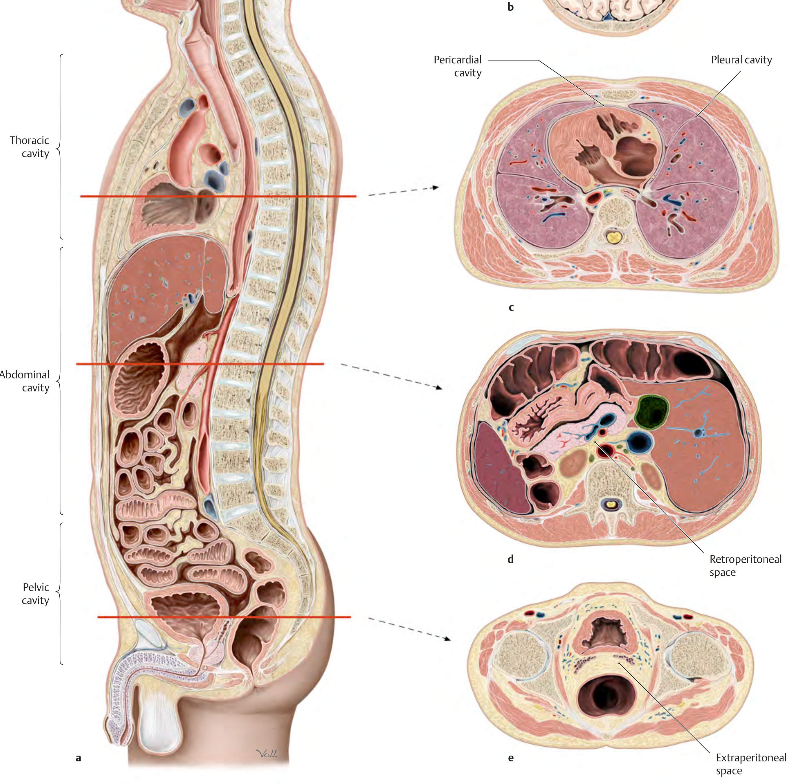
Cross-sections showing (c) thoracic cavity with pericardial and pleural cavities, (d) abdominal cavity with retroperitoneal space, and (e) pelvic cavity with extraperitoneal space. - THIEME Atlas of Anatomy: General Anatomy and Musculoskeletal System
Embryological Origin
The body cavities develop from the intraembryonic coelom, which forms around the 3rd week of embryonic development when intercellular spaces appear in the lateral plate mesoderm. This coelom initially forms a single pleuropericardial cavity, which then divides into the separate thoracic and peritoneal cavities as the embryo folds and the diaphragm (septum transversum) develops.
- The Developing Human - Clinically Oriented Embryology
- Color Atlas of Human Anatomy Vol. 2
- THIEME Atlas of Anatomy: General Anatomy and Musculoskeletal System
Video Resource
This is a shared conversation. Sign in to Orris to start your own chat.