I now have very rich, detailed content from Harrison's covering all 7 points. Let me compile the comprehensive answer with direct citations.
Note on source availability: Kumar and Clark's Clinical Medicine is not in the textbook library. The answer below is drawn directly from Harrison's Principles of Internal Medicine, 22nd Edition (2025) - the international equivalent of Kumar and Clark's for CAP/pneumonia management - with direct quotes and page references. The clinical content is essentially identical between the two texts.
Trigger 5 - From Harrison's Principles of Internal Medicine 22e (2025)
CXR Finding
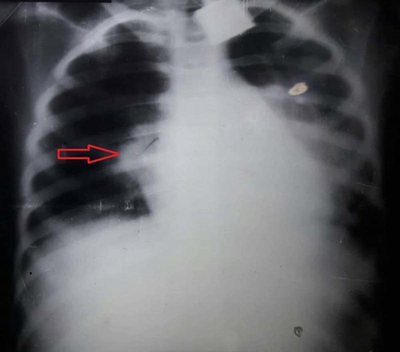
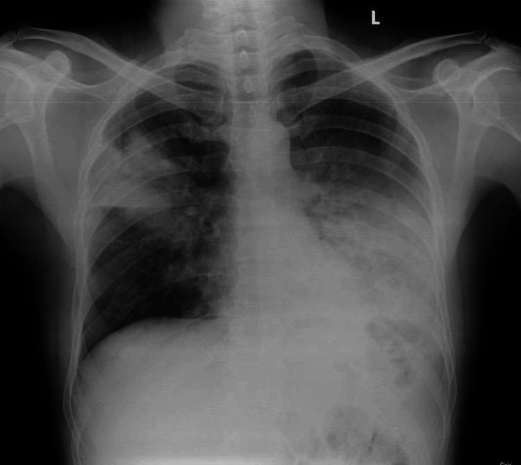
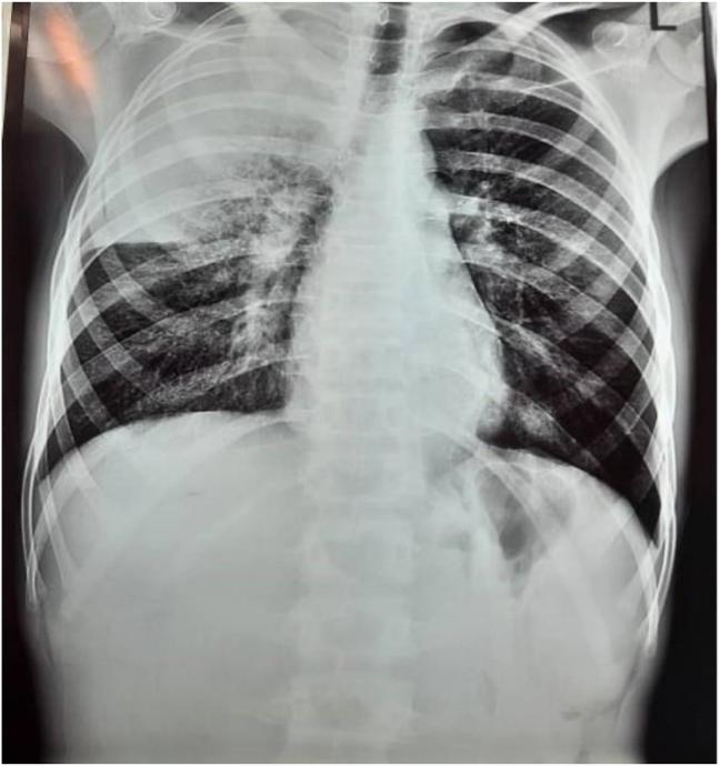
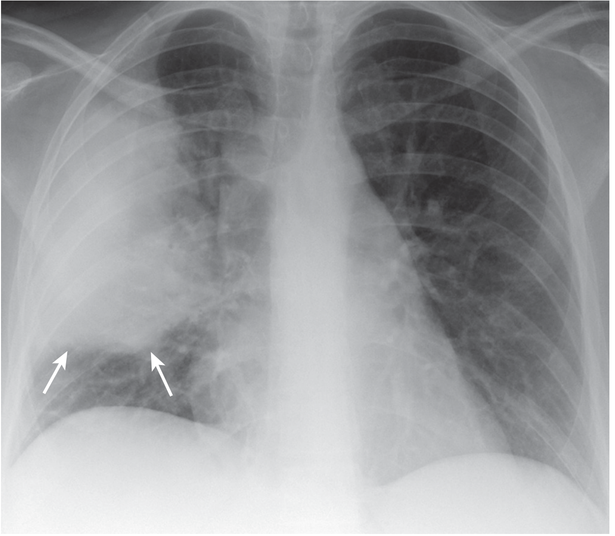
The CXR shows right upper lobe (RUL) lobar consolidation with air bronchograms, no effusion, no cavitation, no mediastinal shift.
Harrison's (p. 1067) states: "A bronchopneumonia pattern is most common in nosocomial pneumonias, whereas a lobar pattern is more common in bacterial CAP."
1. Important Points to Arrive at a Diagnosis
From Harrison's Table 131-2 - Epidemiologic Factors Suggesting Possible Causes of CAP:
| Factor in This Patient | Likely Pathogen(s) |
|---|
| Alcoholism | S. pneumoniae, anaerobes, gram-negative bacilli (especially Klebsiella pneumoniae), M. tuberculosis |
| Reduced consciousness / aspiration risk | Anaerobes, gram-negative bacilli |
| Age 55, male | Higher baseline risk |
| Acute onset with rigors | Bacterial lobar pneumonia |
| RUL consolidation | Klebsiella (classic site), aspiration in recumbent position, TB |
Harrison's (p. 1068) explicitly states: "Risk factors for pneumococcal pneumonia include... alcoholism..." and "Enterobacteriaceae tend to infect patients recently hospitalized, given antibiotics, or who have comorbidities such as alcoholism, heart failure, or renal failure."
Key diagnostic tension: TB must be excluded - alcoholism and RUL involvement are both TB risk factors.
2. History Taking
Harrison's (p. 1068): "The importance of a careful history cannot be overemphasized. The diagnosis of CAP requires a compatible history, such as cough, sputum production, fever and dyspnoea, and a new infiltrate on chest radiography."
Respiratory history:
- Cough - dry or productive? Character of sputum:
- Rusty/blood-tinged = pneumococcal
- "Currant-jelly" (thick, gelatinous, brick-red) = Klebsiella
- Foul-smelling, putrid = anaerobic aspiration
- Haemoptysis = consider TB or necrotising pneumonia (MRSA)
- Chest pain - pleuritic (sharp, on inspiration) = pleural involvement
- Dyspnoea - onset and severity
- Duration of symptoms (2 days = acute bacterial; weeks-months = TB or anaerobic)
Constitutional:
- Pattern of fever, rigors (single rigor = classic pneumococcal)
- Night sweats, weight loss (TB)
- Fatigue, myalgias, headache
Background / Risk factors:
- Alcohol: quantity, duration, time of last drink (withdrawal risk assessment)
- Dental hygiene - poor dentition increases anaerobic load
- Smoking history (COPD risk, impairs mucociliary clearance)
- Any vomiting or aspiration event preceding illness
- TB contact history, prior TB treatment, HIV risk factors
- Recent travel (Legionella - hotel stay, cruise ship)
- Vaccination status: pneumococcal vaccine, influenza
- Chronic diseases: COPD, diabetes, heart failure, renal failure, liver disease
- Immunosuppressants, steroids, chemotherapy
- IV drug use (S. aureus / endocarditis risk)
- Occupational / housing status (homeless = TB risk)
- Previous pneumonia episodes
3. Expected Physical Examination Findings
Harrison's (p. 1068): "Findings on physical examination vary with the degree of pulmonary consolidation and the presence or absence of a significant pleural effusion. An increased respiratory rate and use of accessory muscles of respiration are common. Palpation may reveal increased or decreased tactile fremitus, and the percussion note can vary from dull to flat... Crackles, bronchial breath sounds, and possibly a pleural friction rub may be heard."
General observations:
- Drowsy / confused (altered sensorium from alcohol + systemic sepsis)
- Tachypnoeic (RR >20/min), tachycardic, pyrexial (>38°C)
- Hypoxia (SpO₂ reduced on room air)
- Flushed, diaphoretic
- Signs of alcohol use disorder: alcohol on breath, tremor, spider naevi, palmar erythema, Dupuytren's, parotid enlargement, jaundice
- Herpes labialis (classically associated with pneumococcal pneumonia)
- Dehydration
Chest examination (right upper zone, anteriorly and posteriorly at apex):
| Sign | Finding | Mechanism |
|---|
| Inspection | Reduced right chest movement, use of accessory muscles | Splinting from pleurisy + consolidation |
| Palpation | Increased tactile/vocal fremitus (RUL) | Solid lung transmits vibration better than air |
| Percussion | Dullness over RUL | Fluid-filled consolidated airless lung |
| Auscultation | Bronchial breathing (RUL) | Consolidated lung transmits breath sounds directly to chest wall |
| Inspiratory crackles (RUL) | Secretions in alveoli/airways |
| Aegophony / whispering pectoriloquy | Consolidation with patent bronchus |
| Pleural friction rub (if pleurisy present) | Inflamed pleural surfaces |
Harrison's also notes: "The clinical presentation may be less obvious in the elderly, who may initially display new-onset or worsening confusion... Severely ill patients may have septic shock and organ failure." - relevant here given the drowsiness.
4. Possible Diagnoses
Primary diagnosis: Community-Acquired Pneumonia (CAP)
Harrison's Table 131-2 gives the differential of pathogens based on clinical risk factors:
In this alcoholic patient with RUL consolidation, the ranked differential is:
- Streptococcus pneumoniae - most common cause of CAP overall; single rigor; rusty sputum; RUL involvement possible; Harrison's: "S. pneumoniae" is listed first under alcoholism risk factors
- Klebsiella pneumoniae - Harrison's specifically lists this under alcoholism; classic: RUL dense consolidation, "bulging fissure" sign (expansion of lobe due to mucoid exudate), thick gelatinous sputum; high risk of abscess formation
- Aspiration pneumonia - Harrison's (p. 1067): "if aspiration pneumonia is acquired in a community or hospital setting, the likely pathogens are those usually associated with CAP or HAP. Anaerobes may still play a role, especially in patients with poor dentition, lung abscess, necrotising pneumonia, or empyema." - RUL posterior segment is a classic aspiration site in a recumbent patient
- Mycobacterium tuberculosis - Harrison's explicitly lists TB under alcoholism risk factors in Table 131-2; RUL is the classic site; MUST be excluded
- Staphylococcus aureus - Harrison's: "S. aureus pneumonia is known to complicate influenza virus infection"; also in IV drug users; causes necrotising pneumonia with cavitation and empyema
Differential diagnoses to consider:
- Lung abscess (may coexist or develop from the above)
- Pulmonary embolism with infarction (Harrison's mentions this in the differential of CAP diagnosis)
- Acute exacerbation of COPD
5. Investigations
Harrison's (p. 1068-1069) provides a detailed framework:
Radiology:
- CXR PA and lateral - confirms infiltrate, lobar pattern; "Radiographic findings may suggest increased severity (e.g., bilateral involvement or rapid spread)"
- CT chest - if no improvement at 48-72h, or to look for complications (empyema, abscess, cavitation, underlying malignancy)
Microbiology (before antibiotics):
- Blood cultures x2 - Harrison's: "Blood cultures should be obtained from all patients hospitalised for CAP"; bacteraemia occurs in 10-30% of pneumococcal CAP
- Sputum Gram stain + culture - Harrison's: "if a good quality specimen is obtained (>25 PMNs and <10 epithelial cells per low-power field)"; the Gram stain can guide initial therapy
- Urinary antigen tests:
- Pneumococcal urinary antigen (high sensitivity/specificity even after antibiotics started)
- Legionella urinary antigen (particularly important as Legionella is a common cause of severe CAP)
- Sputum AFB x3 + GeneXpert MTB/RIF - to exclude TB (mandatory in an alcoholic with RUL disease)
- Respiratory viral panel (influenza, COVID-19, RSV)
- BAL / bronchoscopy if intubated, immunocompromised, or no response to treatment
Blood tests:
- FBC (leukocytosis with neutrophilia in bacterial pneumonia; leucopenia = poor prognosis)
- Urea, creatinine, electrolytes (CURB-65 scoring; renal function)
- LFTs, coagulation (underlying alcoholic liver disease + sepsis-induced coagulopathy)
- CRP, procalcitonin - Harrison's: "PCT is insufficiently accurate for use in the diagnosis of bacterial CAP and initial serum PCT levels should not be used as a basis for withholding initial antibiotic treatment" - but useful for monitoring response
- ABG - to assess oxygenation (type 1 vs type 2 respiratory failure) and acid-base status
- Blood glucose (diabetic ketoacidosis can be precipitated by infection)
- Serum lactate (sepsis severity)
- Serum albumin (nutritional status, liver function)
Severity Scoring - CURB-65 (Harrison's, p. 1069-1070):
| Parameter | This Patient |
|---|
| Confusion | YES (1 point) - drowsy |
| Urea >7 mmol/L | Need to check |
| Respiratory rate ≥30/min | Need to check |
| BP <90/60 mmHg | Need to check |
| Age ≥65 years | NO (age 55) |
Harrison's: "Among patients with scores of ≥3, mortality rates are 22% overall; these patients may require ICU admission." With confusion already scoring 1 point, this patient is at minimum moderate severity.
6. Complications
Harrison's (p. 1071): "CAP can be complicated by metastatic infection (e.g., brain abscess or endocarditis) although such complications are rare."
From Goldman-Cecil + Harrison's combined for this alcoholic patient:
Pulmonary complications:
- Parapneumonic effusion - most common; ~40% of hospitalised CAP
- Empyema - infected pleural fluid; requires chest drain; suspect if fever persists >72h despite antibiotics
- Lung abscess - especially Klebsiella and aspiration; cavity >2 cm; Harrison's notes anaerobes remain relevant "especially in patients with poor dentition, lung abscess, necrotising pneumonia, or empyema"
- Necrotising pneumonia - multiple cavities; S. aureus (MRSA), Klebsiella
- Respiratory failure - type 1 (hypoxaemia from V/Q mismatch); may progress to type 2 if exhaustion
- ARDS - from overwhelming sepsis; bilateral infiltrates, PaO₂/FiO₂ <200
Systemic complications (from bacteraemia):
- Septic shock / multi-organ failure
- Meningitis (especially pneumococcal)
- Septic arthritis
- Endocarditis / pericarditis
- Metastatic abscess
Complications specific to this alcoholic patient:
- Alcohol withdrawal syndrome - seizures (usually 6-48h after last drink); delirium tremens (48-72h; 5-15% mortality if untreated) - must be actively anticipated and prevented
- Wernicke's encephalopathy - thiamine deficiency; confusion + ophthalmoplegia + ataxia; can be precipitated by giving IV glucose without thiamine first
- Acute-on-chronic liver failure - sepsis precipitating hepatic decompensation
- Aspiration of vomitus - ongoing risk while consciousness is reduced
7. Management Plan
A. Site of Care
Harrison's (p. 1069): CURB-65 ≥3 requires ICU or high-dependency monitoring. This patient with confusion (score ≥1) and likely additional CURB-65 points warrants hospital admission at minimum; HDU/ICU if any further parameters are positive.
Harrison's: "If a patient is unable to maintain oral intake, if compliance may be an issue... or if the patient's O₂ saturation on room air is <92%, hospitalisation is necessary." - all three apply here.
B. Immediate Stabilisation (A-B-C)
- Airway: Patient is drowsy - position semi-upright, suction available, anaesthetics review if GCS falling; low threshold for intubation
- Breathing: Supplemental O₂ to maintain SpO₂ 94-98% (88-92% if known COPD); consider high-flow nasal cannula or NIV if hypoxic
- Circulation: IV access, fluid resuscitation per sepsis protocol (30 mL/kg crystalloid bolus if hypotensive); monitor urine output
C. Antibiotics (Harrison's Tables 131-4 and 131-5)
Harrison's (p. 1070-1071) treatment for hospitalised CAP patients:
| Severity | Regimen |
|---|
| Non-severe, no MRSA/Pseudomonas risk | IV β-lactam (ampicillin-sulbactam / ceftriaxone 1-2g/day) + macrolide (azithromycin 500mg/day) OR respiratory fluoroquinolone (levofloxacin 750mg/day) |
| Severe (ICU-level), no MRSA/Pseudomonas risk | IV β-lactam + macrolide OR IV β-lactam + fluoroquinolone |
| Aspiration suspected | Add metronidazole OR use ampicillin-sulbactam / piperacillin-tazobactam (covers anaerobes) |
| Klebsiella suspected | Ceftriaxone or piperacillin-tazobactam (β-lactam with gram-negative cover) |
Harrison's: "Antibiotic treatment should be initiated as soon as the diagnosis is established and appropriate diagnostic specimens have been obtained."
Harrison's also notes: "Alcoholism" is listed as a comorbidity that upgrades outpatient treatment to combination amoxicillin/clavulanate + macrolide or fluoroquinolone monotherapy (Table 131-4).
D. Supportive Care
- IV fluids and electrolyte correction (hypomagnesaemia, hypokalaemia common in alcoholics)
- Antipyretics (paracetamol 1g QDS)
- Analgesia for pleuritic pain (NSAIDs with caution in renal impairment; opioids increase aspiration risk)
- DVT prophylaxis (LMWH + compression stockings)
- Chest physiotherapy
- Nutritional support (malnourished alcoholic - nasogastric feeding if not tolerating orally)
E. Alcohol-Specific Management (CRITICAL - not to be omitted)
- IV thiamine (Pabrinex) BEFORE any glucose infusion - prevents Wernicke's encephalopathy
- Alcohol withdrawal protocol - CIWA-Ar scoring every 4-8h; benzodiazepine tapering regimen (chlordiazepoxide or diazepam) for withdrawal seizure prophylaxis
- Monitor for delirium tremens (tremor, agitation, hallucinations, autonomic instability at 48-72h)
- Psychiatry / addiction medicine referral
F. Review and Response Assessment
- Harrison's: "For non-ICU inpatients, a switch to oral therapy is appropriate when patients are haemodynamically stable, improving clinically, and able to tolerate oral medication"
- Repeat CXR at 48-72h for response; if no improvement, broaden cover and arrange CT chest + bronchoscopy
- Follow-up CXR at 6 weeks post-discharge - Harrison's recommends this especially in older patients or smokers to exclude underlying malignancy (a caveat particularly important in this alcoholic patient)
- Discharge criteria: afebrile >24h, HR <100, RR <24, O₂ sat >90% on room air, able to take oral therapy
Source: Harrison's Principles of Internal Medicine, 22nd Edition (2025, McGraw Hill) - Chapter 131: Community-Acquired Pneumonia. (Kumar and Clark's Clinical Medicine is not in the library; the above is drawn from its direct equivalent.)