
Classification of firearm wounds management of gun shot injury from front of chest
"gunshot wound" AND "chest" AND management
Classification of Firearm Wounds and Management of Gunshot Injury to the Front of the Chest
I. Classification of Firearm (Gunshot) Wounds
A. By Wound Morphology
- Typically smaller, round/oval with inverted (indrawn) edges
- Surrounded by an abrasion collar (zone of contusion/friction from bullet spin)
- Edges relatively clean and regular
- May have tattooing (stippling) from unburnt gunpowder in close-range shots
- Burn/blackening/singed hair in contact/near-contact wounds
- Usually larger, irregular, stellate or cruciate, with everted (outturned) edges
- No abrasion collar
- More tissue destruction
- Note: Exit wound may be absent if bullet is retained (low-velocity, dissipated energy)
- Bullet skims across surface without full penetration
- Elongated, boat-shaped trough
- Beveled edges
B. By Range of Fire
| Range | Features |
|---|---|
| Contact | Muzzle placed against skin; star-shaped laceration; blackening, burning, carbon tattooing inside wound |
| Close range (<60 cm) | Gunpowder tattooing/stippling on skin; singeing |
| Intermediate (60 cm - 1 m) | Some tattooing but no burning |
| Distant (>1 m) | Neat entry wound, abrasion collar only; no tattooing |
C. By Ballistic Type (Weapon/Bullet Velocity)
- Bullet causes damage primarily by laceration and crushing along its path
- Relatively limited tissue destruction
- Wound track = caliber of bullet
- Significant cavitation effect: temporary cavity much larger than bullet (up to 30x diameter)
- Massive tissue destruction beyond the wound track
- Yaw and tumbling: bullet becomes unstable inside tissue - greatly increases tissue damage
- Fragmentation possible
- Entry wound often deceptively small compared to internal damage
- Contact/close range: devastating single large wound, wad/pellets + powder inside wound
- Intermediate: pellets begin to scatter, multiple puncture wounds
- Distant: diffuse pellet pattern, each wound individually minor
D. By Depth of Penetration
- Perforating - bullet enters AND exits (both entry + exit wound present)
- Penetrating - bullet enters and is retained within body (entry wound only)
- Avulsive/Tangential - bullet grazes surface without full penetration
II. Injuries Caused by Gunshot to the Front of the Chest
| Zone | Structures at Risk |
|---|---|
| Cardiac box (sternal notch to xiphoid, nipple-to-nipple) | Heart, great vessels, trachea, esophagus |
| Right hemithorax | Right lung, right hilum, liver (low), IVC |
| Left hemithorax | Left lung, left hilum, heart, aorta |
| Upper chest | Trachea, subclavian vessels, brachial plexus |
| Thoracoabdominal (below 4th ICS anteriorly) | Diaphragm, liver, spleen, stomach |
III. Specific Injury Patterns and Management
1. Simple/Tension Pneumothorax
- Respiratory distress, hypoxia, tachypnea
- Absent unilateral breath sounds + hyperresonance
- Tracheal deviation away from affected side
- Distended neck veins, hypotension, tachycardia
- Differentiate from tamponade: tamponade has muffled heart sounds, NO tracheal deviation
- Tension PTX = clinical emergency, do NOT wait for CXR
- Immediate needle decompression: 2nd intercostal space, midclavicular line (or 5th ICS, anterior axillary line in obese)
- Followed by chest tube (intercostal drain) in the 4th-5th ICS, anterior axillary line ("safe triangle")
- Simple PTX: chest tube only; occult PTX (CT only, no CXR finding) may be observed if patient is stable and not on positive pressure ventilation
2. Open Pneumothorax (Sucking Chest Wound)
- Apply three-sided occlusive dressing (allows air egress but prevents inflow) - Asherman chest seal or petroleum gauze
- Insert a chest tube remote from the wound - BEFORE closing the defect (to prevent converting to tension PTX)
- Wound debridement and formal surgical closure once the patient is stable
3. Hemothorax
- Small: <300 mL
- Moderate: 300-1500 mL
- Massive: >1500 mL (immediately life-threatening)
- >85% of cases can be definitively managed with chest tube alone (28-32 Fr intercostal drain, 4th-5th ICS, anterior axillary line) - Schwartz's Principles of Surgery, 11e
- Emergency Department Thoracotomy / Surgical indications:
- Initial chest tube output >1500 mL (20% estimated blood volume in children)
- Ongoing output >150-200 mL/hr for 4 hours (or 2-3 mL/kg/hr in children)
- Hemodynamic instability not responding to resuscitation
- Patient physiology should guide surgical decision over absolute blood volumes
- Retained hemothorax: Video-assisted thoracoscopic surgery (VATS) preferred in stable patients
4. Cardiac Tamponade
- Elevated CVP / distended neck veins
- Hypotension + tachycardia
- Muffled heart sounds
- Pericardiocentesis has NO role in penetrating cardiac tamponade (clot prevents aspiration)
- Correct treatment is operative:
- Subxiphoid pericardial window (if patient is relatively stable)
- OR left anterolateral thoracotomy / sternotomy for direct cardiac repair
- If in ED with witnessed cardiac arrest from penetrating chest injury: Emergency Department Resuscitative Thoracotomy (EDRT)
5. Emergency Department Resuscitative Thoracotomy (EDRT)
| Signs of Life in ED | Mechanism | Location | Recommendation |
|---|---|---|---|
| Yes | Penetrating | Thoracic | Strongly yes |
| Yes | Penetrating | Non-thoracic | Yes |
| No (witnessed arrest) | Penetrating | Thoracic | Yes |
| No (unwitnessed) | Blunt | Any | Not recommended |
6. Pulmonary Parenchymal Injury
- Most are managed with tube thoracostomy alone
- Persistent air leak: bronchoscopy-directed fibrin glue or VATS
- Central injuries during thoracotomy: pulmonary tractotomy (GIA stapler inserted into injury track, filleted open, individual vessels and bronchi ligated) - avoids formal lobectomy
- Peripheral lacerations: stapled wedge resection
- Formal lobectomy only for proximal vascular/bronchial injury found on tractotomy
- Pneumonectomy is almost always fatal due to right heart decompensation
7. Tracheobronchial Injury
- Pass ETT beyond the injury or into contralateral mainstem bronchus for ventilation
- Primary end-to-end repair (3-0 PDS, single layer interrupted)
- Suture line buttressed with vascularized tissue (pericardium, intercostal muscle, pleura)
- Bronchial injuries <1/3 circumference with no major air leak: expectant management
8. Thoracoabdominal Wounds (Below 4th ICS Anteriorly)
- Must exclude diaphragmatic injury (diaphragm rises to 4th ICS anteriorly in expiration)
- Diaphragmatic wounds may not be apparent initially - diagnose by laparoscopy
- All injuries to the diaphragm require repair (staple/suture)
- Must also assess for injury to liver, spleen, stomach
IV. ATLS Primary Survey Framework for Penetrating Chest GSW
- A - Airway: Secure; cricothyroidotomy if cannot intubate
- B - Breathing: Identify/treat tension PTX, open PTX, hemothorax, flail chest
- C - Circulation: FAST for tamponade; control hemorrhage; IV access; permissive hypotension (target SBP 80-90 mmHg in penetrating torso trauma) until surgical hemorrhage control - AVOID fluid overload
- D - Disability: GCS, pupils
- E - Exposure: Full exposure, look for exit wound, thoracoabdominal involvement
Classification of firearm wounds management of gun shot injury from front of chest
Classification of Firearm Wounds and Management of Gunshot Injury to the Front of the Chest
PART 1: CLASSIFICATION OF FIREARM WOUNDS
A. Classification by Wound Morphology
1. Entry Wound
- Typically smaller, round or oval with inverted (indrawn) edges
- Has an abrasion collar (graze contusion ring) - caused by bullet spin and friction against skin
- Central defect = approximately bullet caliber
- Surrounding skin intact / relatively clean edges
- At contact range: star-shaped laceration from gas blast, blackening, burning, carbon tattooing inside the wound canal
- At close range (<60 cm): gunpowder stippling/tattooing on skin, possible singeing
2. Exit Wound
- Usually larger, irregular, stellate or cruciate shape
- Edges are everted (outturned)
- No abrasion collar
- Greater tissue destruction
- May be absent if bullet is retained (low-velocity, energy dissipated, or bullet fragmented)
3. Tangential / Graze Wound
- Bullet skims the surface without full penetration
- Elongated, boat-shaped trough with beveled edges
- Both ends have features of entry (abrasion collar at entry end)
B. Classification by Range of Fire
| Range | Wound Features |
|---|---|
| Contact (muzzle touches skin) | Star-shaped laceration; burning, blackening, carbon tattooing inside wound; muzzle imprint on skin |
| Close range (<60 cm) | Gunpowder stippling/tattooing on surrounding skin; singeing of hair |
| Intermediate (60 cm - 1 m) | Faint tattooing present; no burning |
| Distant (>1 m) | Clean entry wound with abrasion collar only; no tattooing |
C. Classification by Ballistic Type (Velocity)
1. Low-Velocity Wounds (<600 m/s - handguns, pistols)
- Damage by laceration and crushing along bullet track
- Wound track = approximately bullet caliber
- Relatively limited tissue destruction beyond direct path
- Bullet often retained (penetrating, no exit wound)
2. High-Velocity Wounds (>600 m/s - military rifles, assault weapons)
- Cavitation effect: temporary cavity forms that is up to 30x the bullet diameter - massive tissue destruction well beyond the bullet track
- Yaw and tumbling: bullet becomes unstable inside tissue, dramatically increasing damage
- Entry wound is deceptively small compared to internal destruction
- Fragmentation possible - multiple secondary missiles
- Exit wound is large and irregular
3. Shotgun Wounds
| Distance | Pattern |
|---|---|
| Contact/Close | Single devastating wound; wad + pellets + powder may be inside |
| Intermediate | Pellets scatter - multiple puncture wounds |
| Distant | Diffuse pellet pattern; individual wounds minor; pattern useful for range estimation |
D. Classification by Depth of Penetration
- Perforating wound - bullet enters AND exits (entry + exit wound present)
- Penetrating wound - bullet enters and is retained (entry wound only)
- Avulsive/Tangential wound - bullet grazes the surface without full penetration
PART 2: INJURIES FROM GUNSHOT TO THE FRONT OF THE CHEST
Anatomical Zones at Risk - Anterior Chest
| Zone | Structures at Risk |
|---|---|
| Cardiac box (sternal notch to xiphoid, nipple-to-nipple) | Heart, great vessels, trachea, esophagus, thoracic duct |
| Right hemithorax | Right lung/hilum, liver (if low), IVC |
| Left hemithorax | Left lung/hilum, heart, descending aorta |
| Upper anterior chest | Trachea, subclavian vessels, brachial plexus |
| Thoracoabdominal (below 4th ICS anteriorly) | Diaphragm, liver, spleen, stomach |
Critical point: The diaphragm rises to the 4th intercostal space anteriorly during expiration. Wounds at or below the nipple line may injure abdominal viscera. Coincident thoracic penetration occurs in up to 46% of patients with abdominal injuries.
PART 3: MANAGEMENT OF SPECIFIC INJURY PATTERNS
1. Simple Pneumothorax
- Small: <1/3 lung collapse on CXR
- Large: complete lung collapse, no mediastinal shift, no hypotension
- Intercostal drain (chest tube) in 4th-5th ICS, anterior axillary line
- Occult PTX (CT-only finding, not on CXR): may be observed carefully if hemodynamically stable and not on positive-pressure ventilation. Failure of observation rate = 6% overall (14% if on PPV). Factors predicting failure: PTX >7 mm, PPV, respiratory distress, hemothorax, progression on serial CXR
2. Tension Pneumothorax
- Air hunger, hypoxia, tachypnea
- Hyperresonance + absent breath sounds - ipsilateral
- Tracheal deviation away from affected side
- Distended neck veins + hypotension + tachycardia
- Note: Tracheal deviation may be absent if patient is intubated. Neck veins may be flat with concurrent hypovolaemia
- Tamponade: muffled heart sounds, NO tracheal deviation, NO asymmetric breath sounds
- Tension PTX: tracheal deviation, hyperresonance, absent breath sounds
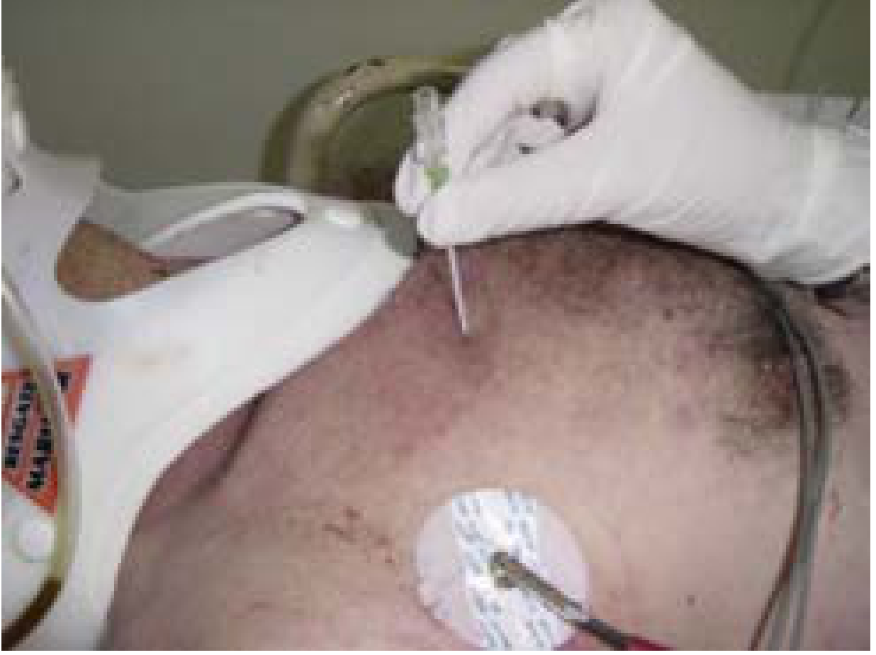
- Immediate needle decompression - 14-16G angiocatheter, 2nd ICS midclavicular line (or 5th ICS anterior axillary line in obese patients). Rush of air = confirms diagnosis, converts to simple PTX
- Followed immediately by chest tube (intercostal drain) in the "safe triangle" (bounded by: lateral border of pectoralis major anteriorly, latissimus dorsi posteriorly, line perpendicular to nipple inferiorly) - Bailey & Love, 28e
3. Open Pneumothorax ("Sucking Chest Wound")
- Apply three-sided occlusive dressing (petroleum gauze / Asherman chest seal) - allows air to exit on expiration but prevents entry on inspiration
- Place intercostal drain remote from the wound - BEFORE closing the defect, to prevent conversion to tension PTX
- Once patient is stable: wound debridement and formal surgical closure in the operating theatre
4. Hemothorax
- Small: <300 mL
- Moderate: 300-1500 mL
- Massive: >1500 mL - may present with tension physiology
- >85% of all hemothoraces are definitively managed with chest tube alone (reexpansion of the lung seals the low-pressure pulmonary laceration)
- One caveat: even if initial output is 1.5 L, if bleeding stops and lung is re-expanded in a hemodynamically stable patient, non-operative management may still be pursued
| Indication | Threshold |
|---|---|
| Initial chest tube output - penetrating | >1000 mL |
| Initial chest tube output - blunt | >1500 mL |
| Ongoing chest tube output | >200 mL/hr for 3 consecutive hours (non-coagulopathic) |
| Caked/retained hemothorax | Despite 2 chest tubes |
| Great vessel injury | Any |
| Pericardial tamponade | Any |
| Oesophageal perforation | Any |
| Air embolism | Any |
- Retained hemothorax: Video-assisted thoracoscopic surgery (VATS) is the preferred approach in stable patients; reduces empyema risk
5. Cardiac Tamponade
- Elevated CVP / distended neck veins
- Hypotension + tachycardia
- Muffled heart sounds
- Pericardiocentesis has NO role in penetrating cardiac tamponade - clot inside pericardium prevents effective aspiration
- Correct treatment is operative:
- Subxiphoid pericardial window - for relatively stable patients (allows drainage and diagnosis)
- Left anterolateral thoracotomy or median sternotomy - for definitive cardiac repair
- Emergency Department Thoracotomy - if patient arrests in the ED
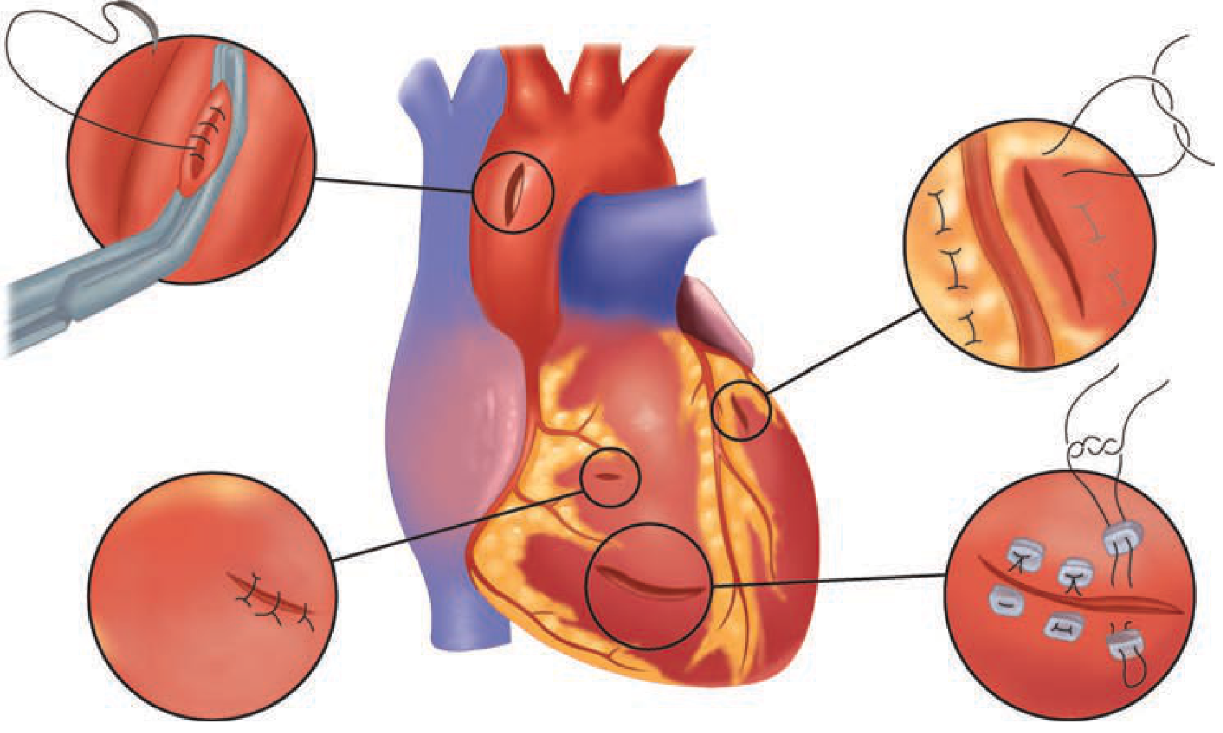
- Atrial injuries: Satinsky vascular clamp for hemorrhage control
- Ventricular injuries: digital pressure initially; skin staples for temporary control of LV lacerations; Foley catheter balloon for large stellate lesions (use cautiously - traction enlarges wound)
- Definitive repair: running 3-0 polypropylene or interrupted pledgeted 2-0 polypropylene sutures; pledgets especially important for thin-walled right ventricle
- Injuries adjacent to coronary arteries: horizontal mattress sutures (running sutures would occlude the coronary artery)
- Stellate/friable GSW wounds: surgical adhesive (BioGlue) may be used if edges cannot be fully approximated
6. Emergency Department Resuscitative Thoracotomy (EDRT)
- Release pericardial tamponade
- Direct hemorrhage control
- Open cardiac massage
- Descending aortic cross-clamping (prioritizes cerebral + coronary perfusion; controls sub-diaphragmatic hemorrhage)
- Internal defibrillation
| Signs of Life in ED | Mechanism | Location | Recommendation |
|---|---|---|---|
| Yes | Penetrating | Thoracic | Strongly YES |
| No | Penetrating | Thoracic | Conditional yes |
| Yes | Penetrating | Extrathoracic | Conditional yes |
| No | Penetrating | Extrathoracic | Conditional yes |
| Yes | Blunt | Any | Conditional yes |
| No | Blunt | Any | Conditional NO |
- Penetrating torso: <15 min prehospital CPR
- Penetrating neck: <5 min prehospital CPR
- Blunt: <10 min prehospital CPR
- Best outcome (~35%): Single penetrating stab wound to cardiac box + tamponade on FAST + witnessed arrest in ED
- GSW to chest: lower survival than stab wounds
- Blunt cardiac arrest: near-zero survival if unwitnessed
- Patient supine, left arm raised
- Incision along 4th-5th ICS (males: along nipple line; females: along inframammary fold)
- Enter just above the superior border of the rib (avoids intercostal neurovascular bundle)
- Open pericardium longitudinally, anterior to phrenic nerve, evacuate clot
- Repair cardiac wound, cross-clamp descending aorta if needed
- Extend to "clamshell" (bilateral) if right-sided injury suspected
7. Tracheobronchial Injury
- Pass ETT beyond the injury or into the contralateral mainstem bronchus to maintain ventilation
- Primary end-to-end repair with 3-0 PDS, single-layer interrupted sutures; buttress suture line with vascularized tissue (pericardium, intercostal muscle, pleura)
- Injuries <1/3 circumference with no major air leak: expectant management
- Peripheral bronchial injuries with persistent leak: bronchoscopically directed fibrin glue
8. Pulmonary Parenchymal Injury
- Majority managed with tube thoracostomy alone
- Pulmonary tractotomy: for central injuries encountered during thoracotomy - GIA-100 stapler inserted into wound track, filleted open, individual vessels and bronchi ligated selectively; avoids formal lobectomy
- Peripheral lacerations: stapled wedge resection
- Formal lobectomy: only when tractotomy reveals proximal injury
- Pneumonectomy: usually fatal (right heart decompensation from acute increase in afterload)
- Bronchovenous fistula (air embolism risk): minimized by prompt control of major air leaks
9. Thoracoabdominal Wounds (Below Nipple Line)
- Must always exclude diaphragmatic injury in anterior wounds below the 4th ICS
- Initial wound may appear thoracic but violate the peritoneum
- Diagnosis: laparoscopy (most sensitive for diaphragmatic lacerations, which can be very small)
- All diaphragmatic injuries require repair
- Must assess for concomitant injury to liver, spleen, stomach
PART 4: ATLS APPROACH - INITIAL RESUSCITATION FRAMEWORK
Primary Survey Priorities (ABCDE)
| Priority | Action |
|---|---|
| A - Airway | Secure airway early; RSI; if cannot intubate - surgical cricothyroidotomy |
| B - Breathing | Identify and treat: tension PTX, open PTX, massive hemothorax, flail chest |
| C - Circulation | eFAST for tamponade; 2x large-bore IV; permissive hypotension (target SBP 80-90 mmHg) until surgical hemorrhage control; massive transfusion protocol if needed (1:1:1 pRBC:FFP:platelets) |
| D - Disability | GCS, pupils |
| E - Exposure | Full undress; find ALL wounds including exit wounds and thoracoabdominal extension |
Approach to aub in reproductive age group
PALM-COEIN classification abnormal uterine bleeding diagram
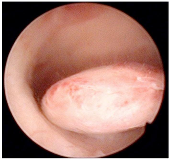
This diagnostic image is an intraoperative hysteroscopic photograph of the uterine cavity. The visual focus is a well-defined, oval-shaped pedunculated endometrial polyp protruding from the posterior uterine wall. The polyp demonstrates a smooth, glistening surface with a pale pink to white color, interspersed with focal areas of erythema and fine vertical striations across its body. Its morphology is slightly lobulated, and it is attached to the uterine wall by a distinct stalk. The surrounding endometrial tissue appears healthy, smooth, and pink, although there is a slight darkening near the base of the pedicle, likely representing minor blood accumulation. This clinical photograph illustrates a common structural cause of abnormal uterine bleeding (AUB), frequently categorized under the 'Polyp' component of the FIGO PALM-COEIN classification system. Such findings are critical in the diagnostic workup of perimenopausal and postmenopausal women to differentiate benign focal abnormalities from endometrial hyperplasia or malignancy.
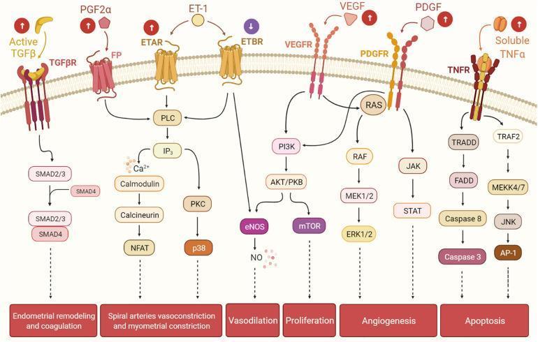
A complex pathophysiology diagram illustrating various cellular signaling pathways in the human endometrium, specifically highlighting mechanisms dysregulated in uterine fibroids. The flow begins with extracellular ligands binding to transmembrane receptors and concludes with functional physiological outcomes. Key pathways include: (1) TGF-β/SMAD signaling leading to endometrial remodeling and coagulation; (2) PGF2α/FP and ET-1/ETAR/ETBR signaling involving PLC, IP3, Calmodulin, and PKC cascades, which result in spiral artery vasoconstriction and myometrial constriction; (3) ETBR/VEGFR/PDGFR cross-talk through PI3K/AKT/mTOR and RAS/RAF/MEK/ERK pathways, driving vasodilation (via NO), proliferation, and angiogenesis; and (4) TNFα/TNFR signaling through Caspase cascades (apoptosis) and JNK/AP-1 pathways. Icons with upward arrows next to ligands like TGF-β, PGF2α, and VEGF indicate molecular upregulation typically seen in the presence of uterine fibroids. This educational diagram is targeted at an advanced level, illustrating how ligand-receptor interactions contribute to heavy menstrual bleeding and abnormal uterine vascular architecture.
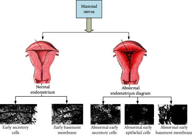
An educational comparison diagram illustrating the differences between normal and abnormal endometrium (specifically endometriosis). The top section displays two anatomical illustrations of a mammal uterus. The 'Normal endometrium' shows a smooth, uniform uterine cavity with an open, Y-shaped internal structure. The 'Abnormal endometrium diagram' depicts a constricted, triangular uterine cavity with thickened, irregular linings and growths, suggestive of ectopic tissue implantation. The bottom section presents five high-magnification grayscale diagnostic images representing cellular pathology. The normal side shows 'Early secretory cells' and 'Early basement membrane' with visible, organized textures and clear cellular arrangements. In contrast, the abnormal side shows 'Abnormal early secretory cells,' 'Abnormal early epithelial cells,' and 'Abnormal early basement membrane,' which exhibit significantly darker, less defined clusters and disrupted structural integrity. This diagram serves as a visual guide to the gross anatomical and microscopic cellular changes associated with endometrial pathology.
Approach to Abnormal Uterine Bleeding (AUB) in the Reproductive Age Group
1. Definition and Terminology
Normal Menstrual Parameters (FIGO)
| Parameter | Normal Range |
|---|---|
| Cycle frequency | 24-38 days |
| Cycle regularity | ± <7-9 days variation |
| Duration of flow | 4-8 days (mean 4.7 days) |
| Volume of blood loss | <80 mL per cycle (mean 35 mL) |
Important: The term "dysfunctional uterine bleeding (DUB)" is obsolete and should no longer be used. AUB with the PALM-COEIN classification has replaced all previous imprecise terms (menorrhagia, metrorrhagia, menometrorrhagia). - Berek & Novak's Gynecology
2. PALM-COEIN Classification (FIGO 2011)
PALM  = Structural causes (diagnosed by imaging or histology)
COEIN = Non-structural/medical causes
PALM - Structural
| Cause | Code | Key Features |
|---|---|---|
| Polyp | AUB-P | Endometrial/endocervical epithelial proliferations; usually benign; most common cause of intermenstrual bleeding in women >35 years; up to 5% malignant transformation |
| Adenomyosis | AUB-A | Endometrial glands within myometrium; presents with heavy, painful periods; enlarged "boggy" uterus |
| Leiomyoma | AUB-L | Most common benign gynecologic tumor (~70% of women by age 50); classified by location (Type 0-8 per FIGO); submucosal types (0, 1, 2) cause most bleeding |
| - Submucosal | AUB-LSM | Most symptomatically significant for bleeding |
| - Other (intramural/subserosal) | AUB-LO | Less directly related to bleeding |
| Malignancy & hyperplasia | AUB-M | Endometrial hyperplasia (simple/complex, with/without atypia); endometrial carcinoma; always rule out in at-risk women |
COEIN - Non-Structural
| Cause | Code | Key Features |
|---|---|---|
| Coagulopathy | AUB-C | Up to 20% of women with HMB have an underlying coagulopathy; Von Willebrand disease is the most common (up to 13% of AUB cases); often presents since menarche |
| Ovulatory dysfunction | AUB-O | Includes anovulation (PCOS, hypothalamic dysfunction, hyperprolactinemia, thyroid disorders, perimenopause); most common cause in reproductive years |
| Endometrial | AUB-E | Primary endometrial disorder - defective local hemostasis, altered prostaglandin balance, impaired endometrial repair; diagnosis of exclusion |
| Iatrogenic | AUB-I | Medications - anticoagulants, hormonal contraceptives (breakthrough bleeding), intrauterine devices, tamoxifen, antipsychotics (via hyperprolactinemia) |
| Not yet classified | AUB-N | AV malformations, myometrial hypertrophy, rare/poorly understood causes |
3. Causes by Age in Reproductive Group
| Sub-group | Most Likely Causes |
|---|---|
| Adolescents (13-19 yr) | Anovulation (HPO axis immaturity), coagulopathy (VWD), PCOS, pregnancy, exogenous hormones |
| Young adults (20-35 yr) | Pregnancy-related (most common), anovulation (PCOS), polyps, leiomyoma, iatrogenic |
| Mid-reproductive (35-45 yr) | Structural causes rise (fibroids, polyps, adenomyosis), PCOS, endometrial hyperplasia, perimenopause begins |
Pregnancy-related complications are the most common cause of AUB during the reproductive years overall.
4. Approach: History
- Cycle length, regularity, duration, volume (flooding, clots, pad/tampon count)
- Intermenstrual bleeding, postcoital bleeding
- Duration of abnormal pattern (acute vs. chronic AUB)
- Pelvic pain/dysmenorrhea (adenomyosis, endometriosis)
- Pressure symptoms (large fibroids)
- Signs of androgen excess: acne, hirsutism (PCOS)
- Galactorrhea (hyperprolactinemia)
- Weight change, cold intolerance (thyroid)
- Easy bruising, epistaxis, family history of bleeding (coagulopathy)
5. Physical Examination
- Speculum: Cervical lesions, polyps protruding through cervical os, signs of infection, source of bleeding (confirm it is uterine)
- Bimanual: Uterine size, shape, regularity, tenderness (enlarged/irregular = fibroids; enlarged, boggy = adenomyosis); adnexal masses
6. Investigations
Essential for All
| Investigation | Purpose |
|---|---|
| Urine/serum βhCG | First step - exclude pregnancy in all reproductive-age women |
| CBC + platelets | Assess anaemia and thrombocytopenia |
| TSH | Thyroid dysfunction (hypothyroidism associated with AUB-O) |
| Pelvic ultrasound (TVUS preferred) | Best initial imaging - uterine contour, fibroid location/size, endometrial thickness, adnexa |
Selective / Based on Clinical Assessment
| Investigation | Indication |
|---|---|
| Prolactin | Irregular cycles, galactorrhea, suspected hyperprolactinemia |
| Coagulation screen (PT, aPTT, platelet function, VWF antigen + activity, Factor VIII, fibrinogen) | Heavy bleeding since menarche; family history of coagulopathy; patient on anticoagulants; signs of systemic bleeding - done in collaboration with haematologist |
| Androgen profile (testosterone, DHEAS), fasting glucose, lipids | Clinical features of PCOS |
| STI screen (Chlamydia, gonorrhoea) | Risk factors for STI or signs of cervicitis |
| Cervical cytology (Pap smear) | If not up to date |
| Sonohysterography (SIS) | Better delineation of intracavitary lesions (polyps, submucosal fibroids) than standard TVUS |
| Hysteroscopy | Gold standard for intracavitary pathology; diagnostic and therapeutic |
| MRI pelvis | Not first-line; useful for adenomyosis, mapping leiomyoma location/depth pre-operatively, staging endometrial cancer |
Endometrial Sampling (Biopsy)
- Age ≥45 years with AUB (including intermenstrual bleeding)
- Any age with:
- Obesity (unopposed oestrogen exposure)
- Chronic anovulation / PCOS
- AUB refractory to medical management
- Persistent AUB despite treatment
- Elevated familial cancer risk (Lynch syndrome)
Endometrial biopsy (Pipelle) has largely replaced D&C as the office procedure of choice. Hysteroscopy + directed biopsy is the gold standard when focal lesion is suspected or office biopsy is unsatisfactory.
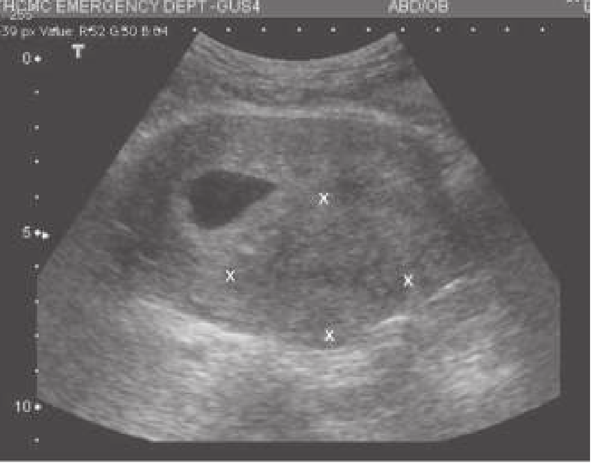
7. Management
Step 1: Assess Haemodynamic Stability
- IV access, fluids, blood products (resuscitation)
- Urgent gynaecology consultation
- Consider urgent surgical options: D&C, uterine artery embolization (UAE), endometrial ablation, hysterectomy
Step 2: Medical Management (First-line for most)
A. Hormonal Therapies
| Agent | Use | Dose/Notes |
|---|---|---|
| Combined oral contraceptive pills (COCPs) | AUB-O (anovulatory); AUB-E; cycle regulation; reduce flow 35-68% | Standard low-dose monophasic pill; contraindicated if thromboembolic history, active smoking >35 yr, breast cancer, liver disease |
| Progestins (oral) | When oestrogen contraindicated; anovulatory AUB | Medroxyprogesterone acetate 5-10 mg days 15-26 of cycle; OR norethindrone 5 mg TDS x 1 week for acute heavy bleeding; 21-day regimen reduces flow but less acceptable than LNG-IUS |
| Levonorgestrel-releasing IUS (LNG-IUS) | Treatment of choice for HMB long-term; comparable to hysterectomy for quality of life; should be offered before hysterectomy | 52 mg LNG-IUS (Mirena); reduces flow up to 90%; also provides contraception |
| Depot medroxyprogesterone acetate (DMPA) | Progestin-only option; causes amenorrhoea over time | Irregular breakthrough bleeding initially (counsel patients); 150 mg IM every 3 months |
| GnRH agonists | Short-term (max 6 months); severe HMB; pre-operative downsizing of fibroids | Leuprolide, goserelin; induce hypo-oestrogenic state; add-back therapy needed if >3-6 months |
| Conjugated equine oestrogen (IV) | Acute heavy bleeding emergency management | 25 mg IV q4-6h; stops acute bleeding by stimulating endometrial growth; always follow with progestin to avoid hyperplasia |
B. Non-Hormonal Therapies
| Agent | Use | Dose/Notes |
|---|---|---|
| Tranexamic acid (antifibrinolytic) | HMB - non-hormonal first choice; FDA-approved 2008 | Oral: 1.3 g q8h x 5 days (during menstruation); IV: 10 mg/kg, max 600 mg for acute bleeding; more effective than NSAIDs for HMB |
| NSAIDs (ibuprofen, mefenamic acid) | HMB + dysmenorrhoea; reduces flow 30-50% | Less effective than tranexamic acid, danazol, or LNG-IUS; avoid in coagulopathy/platelet dysfunction |
| Antibiotics | AUB-I with confirmed STI/endometritis | Directed by culture/sensitivity |
Treatment Efficacy Hierarchy for HMB (Berek & Novak):
LNG-IUS ≈ Danazol > Tranexamic acid > NSAIDs / OCP
Step 3: Management by Specific Cause
| Cause | Specific Management |
|---|---|
| AUB-P (Polyp) | Hysteroscopic polypectomy (definitive); watchful waiting for small asymptomatic polyps |
| AUB-A (Adenomyosis) | LNG-IUS (first-line medical); GnRH agonists; definitive treatment = hysterectomy |
| AUB-L (Leiomyoma) | Submucosal (Type 0-2): hysteroscopic myomectomy; Intramural/subserosal with symptoms: myomectomy (preserves fertility), UAE, hysterectomy; medical: GnRH agonists (pre-op downsizing), ulipristal acetate |
| AUB-M (Malignancy/Hyperplasia) | Hyperplasia without atypia: progestin therapy + surveillance; Atypical hyperplasia/EIN: hysterectomy (high risk of concurrent or progression to cancer); Endometrial cancer: staging hysterectomy + BSO ± lymphadenectomy |
| AUB-O (Ovulatory dysfunction) | Treat underlying cause (PCOS - metformin, weight loss, OCP; thyroid - replace; hyperprolactinemia - dopamine agonist); OCP or cyclic progestins for cycle regulation |
| AUB-C (Coagulopathy) | Manage with haematologist; tranexamic acid, DDAVP for VWD; avoid NSAIDs; LNG-IUS effective; hormonal therapy to suppress menses |
| AUB-I (Iatrogenic) | Modify offending medication; manage breakthrough bleeding with short courses of oestrogen or NSAIDs; counsel about expected bleeding pattern with progestin-only methods |
Step 4: Surgical Options
| Procedure | Indication |
|---|---|
| Endometrial ablation | AUB-O/E without structural cause; no desire for future fertility; effective and cost-efficient alternative to hysterectomy; rates of repeat ablation and eventual hysterectomy increase over time |
| Hysteroscopic polypectomy/myomectomy | AUB-P; AUB-L (submucosal) |
| Abdominal/laparoscopic myomectomy | Symptomatic fibroids, fertility desired |
| Uterine artery embolization (UAE) | Fibroids; alternative to surgery; preserves uterus |
| Hysterectomy | Definitive cure for all causes; adenomyosis (only cure); failed medical therapy; malignancy; patient preference |
8. Special Considerations in Reproductive Age
- Heavy bleeding present since menarche
- Family history of bleeding disorder
- Signs of bleeding from multiple sites
- Failure of routine medical management
9. Summary Algorithm
AUB in Reproductive Age Woman
            ↓
1. EXCLUDE PREGNANCY (βhCG) ← Always first step
            ↓
2. Haemodynamic assessment
   - Unstable → Resuscitate + urgent gynaecology
   - Stable → Systematic workup
            ↓
3. History + Examination
   → Characterise bleeding pattern
   → Risk stratify for malignancy
            ↓
4. Investigations
   CBC, TSH, prolactin, TVUS (± SIS)
   Coag screen (if indicated)
   Endometrial biopsy (if age ≥45 or risk factors)
            ↓
5. PALM-COEIN classification
   Structural (PALM) → targeted surgical/interventional Rx
   Non-structural (COEIN) → medical Rx first
            ↓
6. Medical Management (first-line)
   • LNG-IUS - gold standard for HMB (long-term)
   • OCP - anovulatory AUB, cycle regulation
   • Tranexamic acid - non-hormonal HMB
   • Progestins - oestrogen contraindicated
   • IV conjugated oestrogen - acute emergency
            ↓
7. If medical management fails / no fertility desire
   Endometrial ablation → Hysterectomy
Approach to aub in reproductive age group
Approach to AUB in the Reproductive Age Group
1. Definition & Terminology
- Cycle frequency: 24-38 days
- Duration: 4-8 days
- Blood loss: <80 mL/cycle (mean 35 mL)
The term "dysfunctional uterine bleeding (DUB)" is obsolete. Heavy menstrual bleeding (HMB) replaces "menorrhagia." - Berek & Novak's Gynecology
2. PALM-COEIN Classification (FIGO 2011)
PALM - Structural Causes (diagnosed by imaging or histology)
| Letter | Cause | Code | Key Features |
|---|---|---|---|
| P | Polyp | AUB-P | Endometrial/endocervical proliferations; usually benign; up to 5% malignant transformation; causes intermenstrual bleeding in women >35 yr |
| A | Adenomyosis | AUB-A | Endometrial glands within myometrium; heavy, painful periods; boggy uterus |
| L | Leiomyoma | AUB-L | Most common benign gynecologic tumor (~70% by age 50); submucosal types (Type 0, 1, 2) cause most bleeding |
| M | Malignancy & Hyperplasia | AUB-M | Endometrial hyperplasia (with/without atypia); endometrial carcinoma - must always be excluded |
COEIN - Non-Structural/Medical Causes
| Letter | Cause | Code | Key Features |
|---|---|---|---|
| C | Coagulopathy | AUB-C | Up to 20% of HMB have underlying coagulopathy; Von Willebrand disease most common (up to 13%); often presents since menarche |
| O | Ovulatory Dysfunction | AUB-O | PCOS (most common), hypothalamic dysfunction, hyperprolactinemia, thyroid disorders, perimenopause |
| E | Endometrial | AUB-E | Primary endometrial disorder - defective local haemostasis, altered prostaglandins; diagnosis of exclusion |
| I | Iatrogenic | AUB-I | Anticoagulants, hormonal contraceptives (breakthrough bleeding), IUDs, tamoxifen, antipsychotics |
| N | Not yet classified | AUB-N | AV malformations, myometrial hypertrophy, rare causes |
3. Causes by Age - Reproductive Group
| Sub-group | Most Likely Causes |
|---|---|
| Adolescents (13-19 yr) | Anovulation (HPO axis immaturity), coagulopathy (VWD), PCOS, pregnancy |
| Young adults (20-35 yr) | Pregnancy-related (most common overall), anovulation/PCOS, polyps, fibroids, iatrogenic |
| Mid-reproductive (35-45 yr) | Structural causes (fibroids, polyps, adenomyosis), endometrial hyperplasia, perimenopause, malignancy risk rises |
4. History
- Frequency, regularity, duration, volume (pad count, clots, flooding)
- Intermenstrual bleeding, postcoital bleeding
- Acute vs. chronic AUB
- Heavy periods since menarche → coagulopathy (VWD)
- Irregular cycles + obesity + hirsutism → PCOS (AUB-O)
- Pelvic pain/dysmenorrhea → adenomyosis or endometriosis
- Pressure symptoms → large fibroids
- Galactorrhea + irregular cycles → hyperprolactinemia
- Weight change, cold intolerance → thyroid
- Easy bruising, epistaxis, family history of bleeding → coagulopathy
- Medication history: anticoagulants, COC, progestins, antipsychotics, tamoxifen
- Prior cesarean section → scar defect/niche (AUB-N)
- Risk factors for endometrial malignancy: obesity, diabetes, nulliparity, PCOS, family history (Lynch syndrome)
- Sexual history: STI risk (Chlamydia causes endometrial inflammation → AUB)
5. Physical Examination
| System | Findings to Look For |
|---|---|
| General | Pallor, tachycardia (anaemia/haemodynamic compromise) |
| Body habitus | Obesity, acanthosis nigricans, acne, hirsutism → PCOS |
| Thyroid | Nodules, goitre |
| Skin | Petechiae, ecchymosis → coagulopathy |
| Abdomen | Uterine/pelvic mass |
| Speculum | Cervical lesion, polyp protruding through os, signs of infection, confirm uterine source |
| Bimanual | Uterine size/regularity/tenderness (irregular/firm = fibroids; enlarged/boggy = adenomyosis); adnexal masses |
6. Investigations
Mandatory in All
| Test | Purpose |
|---|---|
| Urine/serum βhCG | Always first - exclude pregnancy |
| CBC + platelets | Anaemia, thrombocytopenia |
| TSH | Thyroid dysfunction |
| Pelvic ultrasound (TVUS preferred) | Uterine contour, endometrial thickness, fibroid mapping, ovaries |
Selective (Guided by History/Examination)
| Test | Indication |
|---|---|
| Prolactin | Irregular cycles, galactorrhea |
| Coagulation screen (PT, aPTT, VWF antigen + activity, Factor VIII, fibrinogen, platelet function) | HMB since menarche; family history; systemic bleeding signs; in collaboration with haematologist |
| Androgen profile (testosterone, DHEAS), fasting glucose | PCOS features |
| STI screen (Chlamydia, gonorrhoea) | Risk factors or signs of cervicitis |
| Cervical cytology | If not up to date |
| Sonohysterography (SIS) | Better delineation of polyps, submucosal fibroids than standard TVUS |
| Hysteroscopy | Gold standard for intracavitary pathology - diagnostic and therapeutic |
| MRI pelvis | Not first-line; for adenomyosis, fibroid mapping pre-operatively, endometrial cancer staging, adnexal pathology |
Endometrial Biopsy - Indications
- Age ≥45 years with AUB (including intermenstrual bleeding)
- Any age with:
- Obesity (chronic anovulation/unopposed oestrogen)
- AUB refractory to medical management
- Persistent AUB despite treatment
- Chronic anovulation / PCOS
- Elevated familial cancer risk (Lynch syndrome)
Pipelle endometrial biopsy has largely replaced D&C as the office procedure of choice. Hysteroscopy + directed biopsy is the gold standard when a focal lesion is suspected or office biopsy yields inadequate tissue.
7. Management
Step 1: Haemodynamic Assessment
- IV access, resuscitation (fluids, blood products)
- Urgent gynaecology consultation
- Surgical options: D&C, UAE, endometrial ablation, hysterectomy
Step 2: Medical Management (First-line for most)
Medical therapy should be attempted before surgical management in most cases. - Berek & Novak
Hormonal Therapies
| Agent | Indication | Dose/Notes |
|---|---|---|
| LNG-IUS (levonorgestrel intrauterine system) | Gold standard for HMB (chronic); AUB-O, AUB-E, AUB-A | 52 mg (Mirena); reduces flow up to 90%; comparable to hysterectomy for QoL; should be offered before hysterectomy; also provides contraception |
| Combined OCP | AUB-O (anovulatory); cycle regulation; reduces flow 35-68% | Low-dose monophasic pill; contraindicated if thromboembolism, smoking >35 yr, breast cancer, liver disease |
| Progestins (oral) | Oestrogen contraindicated; anovulatory AUB | MPA 5-10 mg days 15-26 of cycle (luteal phase); norethindrone 5 mg TDS x 1 week for acute heavy bleeding; 21-day regimen more effective than luteal phase only |
| DMPA | Progestin-only option; causes amenorrhoea over time | 150 mg IM every 3 months; counsel about initial irregular bleeding |
| GnRH agonists | Short-term (max 6 months); severe HMB; pre-operative fibroid downsizing | Leuprolide, goserelin; add-back therapy if >3-6 months |
| IV conjugated equine oestrogen | Acute emergency bleeding | 25 mg IV q4-6h; stops acute bleeding; always follow with progestin to prevent hyperplasia |
Non-Hormonal Therapies
| Agent | Indication | Dose/Notes |
|---|---|---|
| Tranexamic acid (antifibrinolytic) | HMB - non-hormonal first choice; FDA-approved 2008 | Oral: 1.3 g q8h x 5 days during menses; IV: 10 mg/kg max 600 mg for acute bleeding; more effective than NSAIDs |
| NSAIDs (ibuprofen, mefenamic acid) | HMB + dysmenorrhoea; reduces flow 30-50% | Less effective than tranexamic acid, danazol, or LNG-IUS; avoid in coagulopathy/platelet dysfunction |
| Antibiotics | Confirmed STI/endometritis | Directed by culture |
LNG-IUS ≈ Danazol > Tranexamic acid > NSAIDs / OCP
Step 3: Cause-Specific Management
| Cause | Targeted Treatment |
|---|---|
| AUB-P | Hysteroscopic polypectomy (definitive); watchful waiting for small asymptomatic polyps |
| AUB-A | LNG-IUS (first-line medical); GnRH agonists; definitive = hysterectomy (only cure) |
| AUB-L (submucosal) | Hysteroscopic myomectomy; medical: GnRH agonists pre-op |
| AUB-L (intramural/subserosal) | Myomectomy (fertility desired); UAE; hysterectomy (definitive) |
| AUB-M (Hyperplasia, no atypia) | Progestin therapy + surveillance endometrial biopsy |
| AUB-M (Atypical hyperplasia/EIN) | Hysterectomy (high risk of concurrent or progressing cancer) |
| AUB-M (Endometrial cancer) | Staging hysterectomy + BSO ± lymphadenectomy |
| AUB-O (PCOS) | Weight loss, metformin, OCP for cycle regulation; progestin for endometrial protection |
| AUB-O (Thyroid) | Thyroid replacement/treatment |
| AUB-O (Hyperprolactinaemia) | Dopamine agonist (cabergoline/bromocriptine) |
| AUB-C (Coagulopathy) | Haematology co-management; tranexamic acid; DDAVP for VWD; LNG-IUS; hormonal suppression of menses; avoid NSAIDs |
| AUB-I (Iatrogenic) | Modify offending drug; breakthrough bleeding with progestins → short course oestrogen or NSAIDs |
Step 4: Surgical Options (When Medical Therapy Fails / No Fertility Desire)
| Procedure | Indication |
|---|---|
| Endometrial ablation | AUB-O/E, no structural cause, no future fertility desired; effective alternative to hysterectomy; rates of repeat ablation/hysterectomy increase over time |
| Hysteroscopic polypectomy | AUB-P (definitive) |
| Hysteroscopic myomectomy | Submucosal fibroids (Type 0-2) |
| Laparoscopic/open myomectomy | Intramural/subserosal fibroids, fertility desired |
| Uterine artery embolization (UAE) | Fibroids; uterus-preserving; alternative to surgery |
| Hysterectomy | Definitive for all causes; only cure for adenomyosis; failed medical therapy; malignancy/atypical hyperplasia; patient preference |
8. Special Considerations
- Pregnancy exclusion is always the first step - ectopic pregnancy and miscarriage are the leading causes of AUB in reproductive-age women overall
- PCOS is the most common endocrine cause of chronic anovulatory AUB; these women need progestin protection of the endometrium to prevent hyperplasia from chronic unopposed oestrogen
- Coagulopathy screening is warranted when HMB has been present since menarche, there is a family history, or there are signs of bleeding at multiple sites
- LNG-IUS is ideal when a woman with AUB also needs contraception (treats HMB + highly effective contraception in a single device) - copper IUD is contraindicated as it worsens bleeding
- Endometrial malignancy in the reproductive age group is rare but must be actively excluded in high-risk women (obese, diabetic, nulliparous, chronic anovulation, Lynch syndrome)
9. Summary Algorithm
AUB in Reproductive Age
         ↓
1. EXCLUDE PREGNANCY (βhCG) ← ALWAYS FIRST
         ↓
2. Haemodynamic assessment
   Unstable → Resuscitate + urgent gynaecology
   Stable → Systematic workup
         ↓
3. History + Examination
   Characterise bleeding | Risk stratify for malignancy
         ↓
4. Investigations
   CBC, TSH, Prolactin, TVUS
   Coag screen if indicated
   Endometrial biopsy if age ≥45 or risk factors
         ↓
5. PALM-COEIN Classification
   Structural (PALM) → targeted surgical/interventional Rx
   Non-structural (COEIN) → medical Rx first
         ↓
6. Medical Management (first-line)
   LNG-IUS (gold standard for chronic HMB)
   OCP (anovulatory AUB, cycle regulation)
   Tranexamic acid (non-hormonal HMB)
   Progestins (oestrogen contraindicated)
   IV conjugated oestrogen (acute emergency only)
         ↓
7. Medical failure / No fertility desire
   Endometrial ablation → Hysterectomy