
How to differentiate between pansystolic murmur of TR MR and VSD
Quick Answer:
The three pansystolic murmurs - TR (Tricuspid Regurgitation), MR (Mitral Regurgitation), and VSD - can be differentiated by their location, radiation, quality, and response to maneuvers. TR is best heard at the lower left sternal border and increases with inspiration (Carvallo's sign). MR is best at the apex and radiates to the axilla. VSD is loudest at the left sternal border (3rd-4th intercostal space) and is often harsh.
Searching sources...
Reading File
Now let me search the medical library for detailed information on differentiating these murmurs:
Finding Sources
Finding Sources
Finding Sources
Finding Sources
Reading File
Finding Sources
Reading File
Reading File
Reading File
Finding Sources
Reading File
Reading File
Finding Sources
Now let me fetch the diagnostic flowchart image for holosystolic murmurs and search for a relevant diagram:
Loading Image
Finding Sources
I now have comprehensive information from multiple authoritative sources. Here is the full, detailed answer:
Differentiating Pansystolic (Holosystolic) Murmurs: TR vs MR vs VSD
All three produce plateau-shaped holosystolic murmurs because each reflects a sustained, wide pressure gradient throughout systole - between the LV and LA (MR), RV and RA (TR), and LV and RV (VSD). The key differentiators are summarized below.
Diagnostic Flowchart (from Harrison's / Fuster's)
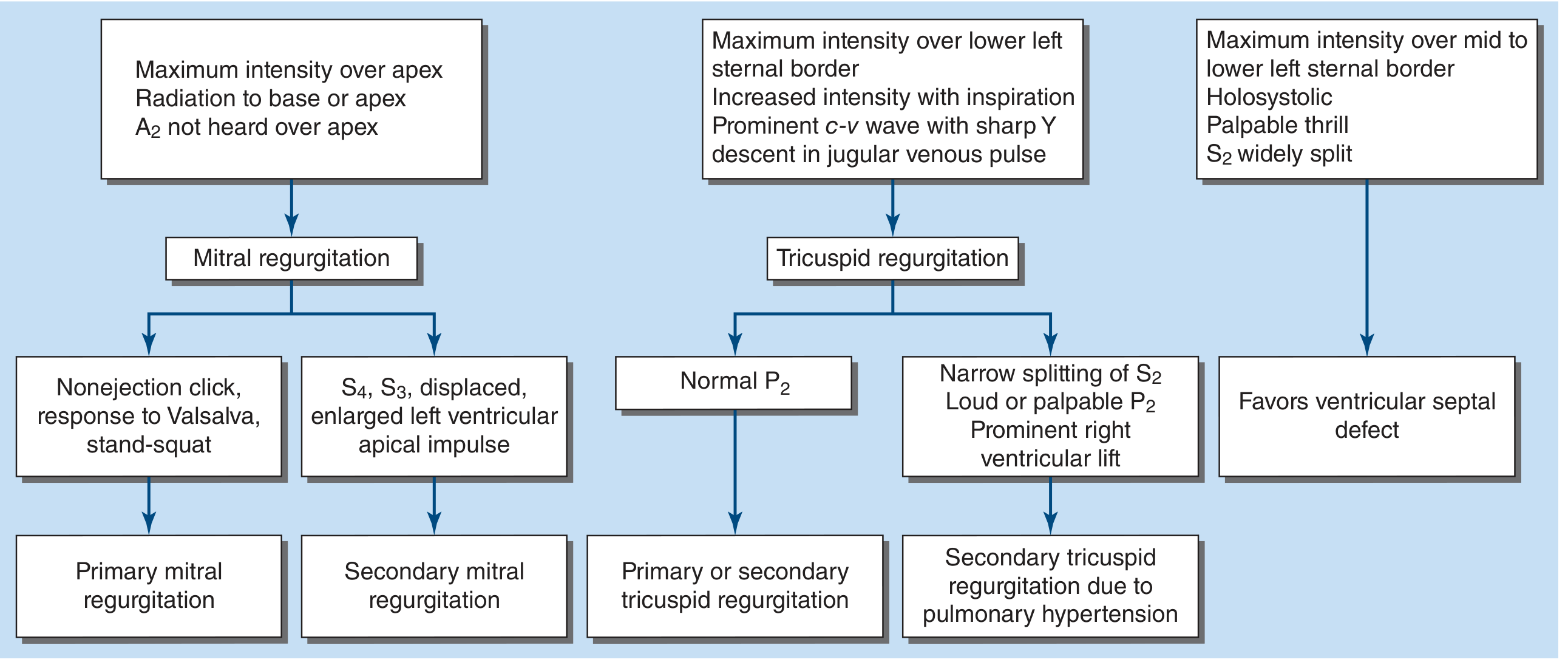
1. Site of Maximum Intensity
| Feature | MR | TR | VSD |
|---|---|---|---|
| Location | Cardiac apex (mitral area) | Lower left sternal border (4th ICS) | Mid- to lower-left sternal border (3rd-4th ICS) |
| Radiation | To left axilla (posterior jet), OR to base/left sternal border (anterior jet) | Minimal; stays at LLSB | Widely across precordium |
- MR: apex is the hallmark. Radiation direction tells you the jet direction - anterior leaflet prolapse/flail -> posteriorly directed jet -> axilla. Posterior leaflet prolapse -> anteriorly directed jet -> base (can mimic AS).
- TR: lower left sternal border, rarely radiates.
- VSD: mid-to-lower left sternal border, radiates widely.
(Harrison's Principles of Internal Medicine 22E, Continuous Murmurs section; Fuster & Hurst's The Heart 15E)
2. Respiratory Variation - The Most Reliable Bedside Sign
| Feature | MR | TR | VSD |
|---|---|---|---|
| With inspiration | No change or decreases | Increases (Carvallo's sign) | No change |
Carvallo's sign (also spelled Carvalho's sign): The holosystolic murmur of TR increases with inspiration due to negative intrathoracic pressure augmenting right ventricular filling, enlarging RV size, increasing regurgitant orifice area, and increasing regurgitant volume into the RA.
Important caveat: Carvallo's sign is NOT sensitive - when the RV is severely failing and cannot augment output with inspiration, the sign may be absent. In that situation, the jugular venous c-v wave pattern becomes the predominant diagnostic clue.
(Harrison's 22E; Braunwald's Heart Disease)
3. Dynamic Auscultation Maneuvers
| Maneuver | MR | TR | VSD |
|---|---|---|---|
| Sustained hand grip (increases LV afterload) | Louder | No significant change | No significant change |
| Valsalva (strain phase) (decreases preload) | Softer | Softer | Softer |
| Leg raising (increases preload) | Louder | Louder | Louder |
| Inspiration | No change | Louder (Carvallo's sign) | No change |
Hand grip is the most useful maneuver for MR - it raises systemic vascular resistance, increasing the LV-to-LA pressure gradient and intensifying the regurgitant murmur.
(Harrison's Principles of Internal Medicine 22E, Cardiac Auscultation section)
4. Associated Clinical Features
| Feature | MR | TR | VSD |
|---|---|---|---|
| Palpable thrill | Uncommon (severe MR) | Absent / rare | Almost always present |
| Jugular venous pulse | Normal or LAP signs (no JVP change) | Prominent c-v wave, sharp Y descent | Normal or widened pulse pressure (large shunt) |
| Apical impulse | Displaced laterally, hyperdynamic | Normal or RV heave | Usually normal or systolic thrill at LSB |
| S2 splitting | Normal or paradoxical (severe) | Normal | Wide splitting (small VSD with increased pulmonary flow) |
| P2 | Normal | Loud if pulmonary hypertension present | Loud if Eisenmenger |
| Liver | Normal | Pulsatile hepatomegaly (severe TR) | Normal |
| Other findings | S3 at apex, displaced LV apex | Ascites, peripheral edema, jugular pulsations | Thrill in 3rd-4th ICS |
(Fuster & Hurst's The Heart 15E; Harrison's 22E)
5. Murmur Quality and Intensity
| Feature | MR | TR | VSD |
|---|---|---|---|
| Quality | High-pitched, blowing | Softer, blowing; often subtle (rarely > grade II) | Harsh, loud |
| Grade | Usually 3-5/6 | Often 1-2/6 (soft) | Usually 3-5/6; small defects = LOUDER (maladie de Roger) |
The "maladie de Roger" principle: small, restrictive VSDs create a very loud murmur because the high pressure gradient is maintained throughout systole. Large VSDs equalize ventricular pressures -> soft or absent murmur.
6. Special Scenarios That Cause Diagnostic Confusion
| Scenario | Clue |
|---|---|
| Post-MI VSD vs post-MI MR | VSD: thrill present, bilateral cath shows step-up in O2 at RV; MR: thrill absent, echocardiography shows flail/papillary muscle rupture |
| Acute severe MR | Murmur may be very SHORT (early systolic only) or absent because LV and LA pressures equalize rapidly due to non-compliant LA |
| TR with severe RV failure | Carvallo's sign absent; diagnosis rests on prominent c-v wave in jugular venous pulse + pulsatile liver |
| MR of MVP | May be late-systolic (not holosystolic) unless chordal rupture/flail develops |
7. Key Summary Table
| Feature | MR | TR | VSD |
|---|---|---|---|
| Site | Apex | LLSB (4th ICS) | Mid/lower LSB (3rd-4th ICS) |
| Radiation | Left axilla or base | Minimal | Widely |
| Respiration | No change | Increases (Carvallo's) | No change |
| Hand grip | Louder | No change | No change |
| Thrill | Rare | Absent | Yes (usually) |
| JVP | Normal | c-v wave prominent | Normal |
| Liver | Normal | Pulsatile | Normal |
| S2 | Normal | Normal | Wide split |
| Intensity | Loud, blowing | Soft | Loud (harsh), esp. small VSD |
| Cardiac chamber enlarged | LV + LA | RV + RA | LV (large shunt) or RV |
Sources:
- Harrison's Principles of Internal Medicine 22E (2025) - Cardiac Auscultation, Continuous Murmurs sections
- Fuster & Hurst's The Heart 15E - Atrioventricular Valve Regurgitation: Holosystolic Murmurs; Dynamic Auscultation
- Braunwald's Heart Disease - Clinical Presentation of Tricuspid Regurgitation
- Frameworks for Internal Medicine - Heart failure and endocarditis sections
65 year old women with pneumonia has purulent sputum culture shows catalase negetive coagulase positive organism like cause is A staph auerues B staph epidermidis C streptococua pneuminiae
Quick Answer
The question contains a deliberate trick. The organism described is catalase-negative AND coagulase-positive — this combination does not match Staph aureus (which is catalase-positive). The answer that fits the clinical picture of pneumonia with purulent sputum is C - Streptococcus pneumoniae, but let me verify this carefully through the medical library.
Reasoning...
Finding Sources
Finding Sources
Your credits are exhausted. You can purchase a top-up or subscribe to a plan to continue using Orris. If you're already subscribed, your daily credits will reset tomorrow.
This is a shared conversation. Sign in to Orris to start your own chat.