
How posterior cervix fibroid exert pressure on urethra
posterior cervical fibroid pressure on urethra mechanism anatomy
https://www.cureus.com/articles/132192-large-cervical-leiomy…
Anatomical Basis
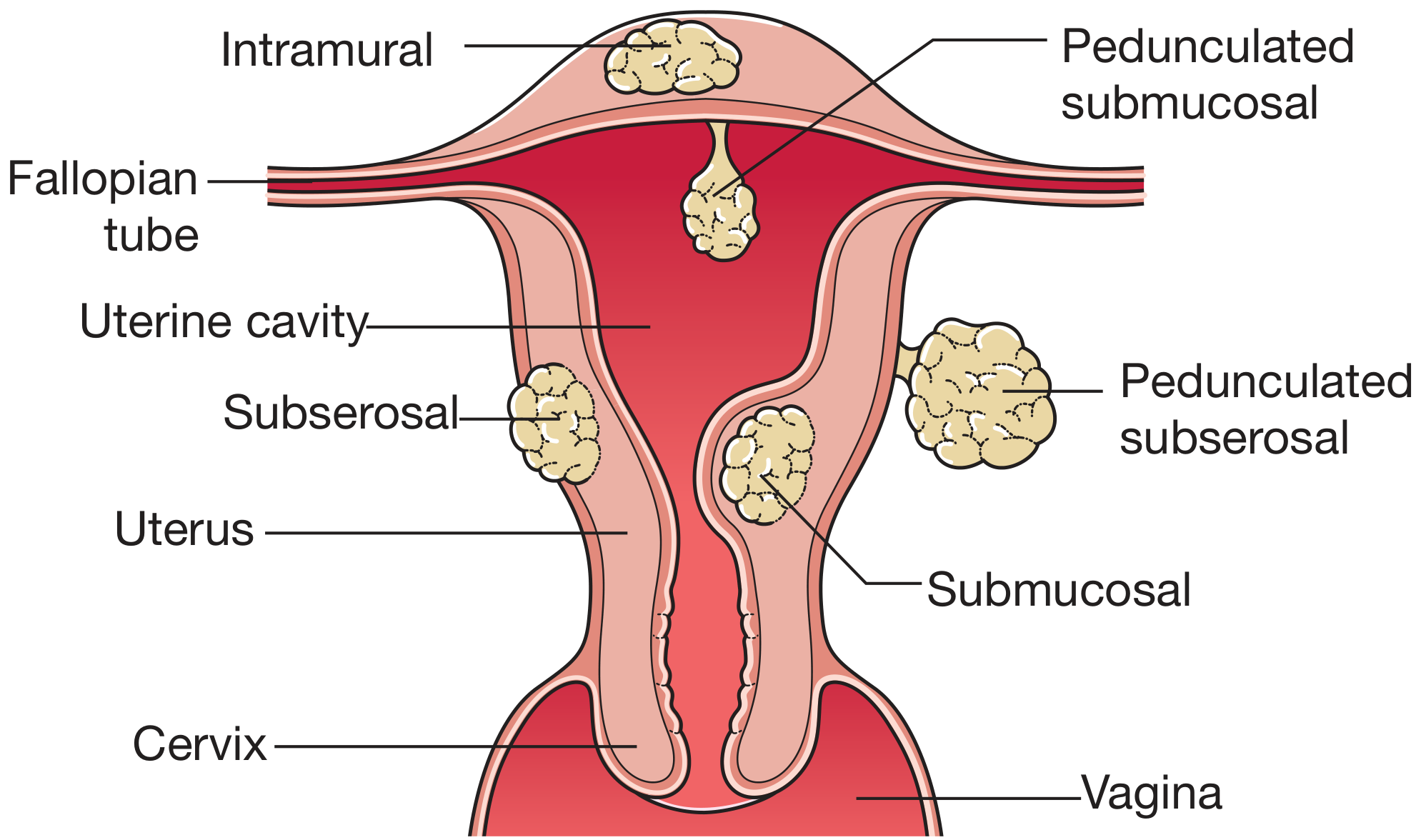
- Anteriorly: The bladder base and urethra lie directly in front of the cervix, separated only by loose areolar tissue (vesicovaginal and vesicervical fascia).
- Posteriorly: The rectum and pouch of Douglas (rectouterine pouch) lie behind it.
- Laterally: The ureters run just lateral to the cervix (within 1-2 cm) before entering the bladder.
How a Posterior Cervical Fibroid Reaches the Urethra
1. Anterior Displacement of the Cervix and Uterus
2. Elevation and Distortion of the Bladder Base
3. Mass Effect in the Confined Pelvic Space
4. Downward Prolapse / Lower Segment Location
Resulting Urological Symptoms
| Symptom | Mechanism |
|---|---|
| Urinary frequency | Reduced bladder capacity from external compression |
| Dysuria | Urethral irritation/partial obstruction |
| Acute urinary retention | Complete urethral obstruction by forward-displaced cervix |
| Urinary incontinence | Overflow incontinence when chronic retention develops; or stress incontinence from altered bladder neck support |
| Hydroureteronephrosis | Lateral ureteral compression (more typical of large or laterally extending cervical fibroids) |
Key Point: Why Posterior > Anterior for Urethral Pressure
- An anterior fibroid directly compresses the bladder but tends to push the urethra away (anteroinferiorly).
- A posterior fibroid forces the entire cervix/lower uterine segment to rock forward, concentrating pressure directly onto the bladder neck and urethra, where they are fixed by the pubourethral ligaments and pelvic floor.
Reason for hmb, polymenorhea in fibroid
fibroid heavy menstrual bleeding polymenorrhea mechanism pathophysiology submucosal intramural
https://pmc.ncbi.nlm.nih.gov/articles/PMC5779569
Reasons for HMB and Polymenorrhoea in Uterine Fibroids
A. Heavy Menstrual Bleeding (HMB / Menorrhagia)
1. Increased Endometrial Surface Area
2. Endometrial Ulceration (Submucosal Fibroids)
3. Venous Plexus Compression and Venous Lakes
4. Aberrant Angiogenesis
5. Impaired Myometrial Contractility
6. Hormonal Mechanisms - TGF-β3 and Coagulation Defects
- Downregulates plasminogen activator inhibitor-1 (PAI-1) and antithrombin in endometrial tissue, impairing local clot formation
- Causes BMP-2 resistance and defective decidualization, leaving the endometrium poorly prepared to limit bleeding
- This molecular mechanism creates a local anticoagulant environment at the endometrial surface
7. Estrogen Excess / Endometrial Hyperplasia
B. Polymenorrhoea (Shortened Cycle / Bleeding More Frequently)
1. Disruption of the Junctional Zone
2. Submucosal Fibroid as a Foreign Body
3. Anovulation / Hormonal Imbalance
4. Congestion and Venous Hypertension
Summary Table
| Mechanism | HMB | Polymenorrhoea | Fibroid Type Most Involved |
|---|---|---|---|
| Increased cavity surface area | ✓ | - | Intramural, submucosal |
| Endometrial ulceration | ✓ | ✓ | Submucosal |
| Venous plexus compression / venous lakes | ✓ | - | Intramural |
| Aberrant angiogenesis (pseudocapsule) | ✓ | - | Intramural |
| Impaired myometrial contractility | ✓ | ✓ | Intramural |
| TGF-β3 anticoagulant effect | ✓ | - | Any |
| Estrogen excess / endometrial hyperplasia | ✓ | - | Any |
| Junctional zone disruption | - | ✓ | Intramural (near JZ) |
| Foreign body prostaglandin effect | - | ✓ | Submucosal |
Ovarian hyperaemia in fibroid and causing polymenorrhea
ovarian hyperemia fibroid polymenorrhea mechanism short cycle
ovarian hyperaemia fibroid mechanism rapid follicle maturation short luteal phase polymenorrhea gynaecology textbook
"ovarian hyperaemia" OR "ovarian congestion" fibroid "polymenorrhea" OR "epimenorrhoea" "rapid follicular" maturation
fibroid "ovarian hyperaemia" "accelerated follicular maturation" polymenorrhea mechanism classic teaching
Ovarian Hyperaemia in Fibroid and Its Role in Polymenorrhoea
Step 1: The Anatomical Basis - Uterine-Ovarian Vascular Connection
- The uterine artery (from internal iliac) sends branches to both the uterus AND the ovary via the tubo-ovarian branch
- The ovarian artery (from aorta) descends to supply the ovary and also anastomoses with the uterine artery within the broad ligament (infundibulopelvic ligament)
Step 2: How Fibroids Cause Ovarian Hyperaemia
- Markedly increase uterine blood flow - fibroids divert a large proportion of uterine arterial flow to sustain their growth, leading to hypertrophy of the uterine arteries and high-flow state in the entire uterine vascular bed
- This increased arterial flow spills over through the uterine-ovarian anastomoses into the ovarian circulation
- The result is ovarian hyperaemia - a state of increased blood flow and vascular congestion within the ovary itself
Step 3: How Ovarian Hyperaemia Causes Polymenorrhoea
| Normal Cycle | With Ovarian Hyperaemia |
|---|---|
| Follicular phase: ~14 days | Follicular phase: shortened (7-10 days) |
| FSH stimulates one dominant follicle over ~14 days | Increased vascularity delivers more FSH/LH and estrogen substrate to the follicle MORE RAPIDLY |
| Normal estradiol rise over 14 days triggers LH surge | Rapid estradiol rise triggers premature LH surge |
| Ovulation at day ~14 | Ovulation at day ~7-10 |
| Total cycle: 28 days | Total cycle: 18-21 days = polymenorrhoea |
- Enhanced ovarian blood flow delivers greater quantities of gonadotropins (FSH, LH) and steroidogenic precursors per unit time to the developing follicle
- Granulosa cells, bathed in more substrate, produce estradiol more rapidly
- The positive feedback threshold for the LH surge is reached earlier than normal
- Ovulation is advanced - the follicular phase is shortened
- The subsequent luteal phase may be of normal length but the whole cycle starts again sooner
- Net result: cycle length < 21 days = polymenorrhoea
Step 4: Additional Ovarian Effects
- Stimulates endometrial proliferation between cycles, contributing to heavier bleeding when that cycle's period arrives
- May cause luteal phase deficiency if the corpus luteum (supplied by the same hyperaemic vessels) undergoes premature regression - further shortening the cycle from the other end
- In large fibroids, pelvic venous congestion from the mass adds to the overall ovarian venous outflow impairment, compounding the hyperaemic state
Summary Diagram of the Mechanism
Fibroid (highly vascular)
        |
        ↓
Increased uterine arterial blood flow
(uterine artery hypertrophy)
        |
        ↓ (via uterine-ovarian anastomoses in broad ligament)
        |
OVARIAN HYPERAEMIA
(increased ovarian blood flow)
        |
        ↓
More FSH/LH/steroidogenic substrate delivered to follicle per unit time
        |
        ↓
Rapid follicular maturation → early estradiol peak
        |
        ↓
Premature LH surge → Early ovulation
        |
        ↓
SHORT FOLLICULAR PHASE
        |
        ↓
POLYMENORRHOEA (cycle < 21 days)
Clinical Teaching Point
- Regular in timing (cycles are short but predictable, unlike the irregular bleeding of submucosal fibroids)
- Associated with normal flow per cycle (the increased bleeding in HMB is a separate mechanism)
- More common with large or multiple fibroids (greater vascular steal effect)
- Managed by treating the fibroid (myomectomy/UAE restores normal uterine blood flow and resolves the ovarian hyperaemia)
Urinary retention in case of cervical and fibroid impacted in pod
cervical fibroid impacted pouch of Douglas urinary retention mechanism anatomy
https://www.ijrcog.org/index.php/ijrcog/article/download/321…
posterior fibroid incarcerated pouch of Douglas mechanism urinary retention urethra kinking pubic symphysis
Urinary Retention in Cervical Fibroid Impacted in the Pouch of Douglas (POD)
Step 1: Understanding "Impaction in the POD"
- Anteriorly: posterior wall of uterus and posterior fornix of vagina
- Posteriorly: anterior wall of rectum
- Inferiorly: rectovaginal fold
- Fills the POD completely
- Gets wedged (impacted) between the sacrum/sacral hollow posteriorly and the cervix anteriorly
- Cannot rise above the pelvic brim - it is trapped in the pelvis = incarceration
Step 2: The Cascade Leading to Urinary Retention
Mechanism 1 - Anterior Deflection of the Cervix (Primary Mechanism)
Fibroid fills POD and impacts into sacral hollow
              ↓
Fibroid pushes POSTERIORLY against sacrum (fixed)
              ↓
Reactive force drives cervix ANTERIORLY and SUPERIORLY
              ↓
Anteriorly deflected cervix presses directly on
the PROXIMAL URETHRA and BLADDER NECK
              ↓
Urethra is compressed/kinked between:
  - Cervix (pushing posteriorly from behind)
  - Pubic symphysis (fixed bony wall anteriorly)
              ↓
URETHRAL OBSTRUCTION → URINARY RETENTION
Mechanism 2 - Bladder Base Elevation and Stretching
- It lifts the entire uterus and cervix anterosuperiorly
- The bladder base, attached to the anterior cervix by the vesicouterine fascia, is dragged upward and stretched
- The bladder assumes an abnormal position, draped over the anterior surface of the uterus
- This stretching of the bladder base angulates and elongates the urethra, creating a functional (non-compressive) obstruction at the bladder neck
- Imaging confirms this: cystoscopy in affected patients shows urethral deviation and bladder dome indentation
Mechanism 3 - Positional Worsening (Supine vs. Upright)
"Such patients usually have more problems during the night (supine position) — when supine, the impacted pelvic mass displaces the cervix superiorly and anteriorly, compressing the lower bladder, leading to obstruction of the internal urethra. During straining, increased abdominal pressure further compresses the lower bladder."
| Position | Effect |
|---|---|
| Upright | Fibroid sinks into pelvis, some urethral relief, patient may void with difficulty |
| Supine (night) | Fibroid shifts, cervix displaced more anteriorly, maximum urethral compression → worse retention |
| Straining/Valsalva | Abdominal pressure pushes fibroid down into POD → worsens compression |
Mechanism 4 - Bladder Incarceration and Overdistension
- The entire bladder becomes stretched across the anterior surface of the uterus
- Detrusor muscle is chronically overstretched → detrusor hypocontractility / acontractile bladder
- Even if the obstruction is relieved, the patient may have persistent voiding difficulty due to detrusor damage
- One documented case showed a bladder stretched to >15 cm with mild hydronephrosis from chronic retention
Mechanism 5 - Ureteral Compression (Additional)
- Compresses the ureters where they cross lateral to the cervix
- Can cause bilateral hydroureteronephrosis and worsening renal function (reported even without significant lower urinary tract symptoms in some large cervical fibroids)
Clinical Picture Summary
| Feature | Detail |
|---|---|
| Symptom onset | Gradual - frequency/urgency → intermittent retention → complete retention |
| Characteristic timing | Worse at night (supine) |
| Examination | Large pelvic/abdominal mass; posterior fornix fullness/bulge; cervix displaced anteriorly and superiorly on PV exam; mass filling POD felt on PR exam |
| Imaging | MRI/CT: fibroid in POD, elongated/angulated urethra, displaced bladder |
| Cystoscopy | Urethral deviation, bladder dome indentation |
| Treatment | Catheterisation (immediate); pessary to lift bladder neck; GnRH agonist to shrink fibroid; definitive - myomectomy or hysterectomy |
The Unifying Concept
Why is there polycythaemia, hypercapcemia, increase prl levels in fibroid .
uterine fibroid polycythemia hypercalcemia raised prolactin mechanism paraneoplastic
https://ecronicon.net/assets/ecgy/pdf/ECGY-10-00667.pdf
uterine fibroid leiomyoma hypercalcemia mechanism PTHrP prolactin secretion case
https://pmc.ncbi.nlm.nih.gov/articles/PMC10038736
Polycythaemia, Hypercalcaemia, and Elevated Prolactin in Uterine Fibroids
1. Polycythaemia (Myomatous Erythrocytosis Syndrome - MES)
What it is
- Erythrocytosis (polycythaemia)
- Uterine fibroid
- Return of haemoglobin to normal after fibroid removal
Proposed Mechanisms (multifactorial)
- Fibroid smooth muscle cells express erythropoietin (EPO) mRNA and protein - confirmed by immunohistochemistry (Suresh & Rizk; Shu et al.)
- This ectopic EPO drives bone marrow to increase RBC production independently of normal renal EPO regulation
- EPO levels in serum are elevated and return to normal post-operatively
- Caveat: Asano et al. found EPO mRNA in 108/114 fibroid patients but only 1 developed MES - suggesting EPO expression alone is insufficient; other co-factors are needed
- A large fibroid compressing the ureters → obstructive uropathy → renal hypoperfusion
- The kidney, sensing reduced perfusion, triggers appropriate EPO secretion (as in renal artery stenosis)
- Supported by: Toyama & Mitus demonstrated increased Hb production in rabbits with increased ureteral pressure
- Supported by the finding that hydroureteronephrosis is common in large cervical fibroids
- Horwitz & McKelway (1955) proposed AV shunting within the fibroid creates functional deoxygenation → EPO stimulation
- Rejected because fibroid histology does not differ between MES and non-MES cases, and uterine AV fistulas exist without erythrocytosis
Summary mechanism diagram
Fibroid tissue
    |
    ├─→ Ectopic EPO production (fibroid cells)
    |                    ↓
    └─→ Ureteral compression → Renal hypoperfusion → Renal EPO
                             ↓
                   ↑ Circulating EPO
                             ↓
                   Bone marrow stimulation
                             ↓
               POLYCYTHAEMIA (raised RBC, Hb, Hct)
Clinical Note
- MES is more common in postmenopausal women (menorrhagia no longer masks the rising Hb)
- Complications: thromboembolic events (DVT, Budd-Chiari), peptic ulcers (histamine from basophils)
- Definitive treatment: myomectomy or hysterectomy → Hb normalises
2. Hypercalcaemia ("Humoral Hypercalcaemia of Benignancy")
What it is
Mechanism
Fibroid uterine smooth muscle cells
            ↓
    Secrete PTHrP ectopically
            ↓
PTHrP acts on PTH/PTHrP receptor (same receptor as PTH)
            ↓
    ┌───────┴───────┐
    ↓               ↓
BONE: osteoclast   KIDNEY: reduced Ca²⁺
activation →       excretion, increased
↑ bone resorption  1,25(OH)₂D₃ production
    └───────┬───────┘
            ↓
    HYPERCALCAEMIA
- PTHrP is normally expressed in myometrium for uterine smooth muscle relaxation
- In fibroids, this expression is upregulated compared to normal myometrium
- It acts systemically via the circulation to raise serum calcium - identical to the mechanism in squamous cell carcinomas and other malignancies
- Resolution of hypercalcaemia after fibroid removal confirms the causal relationship
Clinical clues
- Hypercalcaemia is PTH-independent (PTH is suppressed, not elevated)
- Normal PTH with raised calcium in a woman with a pelvic mass should prompt consideration of fibroid-related PTHrP excess before assuming malignancy
- PTHrP levels are elevated in serum and fibroid tissue
- Rare - only a handful of cases reported
3. Elevated Prolactin (Leiomyoma-Associated Hyperprolactinaemia)
What it is
Mechanism(s)
- Fibroid smooth muscle cells express prolactin and secrete it autonomously
- Prolactin secretion from leiomyomas is not regulated by dopamine (unlike pituitary prolactinomas)
- Hence the key clinical clue: hyperprolactinaemia refractory to cabergoline/bromocriptine with a normal pituitary MRI
- The fibroid-secreted prolactin suppresses anterior pituitary lactotrophs via negative feedback (explaining why prolactin drops to subnormal immediately post-op before recovering to normal)
- Local hormonal dysregulation - fibroid microenvironment activates prolactin gene transcription
- Prolactin-stimulating factors secreted by fibroid → stimulates pituitary prolactin release (indirect)
- Ectopic pituitary tissue within the fibroid mass (rare)
- Lymphocytic infiltration of the fibroid → immune cells secreting prolactin locally
- Mass effect / stress - a very large fibroid causing chronic pelvic pain/stress → stress-induced hyperprolactinaemia via dopamine suppression
Clinical consequences of elevated prolactin in fibroid
- Inhibits GnRH pulsatility → ↓ FSH/LH → anovulation
- Galactorrhoea
- Infertility
- Irregular or absent periods (adds to fibroid-related menstrual disturbance)
Diagnostic clue
| Feature | Pituitary Prolactinoma | Fibroid-associated |
|---|---|---|
| Pituitary MRI | Abnormal (adenoma) | Normal |
| Response to cabergoline | Good | Resistant |
| Fibroid on USS | May or may not have | Present (usually >5cm) |
| Post-myomectomy PRL | Unchanged | Normalises rapidly |
Summary Table
| Phenomenon | Mediator | Mechanism | Diagnostic Confirmation |
|---|---|---|---|
| Polycythaemia | Erythropoietin (EPO) | Ectopic EPO by fibroid cells + renal compression → ↑ RBC production | Hb normalises post-op |
| Hypercalcaemia | PTHrP | Ectopic PTHrP → bone resorption + renal Ca²⁺ retention | Calcium normalises post-op; PTH suppressed |
| ↑ Prolactin | Prolactin (ectopic) | Direct prolactin secretion by fibroid cells, dopamine-resistant | PRL normalises post-myomectomy; resistant to DA agonists |