
Autoimmunity
Reading File
The query "Autoimmunity" is broad. Let me clarify what aspect you need before diving deep.
Asking for Preferences
What aspect of autoimmunity are you interested in?
Finding Sources
Finding Sources
Good. Now let me read the core sections in parallel - the Robbins Pathology autoimmunity section, the Cellular/Molecular Immunology section, and Janeway's Immunobiology section, plus the Firestein Rheumatology section.
Reading File
Reading File
Reading File
Excellent content. Now let me read more from these sections plus search for autoantibodies, specific diseases, and treatment, in parallel.
Reading File
Reading File
Finding Sources
Now let me read more of the Robbins autoimmunity section (central vs peripheral tolerance, specific mechanisms) and fetch the key diagram images, plus search for treatment info.
Reading File
Finding Sources
Finding Sources
Good content building up. Let me now get more on: (1) specific diseases overview from Robbins, (2) peripheral tolerance mechanisms, and (3) fetch the key pathogenesis diagram image.
Reading File
Finding Sources
Loading Image
Excellent. Now let me fetch the infections-in-autoimmunity diagram and read more on specific diseases (RA, T1DM, MS) and treatment.
Loading Image
Finding Sources
I now have rich content from multiple authoritative textbooks. Let me read the Katzung pharmacology autoimmunity treatment section and the table of autoimmune disease examples from Cellular and Molecular Immunology.
Reading File
Reading File
I now have comprehensive material. Let me update the todos and synthesize the full overview.# Autoimmunity - A Comprehensive Overview
1. Definition
Autoimmunity is the failure of the immune system to distinguish self from non-self, leading to immune responses directed against the body's own tissues. It arises from a breakdown of immunological self-tolerance - the normally acquired state of unresponsiveness to self antigens. When these mechanisms fail, self-reactive T and B lymphocytes generate cell-mediated or humoral immune attacks against host tissue, causing autoimmune disease.
- Robbins & Kumar Basic Pathology, p.176
- Janeway's Immunobiology 10e, p.716
2. Self-Tolerance: The Foundation
Normal immunity prevents autoimmunity via two layers of tolerance:
Central Tolerance
Occurs during lymphocyte development in the thymus (T cells) and bone marrow (B cells):
- Developing lymphocytes that recognize self antigens too strongly undergo clonal deletion (apoptosis)
- The AIRE (Autoimmune Regulator) gene in medullary thymic epithelial cells drives expression of peripheral tissue antigens, enabling deletion of organ-specific self-reactive T cells. Mutations in AIRE cause Autoimmune Polyendocrine Syndrome (APS)
- In B cells, receptor editing redirects strongly self-reactive BCRs
Peripheral Tolerance
Catches self-reactive cells that escape the thymus/bone marrow:
-
Anergy - T cells encountering antigen without costimulation become functionally unresponsive
-
Regulatory T cells (Tregs) - FOXP3+ cells suppress effector lymphocytes via IL-10, TGF-β, and CTLA-4. Loss of FOXP3 causes the devastating IPEX syndrome (immune dysregulation, polyendocrinopathy, enteropathy, X-linked)
-
Activation-induced cell death (AICD) via Fas/FasL - mutations cause Autoimmune Lymphoproliferative Syndrome (ALPS)
-
Immune privilege - certain anatomical sites (eye, testis, CNS) sequester antigens from the immune system
-
Cellular and Molecular Immunology (Abbas), p.1019
-
Robbins & Kumar Basic Pathology, p.176
| Gene Defect | Mouse Phenotype | Human Disease |
|---|---|---|
| AIRE | Endocrine organ destruction | APS |
| FOXP3 | Multi-organ lymphocytic infiltrates | IPEX |
| FAS/FASL | Anti-DNA antibodies, lymphoproliferation | ALPS |
| CTLA4 | Lethal lymphoproliferation by 3-4 wks | Systemic inflammatory disease |
| C4 | SLE-like | SLE |
| IL2/IL2R | IBD, anti-erythrocyte autoantibodies | Multi-system autoimmunity |
- Cellular and Molecular Immunology, Table 15.5, p.1019
3. Pathogenesis of Autoimmunity
Autoimmune disease is the product of susceptibility genes interacting with environmental triggers:
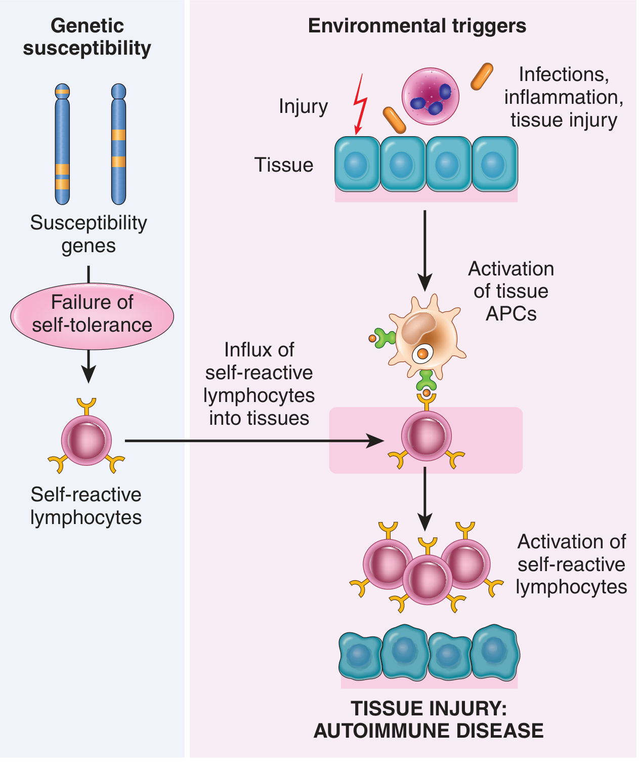
From Robbins & Kumar Basic Pathology - Susceptibility genes cause failure of self-tolerance, while environmental triggers (infection, tissue injury, inflammation) activate self-reactive lymphocytes, resulting in tissue damage.
3a. Genetic Factors
- Autoimmune diseases cluster in families; monozygotic twins have higher concordance than dizygotic
- Strong associations with HLA alleles (particularly HLA-DR and HLA-DQ):
- HLA-B27 in ankylosing spondylitis: relative risk ~100
- HLA-DR4 in rheumatoid arthritis: relative risk ~3-4
- HLA associations with SLE, T1D, MS are weaker
- GWAS has identified many non-HLA polymorphisms - many in non-coding regions affecting immune activation and regulation, and shared across multiple diseases
- Robbins & Kumar Basic Pathology, p.177
3b. Environmental Triggers
Infections are the most important environmental trigger, operating via two main mechanisms:
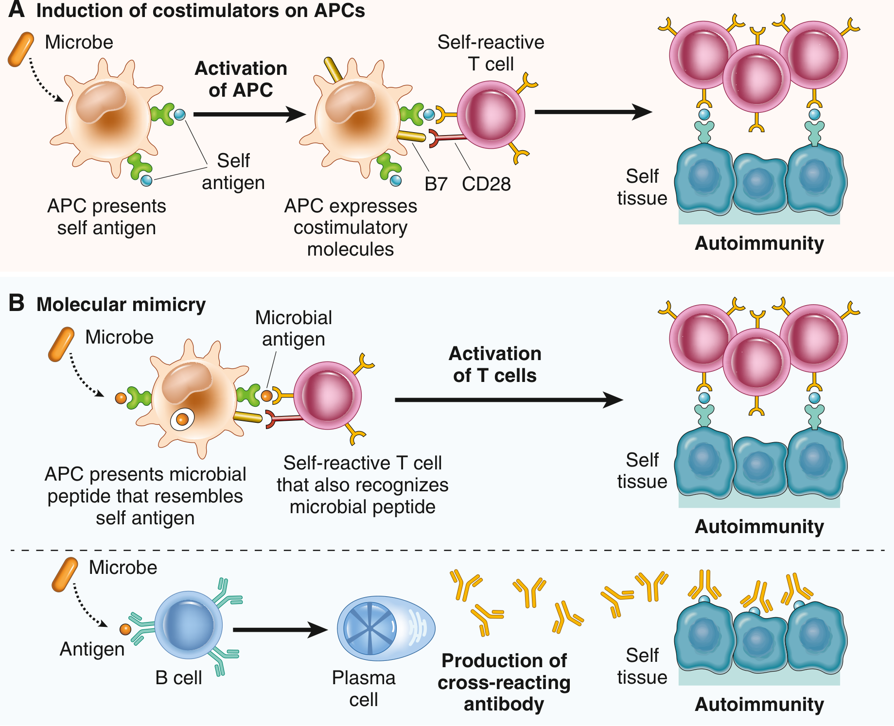
A. Upregulation of costimulators: Infection activates APCs to express costimulatory molecules (B7/CD28 axis) and secrete T-cell-activating cytokines. When these APCs present self antigens in this inflammatory context, self-reactive T cells are activated rather than anergised. Toll-like receptor (TLR) engagement by microbes on dendritic cells also drives type I interferon production (particularly relevant in SLE).
B. Molecular mimicry: Microbial antigens share cross-reactive epitopes with self antigens. The classic example is rheumatic fever - anti-streptococcal antibodies cross-react with myocardial proteins, causing myocarditis.
Other environmental factors:
- UV radiation causes cell death and exposure of nuclear antigens - explains SLE flares with sun exposure
- Smoking (risk factor for RA - possibly through chemical modification of self antigens)
- Release of sequestered antigens (ocular, testicular) following trauma
- Epitope spreading - as autoimmune tissue injury proceeds, new self epitopes are exposed, broadening the response and driving chronicity
Gender bias: Most autoimmune diseases predominate in women (SLE 9:1 F:M). Mechanisms not fully established but likely involve hormonal effects on immune regulation.
- Robbins & Kumar Basic Pathology, p.177-178
- Cellular and Molecular Immunology, p.1020-1021
4. Key Autoimmune Diseases
Systemic Lupus Erythematosus (SLE)
A multi-system autoimmune disease defined by production of antinuclear antibodies (ANAs), particularly anti-dsDNA and anti-Sm. Prevalence up to 400/100,000 in some populations; F:M ratio 9:1 in reproductive years.
Autoantibodies and their significance:
| Antibody | Prevalence | Clinical Significance |
|---|---|---|
| ANA (generic) | ~95% | Screening test |
| Anti-dsDNA | ~70% | Highly specific; correlates with disease activity and nephritis |
| Anti-Sm | ~25% | Virtually diagnostic of SLE |
| Anti-Ro (SSA) | ~30% | Neonatal lupus, SCLE |
| Anti-La (SSB) | ~20% | Neonatal lupus |
| Antiphospholipid | ~50% | Thrombosis, recurrent miscarriage |
| Anti-histone | Varies | Drug-induced SLE |
Pathogenesis: Failure of apoptotic cell clearance leads to persistence of nuclear antigens → nuclear antigen-antibody complexes engage TLRs on B cells and dendritic cells → amplified autoantibody production + type I interferon production → cycle of antigen release and immune activation.
Tissue injury mechanisms:
-
Type III hypersensitivity (immune complex deposition) - glomerulonephritis, vasculitis
-
Type II hypersensitivity (antibody-mediated) - hemolytic anemia, thrombocytopenia, leukopenia
-
Antiphospholipid antibodies - arterial and venous thrombosis, miscarriage
-
Robbins & Kumar Basic Pathology, p.178-183
Rheumatoid Arthritis (RA)
Chronic inflammatory arthritis driven by immune complex formation. Rheumatoid factors are IgM antibodies against the Fc portion of IgG; they form immune complexes that activate complement, causing chronic synovial inflammation. Anti-CCP antibodies are more specific. HLA-DR4 is the key genetic association.
Multiple Sclerosis (MS)
Cell-mediated autoimmune attack on CNS myelin by autoreactive T cells (particularly Th1 and Th17 cells). The primary target antigens include myelin basic protein (MBP) and myelin oligodendrocyte glycoprotein (MOG). Results in demyelination, axonal damage, and cumulative neurological disability.
Type 1 Diabetes Mellitus (T1DM)
Cell-mediated destruction of pancreatic islet β cells by autoreactive CD4+ T cells (Tfh cells) and cytotoxic CD8+ T cells, with macrophage-mediated lytic enzyme production. Autoantibodies (anti-GAD65, anti-IA-2, anti-insulin) are present but do not significantly cause disease - they serve as markers. HLA-DR3 and DR4 are the main risk alleles.
Other Major Autoimmune Diseases
| Disease | Dominant Mechanism | Target | Key Autoantibody |
|---|---|---|---|
| Hashimoto's thyroiditis | T cell-mediated + antibody | Thyroid peroxidase | Anti-TPO, anti-Tg |
| Graves' disease | Stimulatory antibody (Type II) | TSH receptor | Anti-TSHR |
| Myasthenia gravis | Blocking antibody (Type II) | ACh receptor | Anti-AChR |
| Goodpasture syndrome | Anti-GBM antibody (Type II) | Glomerular basement membrane | Anti-GBM/collagen IV |
| Pemphigus vulgaris | Antibody against cell junction | Desmoglein 3 | Anti-desmoglein |
| IBD (Crohn's/UC) | T cell-mediated | Gut mucosa | Various |
| Sjögren's syndrome | T cell + B cell | Exocrine glands | Anti-Ro/La |
- Katzung's Basic & Clinical Pharmacology 16e, p.1527
- Cellular and Molecular Immunology, p.1276
5. Mechanisms of Tissue Injury
Autoimmune damage maps to the Gell-Coombs hypersensitivity classification:
| Type | Mechanism | Examples |
|---|---|---|
| Type I | IgE-mediated (not typical in autoimmunity) | - |
| Type II | Antibody against cell/tissue antigen | Hemolytic anemia in SLE, Graves', Myasthenia, Goodpasture |
| Type III | Immune complex deposition | Lupus nephritis, vasculitis, serum sickness |
| Type IV | T cell-mediated (DTH) | MS, T1DM, Crohn's disease, contact dermatitis |
Th17 cells (producing IL-17) play a major pathogenic role in psoriasis, IBD, RA, MS, and SLE.
6. Diagnostics
Autoantibody Testing
- ANA (antinuclear antibody): the primary screening test. Nearly all SLE patients are ANA-positive, but a positive ANA is not specific (healthy individuals, other diseases). Pattern of immunofluorescence (homogeneous, speckled, nucleolar, rim) gives clues.
- Anti-dsDNA + anti-Sm: virtually diagnostic of SLE
- Rheumatoid factor (RF) + anti-CCP: RA
- Anti-TPO: Hashimoto's thyroiditis
- ANCA (anti-neutrophil cytoplasmic antibody): vasculitides - c-ANCA (anti-PR3) in granulomatosis with polyangiitis; p-ANCA (anti-MPO) in microscopic polyangiitis
- Anti-AChR: Myasthenia gravis
- Anti-GBM: Goodpasture syndrome
Complement levels (C3, C4, CH50) are consumed during active immune complex disease (e.g., lupus nephritis flares), making them useful disease activity markers.
7. Treatment Principles
Current therapy targets immune activation at multiple levels (Cellular and Molecular Immunology, p.1296; Katzung, p.1527):
Non-specific Immunosuppression
- Corticosteroids - broad anti-inflammatory; first-line for most acute disease
- Methotrexate - folate antagonist; antiproliferative; cornerstone of RA therapy
- Azathioprine / mycophenolate mofetil - purine synthesis inhibitors; reduce lymphocyte proliferation
- Cyclophosphamide - alkylating agent; used in severe lupus nephritis, vasculitis
Biologics (Targeted)
- Anti-TNF agents (infliximab, etanercept, adalimumab) - RA, IBD, psoriasis
- Anti-IL-6 (tocilizumab) - RA, cytokine storms
- Anti-IL-17 (secukinumab, ixekizumab) - psoriasis, ankylosing spondylitis
- Anti-IL-12/23, anti-IL-23 (ustekinumab, risankizumab) - IBD, psoriasis
- Anti-CD20 (rituximab) - B cell depletion; SLE, RA, vasculitis
- Anti-CTLA-4-Ig (abatacept) - blocks T cell costimulation (B7-CD28); RA
- Belimumab (anti-BLyS/BAFF) - B cell survival factor; approved for SLE
- Natalizumab - anti-α4 integrin; blocks lymphocyte trafficking to CNS in MS
Future Directions
The ultimate goal is antigen-specific tolerance induction - restoring tolerance specifically to the self antigen driving disease without global immunosuppression. Approaches under investigation include regulatory T cell therapy, tolerogenic dendritic cells, and CAR-Treg strategies.
8. Key Concepts Summary
| Concept | Key Point |
|---|---|
| Self-tolerance | Maintained by central deletion (thymus/BM) + peripheral mechanisms (anergy, Tregs, AICD) |
| Genetic basis | HLA locus (especially DR/DQ) + many non-HLA GWAS variants; multigenic, not monogenic |
| Environmental trigger | Infections (costimulator upregulation, molecular mimicry), UV, smoking, sequestered antigens |
| Epitope spreading | Autoimmune tissue damage releases new antigens, broadening and perpetuating the response |
| Gender bias | ~80% of autoimmune disease patients are women; hormonal mechanisms likely |
| Organ-specific vs. systemic | Organ-specific (Hashimoto's, T1DM, MS) vs. systemic (SLE, RA, Sjögren's) |
| Therapy | Non-specific immunosuppression + targeted biologics; future goal = antigen-specific tolerance |
Sources: Robbins & Kumar Basic Pathology (Elsevier, 2023) | Cellular and Molecular Immunology, Abbas et al. (Elsevier, 2022) | Janeway's Immunobiology 10e | Katzung's Basic and Clinical Pharmacology 16e | Firestein & Kelley's Textbook of Rheumatology | Goldman-Cecil Medicine
This is a shared conversation. Sign in to Orris to start your own chat.