
bone jones protein
Bence Jones protein multiple myeloma clinical significance
Bence Jones protein urine electrophoresis multiple myeloma
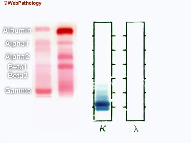
This bipartite laboratory image shows serum protein electrophoresis (SPEP) on the left and urine protein electrophoresis (UPEP) on the right from a patient with plasma cell myeloma. The left panel reveals a sharp monoclonal spike in the gamma region (M component); immunofixation identifies it as IgG kappa. There is suppression of albumin and polyclonal immunoglobulins, producing a disrupted background. The normal serum control lane (red) displays the expected thick albumin band at the top, followed by alpha1, alpha2, beta1, beta2, and a discrete gamma band; by contrast, the patient’s serum lane shows a prominent monoclonal gamma band with diminished other bands. The right panel shows urine electrophoresis with free kappa light chains (Bence-Jones proteins) detected, reflecting light-chain predominance in urine that may be reabsorbed by proximal tubules and may be absent if renal function is intact. In myeloma, the M component is commonly IgG (~50%), followed by IgA (~20%), with rare IgM, IgD, or IgE. About 20% have only free light chains without serum M-protein; ~3% are non-secretory. Excess immunoglobulins can cause hyperviscosity. These patterns aid diagnosis, prognosis, and monitoring response to therapy, including serial SPEP/UPEP and immunofixation, and guide further workup such as bone marrow biopsy and renal assessment.
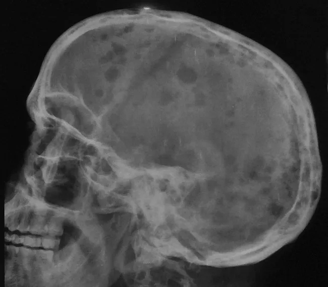
Imaging modality: Skull radiography using a lateral skull view. In a 50-year-old male with weakness, fatigue, backache, weight loss, anemia, and positive Bence Jones protein, the skull radiograph shows multiple small, uniform, sharply demarcated punched-out lytic lesions without sclerotic margins. There is involvement of the calvarial bones with osteolytic foci and mandibular lesions; generalized osteopenia is evident with diffuse reduction in bone density. No periosteal reaction or reactive sclerosis is observed. The lesions originate in the medullary cavity, progressively destroying cancellous bone and eroding toward the cortex, thus increasing the risk of pathologic fracture. This radiologic pattern is highly suggestive of a disseminated plasma cell neoplasm, most consistent with plasma cell myeloma. The imaging findings correlate with systemic disease and can be used for initial assessment and staging, as well as monitoring response to therapy. Radiology findings prompt complementary laboratory and biopsy investigations (serum/urine electrophoresis, bone marrow biopsy) to confirm monoclonal plasma cell proliferation. Clinically, skull involvement contributes to bone pain and skeletal-related events; imaging guides treatment planning, assesses therapeutic response, and monitors disease progression over time. In the context of the described case, the radiographic appearance aligns with classic myeloma hallmarks in the skull and mandible. Case verified clinically.
![Urine test findings
<table><thead><tr><th>Investigation findings and specific features</th><th>Possible cancer</th><th>Recommendation</th></tr></thead><tbody><tr><td>Bence–Jones protein urine results (used if serum free light chain testing is not available)suggest myeloma, plus persistent bone pain, particularly back pain, or unexplained fracture</td><td>Myeloma</td><td>Refer people using a suspected cancer pathway referral [1.10.6]</td></tr></tbody></table>](https://cdn.orris.care/cdss_images/GLGCA_4543562_1766477967690_15261b54-6715-4b19-b3aa-821e812d845d_7b482f70-2cd6-4f94-9a5f-554304473d6c.png)
Urine test findings <table><thead><tr><th>Investigation findings and specific features</th><th>Possible cancer</th><th>Recommendation</th></tr></thead><tbody><tr><td>Bence–Jones protein urine results (used if serum free light chain testing is not available)suggest myeloma, plus persistent bone pain, particularly back pain, or unexplained fracture</td><td>Myeloma</td><td>Refer people using a suspected cancer pathway referral [1.10.6]</td></tr></tbody></table>
Bence Jones Protein
What It Is
- Named after Dr. Henry Bence Jones (1848), who described their unique heat behaviour
- Also called: free light chains, M-proteins, paraproteins
Classic Heat Property (Historical)
- Precipitate at 40-60°C
- Redissolve at 100°C (boiling)
- Reprecipitate on cooling
Clinical Significance
| Condition | Notes |
|---|---|
| Multiple myeloma | Most common cause; present in 50-80% of cases |
| Light-chain myeloma (Bence Jones myeloma) | ~20% of myeloma patients secrete only light chains, no serum M-spike |
| MGUS | Low-level light chains; no end-organ damage |
| Waldenstrom macroglobulinemia | Less common |
| Lymphoma / B-cell malignancies | Occasionally |
| Amyloidosis (AL type) | Light chains deposit as amyloid fibrils |
Renal Toxicity ("Myeloma Kidney")
- Direct tubular toxicity - some light chains are directly toxic to proximal tubular epithelial cells due to their intrinsic physicochemical properties
- Cast nephropathy - Bence Jones proteins combine with Tamm-Horsfall protein under acidic conditions, forming large obstructive tubular casts with a surrounding inflammatory reaction ("light-chain cast nephropathy")
- AL amyloidosis (λ-type light chains) - occurs in 6-24% of myeloma patients
- Light-chain deposition disease (κ-type usually) - nonfibrillar deposits in glomerular basement membrane and tubular basement membranes
Detection Methods
| Method | Notes |
|---|---|
| Classic heat test | Historical; not used routinely now |
| Urine protein electrophoresis (UPEP) | Primary screening method; may miss if low concentration |
| Urine immunofixation electrophoresis (UIFE) | Gold standard - identifies κ or λ type |
| Reagent strip dipstick | Unreliable - often falsely negative for Bence Jones proteins (detects albumin primarily) |
| Serum free light chain (FLC) assay | More sensitive; useful for monitoring response, early relapse, and MGUS risk stratification |
Diagnostic Role in Multiple Myeloma
- Serum protein electrophoresis (SPEP)
- Bone marrow biopsy
- Bence Jones protein in urine (UPEP + immunofixation)