
Can you give me a diagram showing... Um, teres major, teres minor, suprascapularis, subscapularis, all muscles in a single upper limb diagram.
upper limb shoulder muscles diagram teres major teres minor supraspinatus subscapularis rotator cuff anatomy
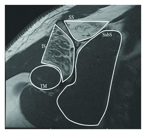
A T1-weighted MRI scan in the sagittal oblique plane demonstrating the rotator cuff musculature of the shoulder. The image highlights the anatomical arrangement and tissue characteristics of the four primary rotator cuff muscles, delineated by white outlines: the supraspinatus (SS), infraspinatus (IS), subscapularis (SubS), and teres minor (TM). Clinically significant findings include increased T1 signal intensity (hyperintensity) within the muscle bellies of the supraspinatus and infraspinatus, indicative of advanced fatty infiltration and muscle atrophy. In contrast, the subscapularis and teres minor maintain a normal, lower signal intensity (isointense to other skeletal muscles). The spatial relationship shows the supraspinatus superiorly, the infraspinatus posteriorly, the subscapularis anteriorly, and the teres minor inferior to the infraspinatus. This diagnostic image is used in orthopedics and radiology to assess chronic rotator cuff tears and associated muscle degeneration, which has implications for surgical prognosis and functional outcomes.
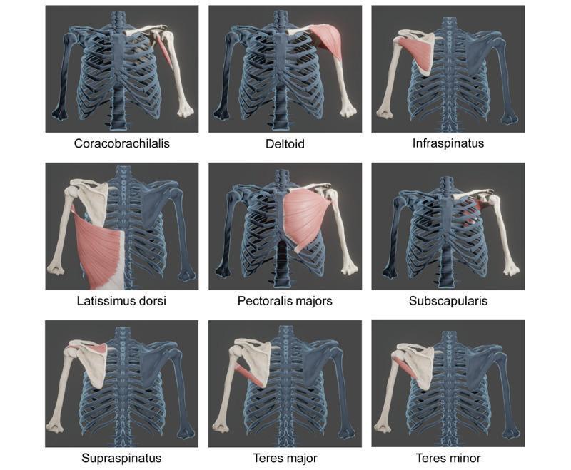
Anatomical diagram consisting of a 3x3 grid of 3D reconstructions showing specific muscles of the shoulder girdle and their skeletal attachments. Each panel highlights a single muscle in pink/red against a blue-gray skeletal model of the rib cage, clavicle, scapula, and humerus. The nine muscles depicted are: Coracobrachialis, Deltoid, Infraspinatus, Latissimus dorsi, Pectoralis major, Subscapularis, Supraspinatus, Teres major, and Teres minor. The models are based on CT-derived DICOM segmentation to illustrate precise origins and insertions. Key anatomical focuses include the rotator cuff group (supraspinatus, infraspinatus, subscapularis, and teres minor) and its relation to the humeral head and scapular fossa. Larger muscles like the deltoid and pectoralis major are shown in their anterior or lateral orientations, while the posterior view is utilized for the infraspinatus and teres muscles. This visual resource is designed for orthopedic and kinesiology education, emphasizing the spatial relationships and morphology of primary movers and stabilizers of the glenohumeral joint.
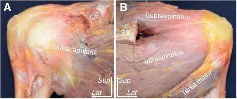
This clinical photograph displays a human cadaveric specimen dissection of a right shoulder, focusing on the rotator cuff musculature and their humeral insertions. The content is presented in two panels: (A) Anterior aspect and (B) Posterior aspect. In panel A, the subscapularis muscle is clearly labeled, demonstrating its broad muscle belly and fibers converging toward the lesser tuberosity of the humerus. The coracoid process (CP) is visible superiorly. In panel B, the posterior rotator cuff muscles are identified in relation to the scapular spine (SS). The supraspinatus is positioned superior to the spine, while the infraspinatus and teres minor are situated inferiorly, showing their tendinous progression toward the greater tuberosity. Spatial orientation is provided by superior (Sup) and lateral (Lat) directional indicators. The specimen has undergone resection of the acromion to provide an unobstructed view of the tendon footprints. This visual resource is intended for medical education regarding shoulder anatomy, surgical landmarks, and musculoskeletal pathology.
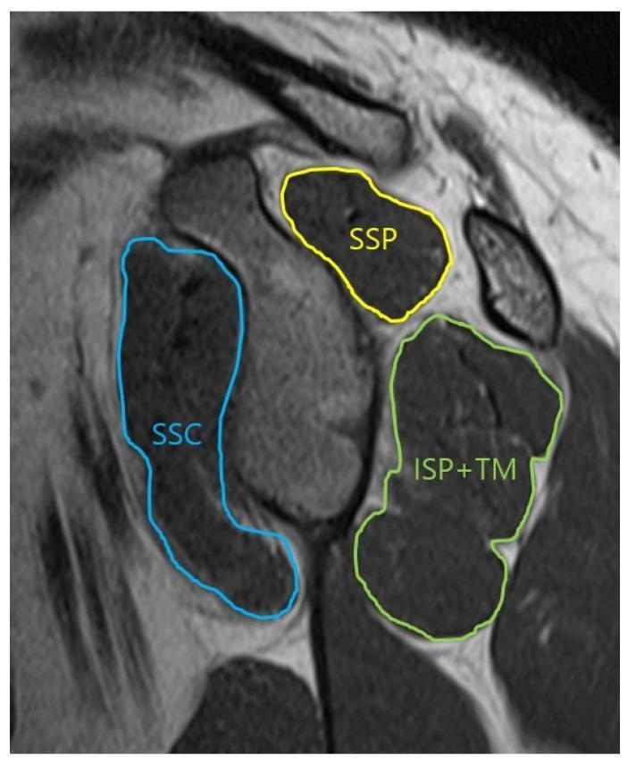
This oblique-sagittal T2 Turbo Spin Echo (TSE) MRI scan demonstrates the musculoskeletal anatomy of the shoulder in a glenoid face view, specifically for assessing the cross-sectional area of the rotator cuff muscles. The image features color-coded annotations outlining the muscular boundaries: the supraspinatus (SSP) is outlined in yellow in the superior position; the subscapularis (SSC) is traced in blue anteriorly; and the infraspinatus combined with the teres minor (ISP+TM) is outlined in green in the posterior-inferior quadrant. The musculature appears as intermediate to dark gray signal intensity, allowing for the evaluation of muscle volume and potential fatty infiltration or atrophy. This imaging view is clinically significant for orthopedic evaluation and preoperative planning in cases of rotator cuff tears or shoulder instability, providing a clear visualization of the muscular support surrounding the glenohumeral joint.
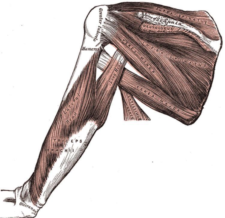
This medical anatomical diagram illustrates the musculature of the human shoulder and upper arm from a posterior perspective. The illustration highlights the rotator cuff complex and its relationship to the scapula and humerus. Key structures identified include the supraspinatus, originating above the spine of the scapula; the infraspinatus, occupying the infraspinous fossa; and the teres minor and teres major muscles positioned along the lateral border of the scapula. The deltoideus is shown partially overlying the posterior joint. On the humerus, the greater tubercle is clearly marked as an insertion point. The triceps brachii is depicted on the posterior arm, specifically identifying the long head—which passes between the teres minor and major—and the lateral head. The distal insertion of the triceps onto the olecranon of the ulna is visible. This diagram serves as a clinical education tool for understanding shoulder stability, biomechanics, and the spatial arrangement of the musculoskeletal system relevant to orthopedics and sports medicine.
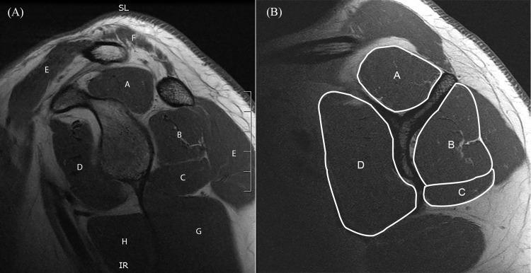
This diagnostic image consists of two panels showcasing sagittal T1-weighted MR-arthrography of the human shoulder, specifically focusing on the musculature of the shoulder girdle at the glenoid level. Panel A identifies anatomical landmarks: supraspinatus (A), infraspinatus (B), teres minor (C), subscapularis (D), deltoid (E), trapezius (F), triceps brachii (G), and teres major (H). The muscles appear as well-defined, intermediate-to-low signal intensity structures (dark gray) against the high-signal intensity (white) of intramuscular and subcutaneous fat. Panel B demonstrates a quantitative clinical assessment of the rotator cuff using cross-sectional area (CSA) measurements. White outlines delineate the CSA for the supraspinatus (A), infraspinatus (B), teres minor (C), and subscapularis (D) at the 'Y-position'—the lateral slice where the scapular spine and body converge. This visual serves as a standardized educational tool for orthopedic radiology and physical therapy to evaluate muscle atrophy, fatty infiltration, and rotator cuff integrity.
- Supraspinatus - above the scapular spine
- Infraspinatus - in the infraspinous fossa
- Teres minor - along the lateral scapular border (superior)
- Teres major - along the lateral scapular border (inferior)
- Deltoid and Triceps brachii (long and lateral heads) also visible