
treatment protocol to follow with drugs and dosage in detail for lung edema in dogs
pulmonary edema dogs treatment protocol furosemide dosage 2024
https://www.acapulco-vet.be/wp-content/uploads/2021/01/emerg…
canine pulmonary edema treatment furosemide
https://www.merckvetmanual.com/pharmacology/systemic-pharmac…
pulmonary edema dog radiograph chest X-ray
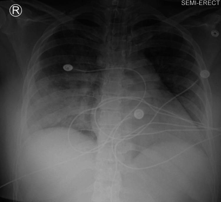
This chest radiograph (X-ray) in a semi-erect position shows diffuse, bilateral, and confluent alveolar opacities consistent with severe pulmonary edema, such as negative-pressure pulmonary edema (NPPE). The opacities are distributed throughout both lung fields, demonstrating a ‘bat-wing’ or ‘butterfly’ pattern typical of acute fluid accumulation. Despite the extensive infiltrates, the cardiac silhouette appears within normal size limits, suggesting a non-cardiogenic etiology for the edema. Several medical devices are visible, including an endotracheal tube (ETT) for airway management and multiple ECG monitoring leads superimposed over the thoracic cavity. Anatomical landmarks including the clavicles, ribs, and diaphragmatic contours are visible but partially obscured by the overlying pulmonary pathology. This diagnostic image serves as an educational example of monitoring the progression and treatment response of acute respiratory distress in a perioperative or intensive care setting.
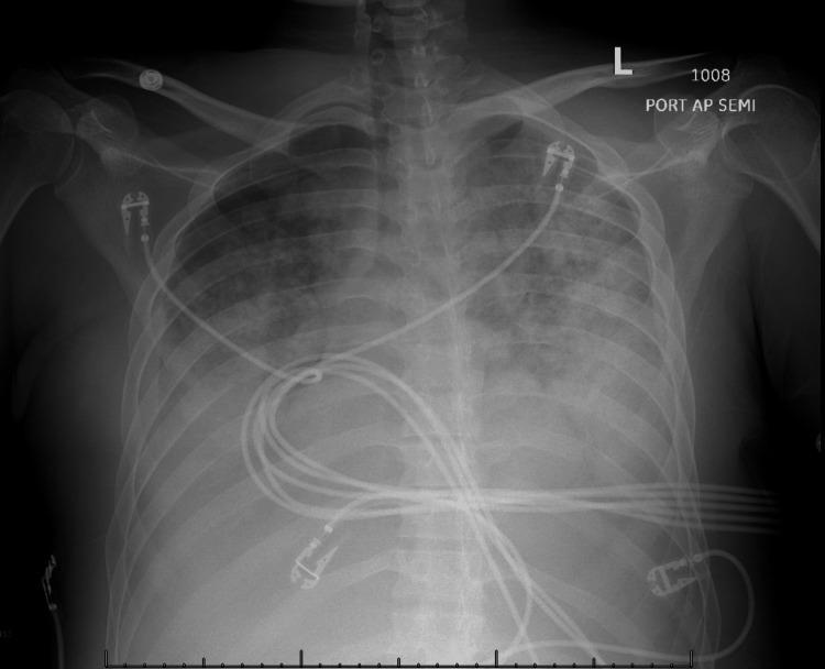
This diagnostic image is a portable anteroposterior (AP) chest X-ray demonstrating significant pulmonary pathology. The radiograph shows diffuse, bilateral patchy opacities throughout both lung fields, consistent with alveolar consolidation or interstitial fluid accumulation. There is a prominent increase in radiographic density and indistinctness of the pulmonary vasculature, particularly in the upper lobes, indicating pulmonary vascular congestion and cephalization. These findings are characteristic of pulmonary edema secondary to volume overload. Several medical devices are visible, including multiple external electrocardiogram (ECG) leads and cables overlying the thoracic cavity. The cardiac silhouette appears partially obscured by the overlying opacities. This image serves as a teaching tool for recognizing the radiographic manifestations of acute pulmonary congestion and parenchymal opacification in a critically ill patient, likely in the context of multi-organ dysfunction or renal failure.
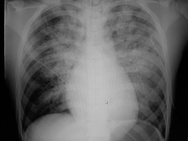
This diagnostic image is an anteroposterior (AP) chest X-ray demonstrating acute bilateral pulmonary edema. The radiograph reveals diffuse, hazy opacities and ill-defined infiltrates throughout both lung fields, exhibiting a characteristic perihilar 'bat-wing' distribution with prominent involvement of the upper and central lung zones. The lung parenchyma shows signs of alveolar consolidation and fluid accumulation. Cardiomegaly is evident, with an enlarged cardiac silhouette and indistinct borders. A thin, radiopaque monitoring line or catheter is visible crossing the right thoracic cavity toward the mediastinum. The mediastinal structures and bony thorax, including the clavicles and ribs, are otherwise unremarkable. This visual is clinically significant for students learning to identify radiological signs of heart failure and pulmonary congestion, as it highlights the hallmark features of cardiogenic pulmonary edema versus acute respiratory distress syndrome (ARDS).
Pulmonary Edema in Dogs - Treatment Protocol
Step 1 - Initial Stabilization (First 0-30 Minutes)
Minimize Stress
Oxygen Supplementation
- Oxygen cage: 40-60% FiO2 - preferred as least stressful
- Flow-by oxygen: 2-3 L/min held near the nose
- Intranasal catheter (humidified O2): 50-100 mL/kg/min - useful but only if dog tolerates it
- Target SpO2 >95%
IV Access
Step 2 - Emergency Drug Therapy
DRUG 1: FUROSEMIDE (Loop Diuretic) - First-Line, Most Critical
| Severity | Route | Dose | Frequency |
|---|---|---|---|
| Life-threatening (acute) | IV (preferred) | 2-4 mg/kg | Every 0.5-2 hours initially |
| Moderate | IM or SC | 2-4 mg/kg | Every 2-4 hours |
| CRI (constant rate infusion) | IV | 0.25-1 mg/kg/hour | Continuous |
| Chronic maintenance (oral) | PO | 1-2 mg/kg | Every 12 hours |
| Severe chronic disease (oral) | PO | up to 4-6 mg/kg | Every 8 hours |
- Bolus dosing is tapered over 12-24 hours as clinical signs resolve.
- IV route is strongly preferred acutely as it also provides venodilator effect.
- Ototoxicity risk at doses >20 mg/kg IV (single dose); irreversible hearing loss at 50-100 mg/kg.
DRUG 2: NITROGLYCERIN (Venodilator) - First-Line Vasodilator in Field Settings
| Form | Dose | Frequency | Notes |
|---|---|---|---|
| 2% ointment (topical) | 2.5 cm per 20 kg body weight | Every 8 hours | Apply to hairless skin (medial pinna or groin) |
| Transdermal patch | 2.5-10 mg/24 hours | Once daily | Easy, practical |
- Wear gloves when applying - can cause headaches in handlers
- Post a warning sign on the kennel about application site
- Use only for 3-5 days - dogs become refractory with continued use
- Avoid in cardiogenic shock
DRUG 3: PIMOBENDAN (Positive Inotrope + Vasodilator) - Start Early in Cardiogenic Edema
| Route | Dose | Frequency |
|---|---|---|
| PO | 0.2-0.3 mg/kg (range 0.20-0.48 mg/kg) | Every 12 hours |
- Give on an empty stomach (at least 1 hour before food)
- Studies show dogs receiving pimobendan survived a median of 267 days vs 140 days without it
- Also reduces recurrence of pulmonary edema
DRUG 4: ACE INHIBITORS - Start After Acute Stabilization
- Dose: 0.5 mg/kg PO every 12-24 hours
- Mechanism: Reduces afterload and preload via RAAS blockade
- Dose: 0.25-0.5 mg/kg PO every 24 hours
DRUG 5: BUTORPHANOL (Sedation/Anxiolysis) - For Severely Distressed Dogs
| Route | Dose |
|---|---|
| IM or IV | 0.2-0.4 mg/kg as a single dose |
- Especially useful in fractious or severely panicked dogs
- Avoid respiratory depressants in dogs with marginal airway protection
DRUG 6: SODIUM NITROPRUSSIDE - ICU Use Only (Hemodynamic Monitoring Required)
| Route | Dose | Notes |
|---|---|---|
| IV CRI | 1-10 µg/kg/min in 5% dextrose | Titrate to effect |
- ONLY use with continuous blood pressure monitoring
- Adverse effects: profound hypotension, tachycardia, nausea
- Risk of cyanide poisoning with prolonged use (>72 hours)
- Light-sensitive: wrap infusion bag in foil
DRUG 7: HYDRALAZINE - Alternative Arteriodilator (When Nitroprusside Unavailable)
- Most useful for pulmonary edema secondary to mitral regurgitation
- Less effective in dilated cardiomyopathy
- Avoid in dogs already on ACE inhibitors (additive hypotension)
- Side effects: first-dose hypotension, anorexia, vomiting, diarrhea
DRUG 8: TORSEMIDE - Alternative Loop Diuretic (Furosemide-Resistant Cases)
| Severity | Dose | Frequency |
|---|---|---|
| Mild CHF / maintenance | 0.13-0.25 mg/kg | Every 24 hours |
| Severe / initial stabilization | 0.26-0.4 mg/kg | Every 24 hours |
- Used when furosemide resistance is suspected
- Also emerging as a first-line alternative to furosemide
DRUG 9: SPIRONOLACTONE - Add-On for Chronic Management
- Potassium-sparing diuretic and aldosterone antagonist
- Used in conjunction with furosemide for chronic CHF
- Reduces cardiac remodeling
Step 3 - Non-Cardiogenic Pulmonary Edema
- Treat the underlying cause first
- Furosemide is used more cautiously (smaller doses, monitor for dehydration)
- Avoid aggressive vasodilation
- Corticosteroids are controversial and generally not recommended for cardiogenic edema, but may be considered for certain inflammatory/allergic causes
- Ventilatory support (positive pressure) may be necessary in severe ARDS
Summary Treatment Timeline
| Time | Action |
|---|---|
| 0-5 min | Oxygen, minimal handling, IV access |
| 5-10 min | Furosemide 2-4 mg/kg IV, nitroglycerin ointment |
| 15-30 min | Reassess breathing - repeat furosemide if no urine output or clinical improvement |
| 0.5-2 hrs | Continue furosemide q 1-2h, butorphanol if distressed, blood pressure monitoring |
| 2-6 hrs | Pimobendan PO (empty stomach), taper furosemide intervals as dog improves |
| 6-24 hrs | Begin ACE inhibitor (enalapril/benazepril) once hemodynamically stable |
| 24-48 hrs | Transition to oral maintenance therapy, cardiac workup (echo, ECG, radiograph) |
| Ongoing | Oral furosemide + pimobendan + ACE inhibitor as chronic CHF protocol |
Monitoring Parameters
- Respiratory rate (goal: <30-40 breaths/min at rest)
- SpO2 (goal: >95%)
- Urine output (confirm diuresis within 30 min of IV furosemide)
- Blood pressure (avoid hypotension <80 mmHg systolic)
- Electrolytes (especially potassium - hypokalemia with aggressive furosemide use)
- BUN/creatinine (azotemia risk with aggressive diuresis)
- Body weight (daily - loss of 0.5-1 kg/day is expected initially)
Chest Radiograph Appearance (Cardiogenic Pulmonary Edema)
Important: This protocol is intended for licensed veterinary professionals. Drug doses and treatment decisions must always be individualized based on the dog's weight, renal function, blood pressure, and underlying cardiac diagnosis.