
Write a five mark short note on Diabetic foot and gangrene Arteial v/s venous v/s diabetic ulcer Make it compatible, compact but high yielding. So that... I can easily remember and, uh, write it in a... Systematic way being a 2nd year resident. make it in a way its interesting and easy to learn visually and Create a high density, high yielding image based(generate image) Flashcard I can use Before exam to revise and remember all the important points regarding this short-note. but it should be in a way so its easy to understand and remember also use flowcharts or mindmaps in the response wherever possible dont use code
diabetic foot ulcer gangrene neuropathic
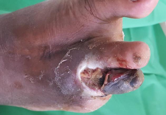
This clinical photograph displays a high-risk diabetic foot with advanced complications, including a deep neuropathic or neuro-ischaemic ulcer and dry gangrene. The primary lesion is a large, deep ulcer located on the dorsal aspect of the forefoot at the base of the second toe. The ulcer bed contains a mixture of yellowish slough and fibrin alongside areas of darker necrotic tissue. Surrounding the ulcer, the skin is markedly macerated with significant purplish-black discoloration, suggesting severe ischaemia and compromised blood flow. The distal portion of the second toe is distinctly cyanotic and black, consistent with gangrene, and shows signs of tissue integrity loss and small areas of haemorrhage. A surgical scar is visible on the midfoot, indicating a previous toe amputation, which classifies this as a high-risk limb. The image serves as a critical educational example of diabetic foot disease progression, emphasizing the confluence of peripheral neuropathy and peripheral arterial disease in clinical practice.
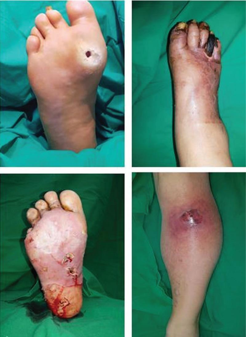
This set of four clinical photographs illustrates various stages and manifestations of diabetic foot complications. (Top-left): A plantar surface view showing a classic neuropathic ulcer. The wound is small, circular, and deep with a hyperkeratotic rim, located over a high-pressure area (the metatarsal head). (Top-right): A dorsal view of a foot exhibiting dry gangrene and critical limb ischemia. The distal phalanges are blackened, shriveled, and necrotic, with proximal cyanotic and erythematous skin changes indicating impaired perfusion. (Bottom-left): A plantar view demonstrating extensive tissue infection with an abscess forming within a large, macerated open wound. It shows pale granulation tissue, purulent exudate, and multiple sinus tracts, suggesting deep-seated infection. (Bottom-right): A lower leg view showing an abscess within a closed wound. This is characterized by localized swelling, a central focus of purulence/necrosis, and a prominent circumferential erythematous halo indicating cellulitis and acute inflammatory response. These images collectively demonstrate the progression from simple ulceration to complex infectious and ischemic complications in diabetic patients.
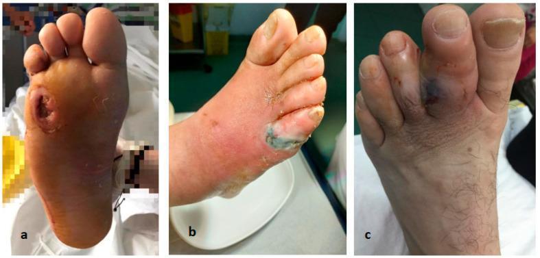
This set of clinical photographs illustrates three common complications of the diabetic foot. Panel A shows a deep plantar ulcer located over the first metatarsophalangeal joint. The lesion is circular with hyperkeratotic, raised edges and a granular base, characteristic of a neuropathic ulcer. Panel B demonstrates toe gangrene and necrosis, featuring significant tissue darkening (purplish-black) and desquamation involving the distal forefoot and toes, accompanied by surrounding erythema suggesting cellulitis. Panel C displays the dorsal aspect of a foot with toe osteitis, characterized by marked swelling (edema), cyanotic discoloration of the third and fourth digits, and signs of inflammatory soft tissue involvement. Collectively, these images represent the clinical spectrum of diabetic foot infections and peripheral vascular complications, ranging from localized ulceration to tissue necrosis and underlying bone inflammation (osteitis), which are critical for surgical triage and management in podiatry and vascular surgery.
arterial venous ulcer leg comparison
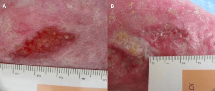
Clinical photograph comparison of a venous leg ulcer on the lower limb of an 85-year-old patient, showcasing therapeutic progression over one month. Image A (Baseline): A large, irregularly shaped ulcer with a dark red, granular wound bed and defined margins. The surrounding periwound skin exhibits significant erythema and signs of chronic venous insufficiency, such as lipodermatosclerosis or post-phlebitic changes. Image B (One-month follow-up): The same anatomical region demonstrates significant wound healing. The ulcer has markedly decreased in size and depth. The wound bed has transitioned to a lighter pink hue, indicating the formation of healthy granulation tissue and re-epithelialization from the periphery. The surrounding skin integrity is improved, with reduced inflammatory erythema and a more uniform texture. Both images include a medical ruler for scale, illustrating the objective reduction in lesion dimensions. This comparison serves as an educational example of secondary intention healing in chronic venous stasis ulcers following targeted topical intervention.
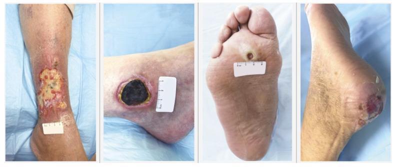
This clinical photograph comparison presents four distinct types of chronic lower extremity wounds, facilitating differential diagnosis in wound care and dermatology. From left to right, the panel demonstrates: 1) Venous Leg Ulcer: A large, irregularly shaped wound on the gaiter area of the lower leg, characterized by a shallow bed with fibrinous slough, granulation tissue, and periwound erythema suggestive of chronic venous insufficiency. 2) Arterial Leg Ulcer: A deeper, more circumscribed wound on the lateral ankle/foot with a dark, necrotic eschar bed and defined borders, indicating severe ischemia. 3) Neuropathic Diabetic Foot Ulcer: Located on the plantar surface (pressure point) of the foot, showing a 'punched-out' circular appearance with a calloused hyperkeratotic rim typical of diabetic neuropathy. 4) Pressure Ulcer: Situated on the posterior heel, displaying deep reddish-purple tissue discoloration and skin breakdown consistent with Stage 2 or deep tissue injury due to prolonged pressure. These images serve as a teaching tool for medical professionals to recognize varied wound etiologies based on anatomical location, wound bed morphology, and periwound skin changes.
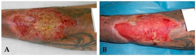
This comparison chart consists of two clinical photographs (A and B) demonstrating the progression of a venous leg ulcer (VLU) through different stages of wound healing. Image A displays a chronic inflammatory ulcer characterized by a significant amount of yellow fibrinous slough and necrotic debris covering the wound bed, surrounded by irregular, erythematous borders indicating localized inflammation. In contrast, image B shows the same anatomical site following therapeutic intervention, now presenting as a granulating ulcer. This stage is marked by a healthy, beefy-red wound bed with visible granulation tissue and a marked reduction in yellow slough. Both images show the ulcer located on the lower leg, with surrounding hyperpigmented skin consistent with chronic venous insufficiency. This visual comparison illustrates the transition from a 'blocked' inflammatory state to an active proliferative phase of wound repair, serving as an educational tool for identifying wound bed characteristics and assessing healing progress in dermatology and wound care specialties.
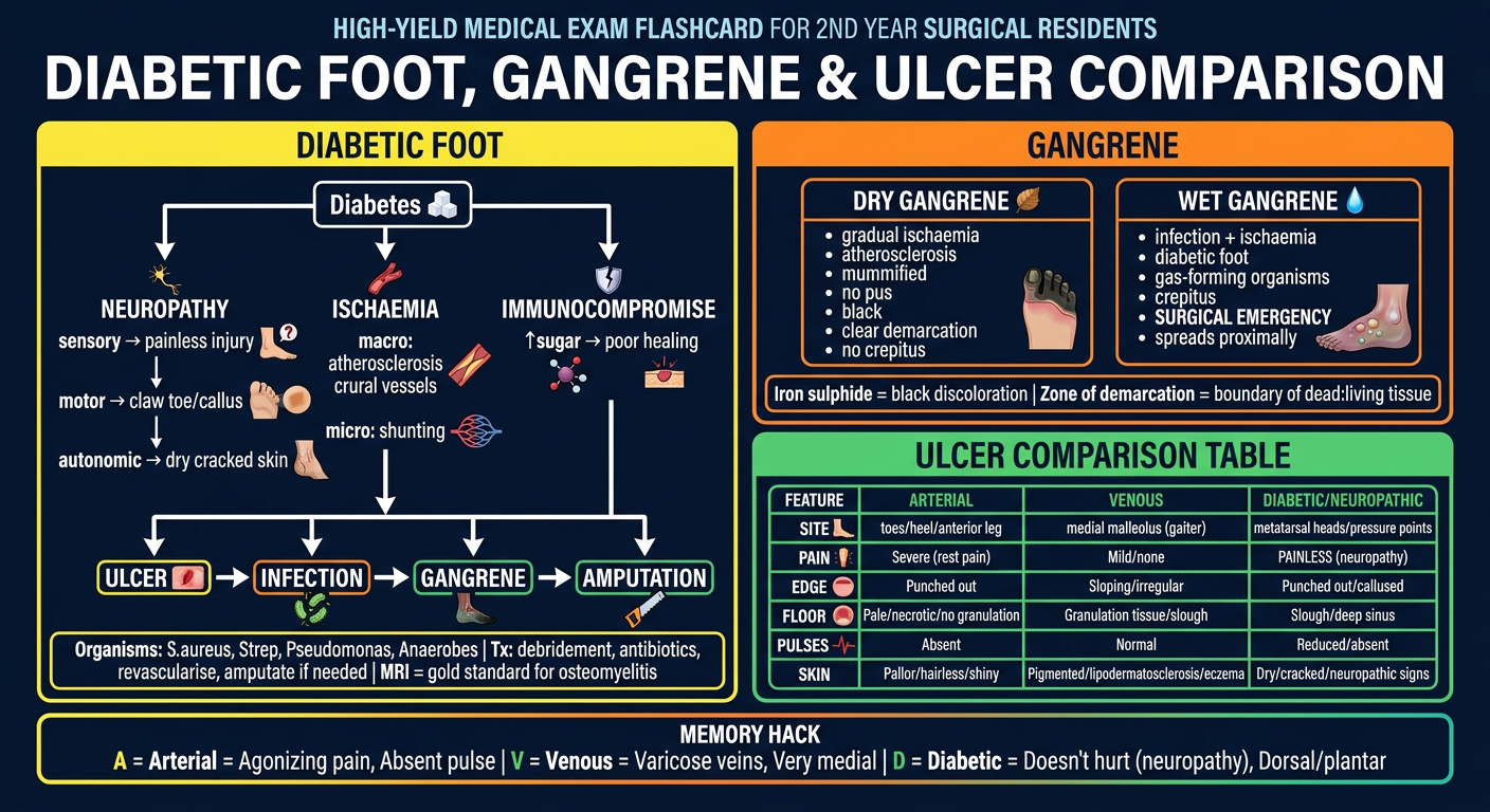
A high-yield medical exam flashcard for 2nd year surgical residents titled "DIABETIC FOOT, GANGRENE & ULCER COMPARISON". Design as a dark background (navy blue) with bright colored text sections. Include three main sections: SECTION 1 - "DIABETIC FOOT" (bright yellow header): Show a mini flowchart/pathway: Diabetes → [3 pathways in columns] NEUROPATHY (sensory→painless injury, motor→claw toe/callus, autonomic→dry cracked skin) | ISCHAEMIA (macro: atherosclerosis crural vessels, micro: shunting) | IMMUNOCOMPROMISE (↑sugar→poor healing). Then arrows converging → ULCER → INFECTION → GANGRENE → AMPUTATION. Bottom box: "Organisms: S.aureus, Strep, Pseudomonas, Anaerobes | Tx: debridement, antibiotics, revascularise, amputate if needed | MRI = gold standard for osteomyelitis" SECTION 2 - "GANGRENE" (orange header): Two side-by-side boxes: DRY GANGRENE: gradual ischaemia, atherosclerosis, mummified, no pus, black, clear demarcation, no crepitus | WET GANGRENE: infection + ischaemia, diabetic foot, gas-forming organisms, crepitus, SURGICAL EMERGENCY, spreads proximally. Bottom: "Iron sulphide = black discoloration | Zone of demarcation = boundary of dead:living tissue" SECTION 3 - "ULCER COMPARISON TABLE" (green header): A compact table with 5 rows and 4 columns. Headers: FEATURE | ARTERIAL | VENOUS | DIABETIC/NEUROPATHIC. Row data: SITE: toes/heel/anterior leg | medial malleolus (gaiter) | metatarsal heads/pressure points | PAIN: Severe (rest pain) | Mild/none | PAINLESS (neuropathy) | EDGE: Punched out | Sloping/irregular | Punched out/callused | FLOOR: Pale/necrotic/no granulation | Granulation tissue/slough | Slough/deep sinus | PULSES: Absent | Normal | Reduced/absent. Extra row: SKIN: Pallor/hairless/shiny | Pigmented/lipodermatosclerosis/eczema | Dry/cracked/neuropathic signs Add a MEMORY HACK box at the bottom: "A = Arterial = Agonizing pain, Absent pulse | V = Venous = Varicose veins, Very medial | D = Diabetic = Doesn't hurt (neuropathy), Dorsal/plantar" Use clean clinical infographic style with white text, colored borders for each section, bold important words, small icons where relevant.
🦶 DIABETIC FOOT & GANGRENE — 5 Mark Short Note
PART 1 — DIABETIC FOOT
Definition
The Triad — Think "NII"
DIABETES
    |
    ├──► NEUROPATHY (peripheral sensorimotor)
    │         ├── Sensory: painless injury, unnoticed trauma
    │         ├── Motor: claw toes, callus formation (↑pressure)
    │         └── Autonomic: dry/cracked skin, AV shunting
    │
    ├──► ISCHAEMIA
    │         ├── Macrovascular: atherosclerosis → crural vessels
    │         └── Microvascular: AV shunting → poor perfusion
    │
    └──► IMMUNOCOMPROMISE
              └── Hyperglycaemia → impaired neutrophil function
                  → poor wound healing

    ↓ All three act SYNERGISTICALLY ↓

ULCER → INFECTION → DEEP FASCIA SPREAD → GANGRENE → AMPUTATION
Infection Profile
| Organism | Notes |
|---|---|
| S. aureus | Most common |
| β-haemolytic Streptococci | Common |
| Pseudomonas aeruginosa | Over-represented - cover in severe infection |
| Anaerobes | Add metronidazole if abscess/devitalised tissue |
Investigations
- Bloods: Inflammatory markers often NORMAL (don't be fooled)
- Plain X-ray: May show osteomyelitis (often normal early)
- MRI: Gold standard for bone involvement
- Probe-to-bone test + ↑CRP + abnormal X-ray = confirms osteomyelitis
- Superficial swabs = unreliable (deep tissue cultures preferred)
Management — "DIVA"
D - Debridement (surgical - collections, necrosis, osteomyelitis)
I - Infection control (antibiotics: cover Pseudomonas in severe cases)
V - Vascular assessment + revascularisation if needed
A - Amputation (if gangrene/unsalvageable - ray/below knee/above knee)
Multidisciplinary team: vascular surgeon + diabetologist + podiatrist + orthopaedic
PART 2 — GANGRENE
Definition
Dry vs Wet Gangrene — Quick Compare
            DRY                          WET
        ────────────               ────────────────
Cause:  Gradual ischaemia          Ischaemia + INFECTION
Cause:  Atherosclerosis            Diabetic foot (most common)
Smell:  No odour                   Foul smelling
Pus:    Absent                     Present
Feel:   Mummified, leathery        Boggy, swollen
Gas:    No crepitus                Crepitus (gas-forming organisms)
Spread: Doesn't spread             SPREADS PROXIMALLY ⚠️
Line:   Clear demarcation          Indistinct demarcation
Urgency: Elective/planned          SURGICAL EMERGENCY
Zone of Demarcation
- Granulation tissue forms between dead and living tissue
- Dry gangrene → clean separation in days (if proximal supply good)
- Wet gangrene → demarcation is proximal and indistinct; may form "skip lesions" up the limb if arterial supply is poor
Diabetic Gangrene = Wet type + crepitus + emergency debridement/amputation
PART 3 — ARTERIAL vs VENOUS vs DIABETIC ULCER
The Comparison Table (HIGH YIELD)
| Feature | Arterial | Venous | Diabetic/Neuropathic |
|---|---|---|---|
| Site | Toes, heel, anterior leg, dorsum | Medial malleolus (gaiter area) | Metatarsal heads, pressure points, plantar |
| Pain | SEVERE (rest pain, ↑on elevation) | Mild/none | PAINLESS (neuropathy) |
| Edge | Punched out, deep | Sloping, irregular | Punched out, callused rim |
| Floor | Pale/necrotic, minimal granulation | Granulation tissue + slough | Slough, sinus tracts, can expose bone |
| Pulses | Absent/↓ (DP, PT) | Normal | Reduced/absent |
| Skin | Pallor, hairless, shiny, cold | Pigmented, lipodermatosclerosis, eczema, warm | Dry, cracked, neuropathic; Charcot foot possible |
| Varicosity | Absent | Often present | Absent |
| ABPI | < 0.5 | Normal (>0.9) | Variable |
| Malignant change | Rare | Marjolin's ulcer (SCC) | Rare |
Memory Mnemonics
A = Arterial = Agonizing pain + Absent pulse + Anterior/distal
V = Venous = Varicosities + Very medial (malleolus) + Venous eczema
D = Diabetic = Doesn't hurt + Dorsal/plantar (pressure points) + Deep punch

PAIN RULE:
Arterial  → Rest pain → WORSE on elevation (gravity helps → hang leg down)
Venous    → WORSE on standing/dependency → better with elevation
Diabetic  → NO PAIN (neuropathy - the silent danger)
Flowchart: Approach to a Leg Ulcer
LEG ULCER
    │
    ├── Pulses absent? → Arterial/Ischaemic ulcer
    │        └── Check ABPI, Arteriogram
    │
    ├── Pulses present + medial + pigmented skin? → Venous ulcer
    │        └── Duplex USS, Compression therapy
    │
    └── Diabetic? Painless? Pressure point? → Neuropathic/Diabetic ulcer
             └── MRI foot, blood sugars, wound swab, vascular review
📸 Clinical Reference Images
🗂️ PRE-EXAM FLASHCARD — Save & Revise
EXAM TIPS (5-mark write-up structure)
- Define diabetic foot (2 lines)
- Triad of pathophysiology (NII - 3 bullets)
- Clinical features (pain/painless, ulcer site, skin changes)
- Complications (ulcer → osteomyelitis → gangrene → amputation)
- Management (DIVA framework)