
Give me clinical features, history points, investigations, and management of COPD.
Reading File
Now let me search the medical library for comprehensive COPD coverage:
Finding Sources
Finding Sources
Excellent. I'll now read the most comprehensive COPD sections in parallel - the Murray & Nadel block27 (full clinical coverage), Goldman-Cecil Medicine block10, and Robbins pathology block5:
Reading File
Reading File
Reading File
Let me now read the comprehensive COPD clinical management section from Goldman-Cecil and the detailed clinical section from Murray & Nadel:
Reading File
Reading File
I have good content from Goldman-Cecil. Let me now read the key clinical features, investigations, and management sections:
Reading File
Searching PubMed
COPD management guidelines GOLD
Excellent content from Goldman-Cecil. I now have comprehensive material. Let me fetch the key GOLD classification image and compile the full answer:
Loading Image
Reading File
I now have comprehensive information. Let me compile the full detailed response on COPD.
Chronic Obstructive Pulmonary Disease (COPD)
Definition
COPD is a common, preventable and treatable disease characterized by persistent respiratory symptoms and airflow limitation due to airway and/or alveolar abnormalities, usually caused by significant exposure to noxious particles or gases. It encompasses two overlapping entities:
- Chronic bronchitis: Productive cough for at least 3 months per year for 2 or more consecutive years
- Emphysema: Permanent enlargement of air spaces distal to terminal bronchioles with destruction of alveolar walls, without significant fibrosis
Airflow limitation is defined as post-bronchodilator FEV1/FVC < 0.7 - Goldman-Cecil Medicine, p. 892
History Points
Presenting Complaints
- Dyspnea on exertion - the cardinal symptom, progressive, gradually worsening over years
- Chronic cough - often productive, worse in the morning ("morning cough")
- Sputum production - mucopurulent sputum, amount varies
- Wheeze - particularly with exertion or during exacerbations
- Chest tightness
Key Risk Factor History
- Smoking history (most important): Ask pack-years (packs/day x years); 35-50% of heavy smokers develop COPD; ~80% of COPD is attributable to smoking
- Biomass fuel exposure: Cooking/heating with wood, dung, or coal in poorly ventilated spaces
- Occupational exposures: Mines (coal/silica dust), grain-handling facilities, cotton mills, chemical fumes
- Environmental air pollution
- Childhood respiratory infections (may impair peak lung development)
- Alpha-1 antitrypsin deficiency (ask for family history of early-onset emphysema, liver disease)
- Recurrent lower respiratory infections in adulthood
Symptom Characterization
- Onset typically in the 6th decade or later
- Symptoms are insidious and slowly progressive (patients often "adapt" and reduce activity)
- Ask about exercise tolerance and how it has changed over time
- Exacerbation history: frequency, severity, need for hospitalization, use of antibiotics/steroids
- Impact on daily activities (use mMRC scale: 0-4 or CAT score)
Red Flags/Exacerbation Triggers
- Increased sputum volume or purulence
- Fever, viral URTI symptoms
- Worse dyspnea than baseline
- New or worsening wheeze
Clinical Features
Symptoms
| Feature | Detail |
|---|---|
| Dyspnea | Progressive exertional breathlessness, later at rest |
| Cough | Chronic, productive, worse in the morning |
| Sputum | Mucoid (stable) or mucopurulent (exacerbation) |
| Wheeze | On exertion or during exacerbations |
| Fatigue | Due to hypoxia, deconditioning, systemic inflammation |
Signs - Stable COPD
Inspection:
- Barrel chest (increased AP diameter from hyperinflation)
- Use of accessory muscles of respiration (sternocleidomastoid, scalenes)
- Pursed-lip breathing (increases intrinsic PEEP, improves gas exchange)
- Tracheal tug
- Central cyanosis (in severe disease)
- Peripheral edema (cor pulmonale - right heart failure from chronic hypoxia)
- Nicotine staining on fingers
Palpation:
- Reduced chest expansion bilaterally
- Trachea may be central (unlike pneumothorax)
- Hyperresonance on percussion (due to air trapping)
Percussion:
- Hyperresonance
- Reduced liver dullness (displaced downward by hyperinflated lungs)
- Loss of cardiac dullness
Auscultation:
- Reduced breath sounds (air trapping, bullae)
- Prolonged expiratory phase
- Wheeze (expiratory, sometimes biphasic)
- Crackles (if mucus in airways)
- Quiet heart sounds (from overinflation)
Classic Phenotypes
| Feature | "Pink Puffer" (Emphysema type) | "Blue Bloater" (Chronic Bronchitis type) |
|---|---|---|
| Build | Thin, cachectic | Overweight |
| Cyanosis | Absent (maintains PaO2 by hyperventilating) | Present (hypoxic, hypercapnic) |
| Sputum | Little | Copious |
| Dyspnea | Severe | Less severe |
| Cor pulmonale | Late | Early |
| OSA risk | Lower (lean, hyperventilator) | Higher (obese, reduced respiratory drive) |
Note: Most patients have mixed features. The pink puffer/blue bloater distinction is conceptual rather than absolute - Murray & Nadel's Textbook of Respiratory Medicine
Signs of Cor Pulmonale (Advanced)
- Elevated JVP
- Loud P2 (pulmonary hypertension)
- Right ventricular heave
- Peripheral pitting edema
- Hepatomegaly (congestive)
- Ascites (severe)
Investigations
Pulmonary Function Tests (Most Important)
- Spirometry (pre and post bronchodilator): Diagnostic cornerstone
- FEV1/FVC < 0.70 confirms obstruction (post-bronchodilator)
- Obstruction is NOT fully reversible (unlike asthma)
- FVC may also be reduced (air trapping)
- Increased RV, FRC, TLC (air trapping/hyperinflation measured by body plethysmography)
GOLD Staging (by FEV1 % predicted, post-bronchodilator):
| Stage | Severity | FEV1 % Predicted |
|---|---|---|
| GOLD 1 | Mild | ≥ 80% |
| GOLD 2 | Moderate | 50-79% |
| GOLD 3 | Severe | 30-49% |
| GOLD 4 | Very Severe | < 30% |
- DLCO (diffusing capacity for CO): Reduced in emphysema (loss of alveolar surface area and capillary beds)
- Flow-volume loop: Shows expiratory flow limitation
The GOLD classification also incorporates symptom burden and exacerbation risk to assign patients to Groups A, B, C, or D:
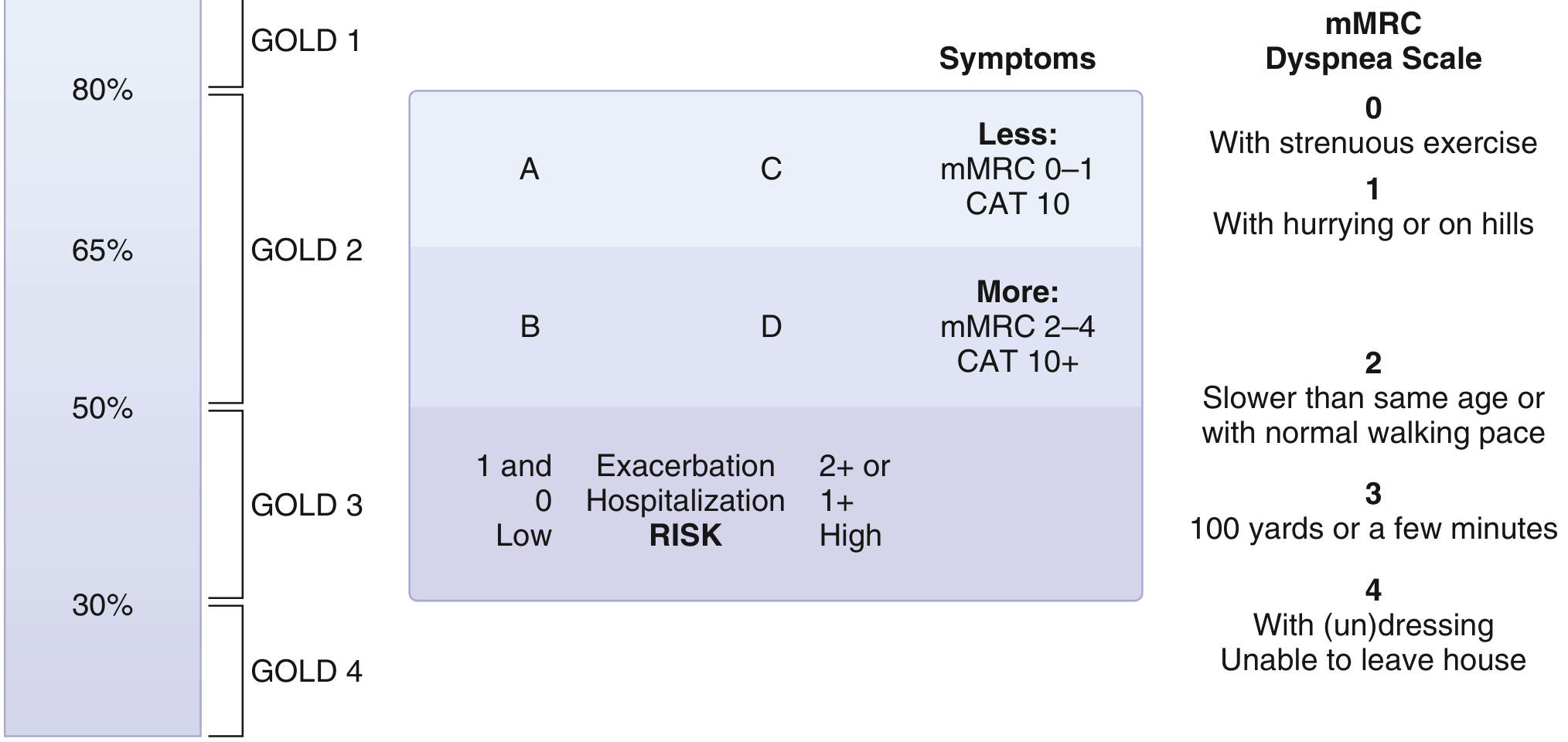
GOLD Classification System - Goldman-Cecil Medicine
Arterial Blood Gas (ABG)
- Indicated when SpO2 < 92% or severe COPD (FEV1 < 50%)
- Chronic stable COPD: May show mild hypoxemia; CO2 normal or mildly elevated
- Severe/end-stage: Hypoxemia (PaO2 < 60 mmHg), hypercapnia (PaCO2 > 45 mmHg)
- Chronic hypercapnia: respiratory acidosis with compensatory metabolic alkalosis (elevated HCO3-)
- Type II Respiratory Failure: PaO2 < 60 + PaCO2 > 50 mmHg
Chest Imaging
Chest X-Ray (CXR):
- Hyperinflation: flattened hemidiaphragms, increased retrosternal airspace (lateral view)
- Increased AP diameter
- Decreased vascular markings (emphysema)
- Bullae (emphysema)
- Tubular heart shadow (narrow, vertical)
- Pulmonary arterial enlargement (PH)
- Low sensitivity for early disease
CT Chest (High Resolution):
- Gold standard for detecting emphysema morphology (centriacinar vs. panacinar)
- Detects bullae, bronchiectasis, air trapping
- Airway wall thickening (chronic bronchitis)
- More sensitive than plain X-ray
- Now the tool of choice for lung cancer screening in at-risk COPD patients with significant smoking history
- Limited role in guiding routine pharmacotherapy (mainly useful when surgical intervention is considered) - Goldman-Cecil Medicine, p. 895
Blood Tests
- FBC (Full Blood Count): Polycythemia (secondary, from chronic hypoxia); eosinophilia may guide ICS therapy
- ABG: As above
- Alpha-1 antitrypsin level: In patients < 45 years, minimal smoking history, strong family history, panacinar emphysema, predominantly lower lobe disease
- BNP/Pro-BNP: If cor pulmonale or cardiac failure suspected
- ECG: Right ventricular hypertrophy (tall R in V1, right axis deviation), P pulmonale (peaked P waves in lead II)
- Echocardiography: To estimate pulmonary artery pressure (tricuspid regurgitation velocity), but accuracy limited by hyperinflation
- Peripheral blood eosinophil count: Guides decision on inhaled corticosteroids (ICS) - Goldman-Cecil Medicine, p. 942
- Sputum culture: During exacerbations (common pathogens: H. influenzae, S. pneumoniae, M. catarrhalis, Pseudomonas in severe disease)
6-Minute Walk Test (6MWT)
- Functional exercise capacity
- Correlates with mortality and QoL
- Component of the BODE index (BMI, airflow Obstruction, Dyspnea, Exercise capacity)
Management
GOLD Groups and Initial Treatment Algorithm
| Group | Characteristics | Initial Treatment |
|---|---|---|
| A | Low symptoms, low risk (0-1 exacerbations/year, no hospitalization, mMRC 0-1 / CAT < 10) | 1 bronchodilator (SABA or LABA or LAMA) |
| B | High symptoms, low risk (mMRC ≥2 / CAT ≥10) | LABA + LAMA (dual bronchodilation) |
| C | Low symptoms, high risk (≥2 exacerbations/year or 1+ hospitalization) | LAMA |
| D | High symptoms, high risk | LABA + LAMA; add ICS if eos ≥300/μL |
Non-Pharmacological Management
1. Smoking Cessation (Most Important Intervention)
- Single most effective intervention to slow FEV1 decline
- Nicotine replacement therapy (NRT): patch, gum, lozenge, inhaler
- Pharmacotherapy: Varenicline (most effective), Bupropion
- Behavioral counseling
2. Pulmonary Rehabilitation
- Exercise training + education + self-management
- Reduces dyspnea, improves exercise tolerance and quality of life
- Indicated for patients with mMRC ≥ 2 or post-exacerbation
- Does NOT improve FEV1 but significantly reduces symptoms and hospital admissions
3. Vaccinations
- Annual influenza vaccine (reduces exacerbations and mortality)
- Pneumococcal vaccine (PCV13 and PPSV23)
- COVID-19 vaccine
- Tdap/pertussis booster
4. Nutritional Support
- BMI < 21 associated with poor prognosis (BODE index)
- Dietary counseling and supplementation in underweight patients
5. Long-term Oxygen Therapy (LTOT)
- Indicated when PaO2 ≤ 55 mmHg (or SpO2 ≤ 88%) at rest
- Also if PaO2 56-59 mmHg + cor pulmonale, polycythemia, or nocturnal desaturation
- Must be used ≥ 15 hours/day (including sleep)
- Only intervention (besides smoking cessation) proven to improve mortality in hypoxemic COPD
Pharmacological Management
Bronchodilators (Cornerstone of Therapy)
| Drug Class | Examples | Route | Notes |
|---|---|---|---|
| SABA (Short-acting beta2-agonist) | Salbutamol (albuterol), Terbutaline | Inhaled | PRN for symptom relief |
| SABA anticholinergic combo | Ipratropium + Salbutamol (Combivent) | Inhaled | For acute relief |
| LABA (Long-acting beta2-agonist) | Salmeterol, Formoterol, Indacaterol | Inhaled | Once or twice daily |
| SAMA (Short-acting muscarinic antagonist) | Ipratropium | Inhaled | 4x daily |
| LAMA (Long-acting muscarinic antagonist) | Tiotropium, Umeclidinium, Aclidinium, Glycopyrronium | Inhaled | Once daily; preferred maintenance agent |
| LABA + LAMA combo | Indacaterol/Glycopyrronium (Ultibro), Vilanterol/Umeclidinium (Anoro) | Inhaled | Preferred in Group B, C, D |
Inhaled Corticosteroids (ICS)
- NOT used as monotherapy in COPD
- Combined with LABA (e.g., budesonide/formoterol, fluticasone/salmeterol)
- Indicated when: blood eosinophils ≥ 300/μL, asthma-COPD overlap, frequent exacerbations despite dual bronchodilation
- Risks: pneumonia (especially high-dose fluticasone), oral candidiasis, osteoporosis
- LABA + LAMA + ICS (triple therapy) for very symptomatic, frequent exacerbators
Other Drugs
- Roflumilast (PDE-4 inhibitor): Add-on for severe COPD with FEV1 < 50%, chronic bronchitis phenotype, frequent exacerbations; reduces exacerbation frequency
- Theophylline: 3rd-line bronchodilator; narrow therapeutic window; monitor levels; rarely used now
- Macrolide antibiotics (Azithromycin): Long-term low-dose in frequent exacerbators; risk of hearing loss, QT prolongation, macrolide resistance
- N-acetylcysteine (NAC): Mucolytic; modest evidence for reducing exacerbations
- Alpha-1 antitrypsin augmentation therapy: For documented deficiency with emphysema
Management of Acute Exacerbations
Definition: Acute worsening of respiratory symptoms beyond normal day-to-day variation requiring a change in medication.
Assessment (Anthonisen Criteria - type of exacerbation)
- Type 1: All 3 - increased dyspnea + increased sputum volume + purulent sputum
- Type 2: Any 2 of the above
- Type 3: Any 1 of the above + URTI/fever/wheeze/cough
Mild-Moderate (outpatient):
- Increase bronchodilator frequency (SABA ± SAMA)
- Oral prednisolone: 30-40 mg/day for 5 days (shortens recovery, improves FEV1)
- Antibiotics: If purulent sputum or clinical signs of infection
- Amoxicillin/doxycycline/clarithromycin (mild-moderate)
- Co-amoxiclav or moxifloxacin (severe or risk factors for Pseudomonas)
Hospitalization Criteria (CURB65 equivalent approach + clinical judgment):
- Severe dyspnea (mMRC 4, unable to speak in sentences)
- Marked hypoxemia or hypercapnia
- Cyanosis, confusion
- Lack of response to initial outpatient therapy
- Significant comorbidities
- Inability to care for self at home
Inpatient Management:
- Controlled oxygen therapy: Target SpO2 88-92% (NOT > 95%) - avoids hypercapnic drive suppression
- Nebulized SABA + SAMA (salbutamol + ipratropium)
- Systemic corticosteroids: Prednisolone/methylprednisolone IV
- Antibiotics: Based on sputum and culture
- Non-invasive positive pressure ventilation (NIPPV/BiPAP): First-line for type II respiratory failure (pH < 7.35, PaCO2 > 6 kPa); reduces intubation and mortality
- Invasive mechanical ventilation: If NIPPV fails or contraindicated
- DVT prophylaxis, nutritional support
Surgical Interventions
- Lung Volume Reduction Surgery (LVRS): Removes most emphysematous areas (usually upper lobe); improves FEV1 and survival in selected patients; high perioperative mortality - not suitable for all
- Bullectomy: For giant bullae causing compression of adjacent lung
- Bronchoscopic lung volume reduction (BLVR): Endobronchial valves (EBV) - less invasive alternative to LVRS in selected patients
- Lung transplantation: End-stage COPD with no other options; improves quality of life; limited by donor availability
COPD Assessment Tools
| Tool | Purpose | Score Range | Threshold |
|---|---|---|---|
| mMRC Dyspnea Scale | Dyspnea severity | 0-4 | ≥ 2 = significant symptoms |
| CAT (COPD Assessment Test) | Symptom impact | 0-40 | ≥ 10 = high symptom burden |
| BODE Index | Prognosis | 0-10 | Higher = worse 5-year survival |
| GOLD A/B/C/D | Risk stratification | - | Guides treatment selection |
Differentiating COPD from Asthma
| Feature | COPD | Asthma |
|---|---|---|
| Age of onset | > 40 years | Often childhood/young adult |
| Smoking | Usually significant (>20 pack-years) | Not required |
| Dyspnea | Progressive, persistent | Variable, episodic |
| Obstruction | Fixed (irreversible) | Variable (reversible) |
| Sputum | Chronic productive | Variable |
| Eosinophils | Less prominent (unless overlap) | Common |
| Response to steroids | Modest (some phenotypes) | Excellent |
| DLCO | Reduced (emphysema) | Normal |
Asthma-COPD Overlap Syndrome (ACOS): Fixed obstruction + features of asthma (atopy, marked bronchodilator reversibility, eosinophilia); treat with LABA + ICS + LAMA.
Sources:
- Goldman-Cecil Medicine, Chapter 76 (COPD), p. 892-898
- Murray & Nadel's Textbook of Respiratory Medicine, 2-Volume (Overlap syndrome, PH in COPD sections)
- Robbins & Kumar Basic Pathology (COPD pathology, emphysema subtypes), p. 447-450
This is a shared conversation. Sign in to Orris to start your own chat.