
Duodenal atreisia features
Reading File
Now let me search the medical library for duodenal atresia content in parallel.
Finding Sources
Finding Sources
Good results. Now let me read the most comprehensive sections in parallel.
Reading File
Reading File
Reading File
Reading File
Excellent - I have rich content from multiple authoritative sources. Let me also fetch the double-bubble ultrasound image and read the treatment section.
Loading Image
Reading File
I now have comprehensive information from multiple authoritative sources. Here is the full answer:
Duodenal Atresia - Features
Embryology and Pathogenesis
The duodenum begins development in the 4th week of fetal life. During the 5th-6th weeks, mucosal proliferation temporarily obliterates the lumen. A gradual process of recanalization (vacuolization) normally follows at 8-10 weeks gestation. Duodenal atresia (DA) results from failure of this recanalization.
- The Developing Human (Clinically Oriented Embryology) notes that if complete recanalization fails, a short segment of duodenum remains occluded
- Investigation of familial cases suggests autosomal recessive inheritance
Distinction from jejunoileal atresia: DA results from recanalization failure; jejunal/ileal atresia results from intrauterine vascular accidents - a fundamentally different mechanism.
Anatomic Types
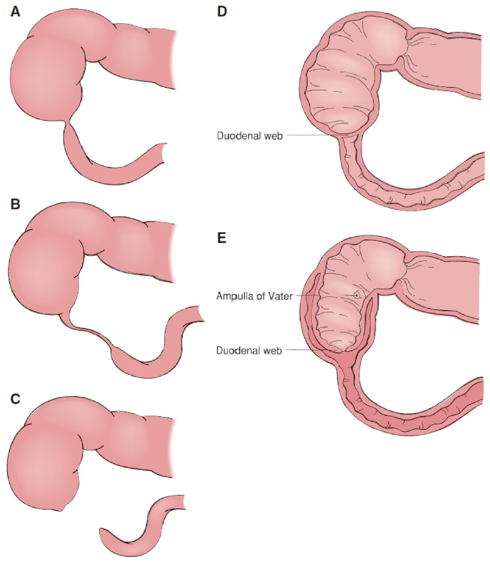
Atresias occur in three configurations:
- Blind-ending pouch with no connection to distal duodenum (least common)
- Pouch with fibrous cord connecting to the distal duodenum
- Complete intraluminal membrane obstructing the lumen (most common)
Additionally, duodenal webs can be perforated (causing stenosis rather than complete atresia). A special variant is the "wind-sock" deformity - a periampullary web projecting distally into the duodenal/jejunal lumen. This is surgically important because the apparent transition point from dilated to narrow duodenum is several centimeters distal to the actual base of the web.
Key Epidemiology
| Feature | Detail |
|---|---|
| Incidence | ~1 in 6,000-10,000 births |
| Sex | Slight female predilection |
| Down syndrome (Trisomy 21) | 30% of cases - mandatory karyotyping |
| Prematurity | ~20% are premature |
| Location | 80% distal to ampulla of Vater (2nd/descending duodenum) |
| Proximal to ampulla | 5-10% of cases |
Associated Anomalies (>50% of cases have at least one)
- Down syndrome (Trisomy 21) - 25-50% (most common association)
- Congenital heart disease - ~20%
- Malrotation of the gut - 20-30%
- Annular pancreas - extrinsic compression component
- Esophageal atresia / VACTERL association - oesophageal atresia in up to 10%
- Anorectal defects
- Genitourinary malformations
- Meckel's diverticulum
- Central nervous system lesions
Clinical Presentation
Prenatal:
- Polyhydramnios (33-50% of cases) - DA prevents normal intestinal absorption of swallowed amniotic fluid
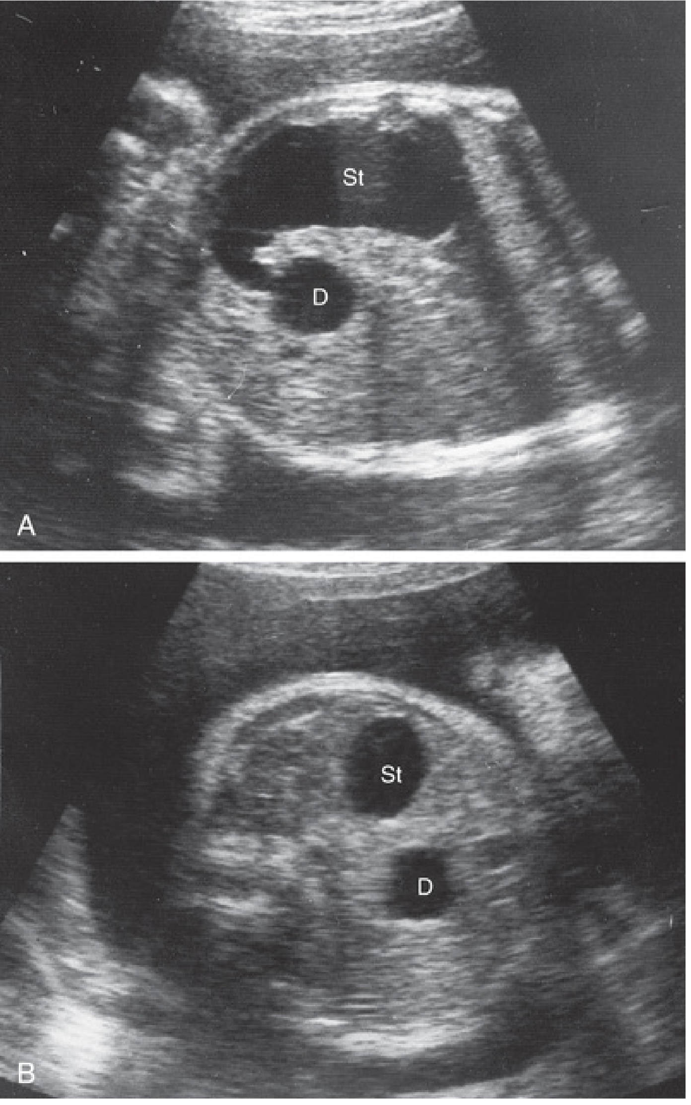
- Fetal ultrasound showing dilated, fluid-filled stomach and duodenal cap
Postnatal (within first 24-48 hours):
- Bilious vomiting (most common) - because obstruction is usually distal to the ampulla of Vater
- Non-bilious emesis in 15-20% - when obstruction is proximal (pre-ampullary) to the ampulla
- Upper abdominal / epigastric distension - due to overfilled stomach and proximal duodenum
- Palpable epigastric mass
- Visible gastric peristaltic waves
- Collapsed distal bowel - typically no diffuse abdominal distention
- Partial obstruction (fenestrated web) may be asymptomatic in neonates and present later in childhood or even adulthood
Diagnosis
Prenatal Ultrasound
The classic "double-bubble" sign can be detected prenatally - showing the dilated fluid-filled stomach and proximal duodenum:
Plain Abdominal Radiograph (Postnatal)
- "Double bubble" sign - two air-filled bubbles: the distended stomach and the proximal duodenum
- No gas distal to the duodenum in complete obstruction - this is sufficiently diagnostic; no further GI imaging is needed in a stable child
- Gas seen distally = partial obstruction (fenestrated web or malrotation/volvulus must be excluded)
Upper GI Contrast Study
- Indicated when partial obstruction is suspected (to exclude malrotation with acute volvulus - a surgical emergency)
- Shows duodenal narrowing in stenosis
- A duodenal web appears as a thin filling defect with pre-stenotic dilatation
Important Radiologic Caveat
The plain film alone may not always differentiate DA from malrotation or stenosis. If there is concern for malrotation, an urgent upper GI series is required given the risk of an acute volvulus requiring emergent surgery. - Mulholland & Greenfield's Surgery, 7e
Treatment
Surgical - duodenoduodenostomy (gold standard):
- A transverse incision in the dilated proximal duodenum is anastomosed to a longitudinal incision in the unused downstream duodenum, creating a diamond-shaped anastomosis
- Downstream patency must be confirmed (catheter/saline/air) to exclude synchronous distal atresia
- Laparoscopic repair is also well established
Annular pancreas with DA: Treated by duodenoduodenostomy - never by direct division of the annular pancreas (risks injury to accessory pancreatic duct and doesn't address the intraluminal obstruction)
Duodenal webs: Excised through longitudinal duodenotomy; the ampulla of Vater must be unambiguously identified before excision (especially in wind-sock deformities) to avoid bile duct injury
Summary Mnemonic
DUODENAL ATRESIA = "Double Bubble + Down's + Distal-to-ampulla + Bilious vomiting"
- Double bubble sign (X-ray/US)
- Down syndrome in 30%
- Distal to ampulla in 80%
- Bilious vomiting (or non-bilious if pre-ampullary)
- Polyhydramnios prenatally
- Recanalization failure (embryologic basis)
Sources: The Developing Human - Clinically Oriented Embryology; Mulholland & Greenfield's Surgery 7e; Sleisenger & Fordtran's Gastrointestinal and Liver Disease; Grainger & Allison's Diagnostic Radiology
Chocking in duod3nal atresia?
The user is asking about "chocking" in duodenal atresia - this likely refers to "choking" as in the wind-sock/web deformity, or possibly the radiological/surgical concept of a "sock" or "windsock" deformity. But it could also mean the clinical concept of aspiration/choking from vomiting. Let me clarify.
Asking for Preferences
What do you mean by 'chocking' in duodenal atresia?
Choking
"Choking" in duodenal atresia most likely refers to the "wind-sock" deformity - a specific type of duodenal web where the obstruction point is not where it appears to be. This is a well-known surgical pitfall. Let me look it up properly.
Searching sources...
Finding Sources
Finding Sources
Reading File
Loading Image
Wind-Sock (Choking) Deformity in Duodenal Atresia
"Choking" in duodenal atresia refers to the wind-sock deformity - a specific and surgically important variant of a duodenal web.
What is it?
A periampullary intraluminal web that is attached at its base near or just distal to the ampulla of Vater, but then balloons out distally into the duodenal or jejunal lumen - like a wind sock or stocking being pushed outward by the flow of contents.
(Panel E in the diagram above shows the wind-sock deformity - note how the web originates near the Ampulla of Vater but projects distally into the lumen)
Why is it Surgically Important (The "Trap")?
This is the key clinical point:
The visible transition from dilated proximal duodenum to narrow downstream duodenum may be several centimeters DISTAL to the actual base of the web.
In other words:
- The surgeon sees the duodenum narrow at point X
- But the real obstruction (web base) is at point Y, which is several cm proximal to X
- If the surgeon incises at the apparent narrowing, they will miss the actual web and may injure the ampulla of Vater
Key Surgical Steps for Wind-Sock Web
- Longitudinal duodenotomy is made to enter the lumen
- The web must be carefully traced proximally to its true base - not assumed to be at the level of luminal narrowing
- The ampulla of Vater must be unequivocally identified before excision - it is in close proximity and must be protected
- After web excision, the duodenotomy is closed transversely (to avoid narrowing)
Summary
| Feature | Detail |
|---|---|
| What it is | Intraluminal duodenal web ballooning distally |
| Location | Periampullary (near Ampulla of Vater) |
| The trap | Apparent narrowing is distal to the actual web base |
| Risk if missed | Wrong incision site, ampullary injury |
| Treatment | Longitudinal duodenotomy + web excision, with ampulla identification first |
- Mulholland & Greenfield's Surgery, 7e
Does choking happen in duodenla atresia?
The user is now asking whether actual choking (aspiration/airway choking) occurs in duodenal atresia - as a clinical complication from vomiting. Let me search for this properly.
Searching sources...
Finding Sources
Finding Sources
Reading File
Reading File
Yes - aspiration/choking is a real and recognized complication of duodenal atresia, particularly from the persistent bilious vomiting. Here is the full picture:
Does Choking (Aspiration) Happen in Duodenal Atresia?
Yes. Choking and aspiration are recognized complications of duodenal atresia and are one of the primary reasons for urgent neonatal management.
Why It Happens
In duodenal atresia, the duodenum is obstructed - usually distal to the ampulla of Vater. This means:
- The stomach and proximal duodenum fill with gastric secretions, swallowed amniotic fluid (prenatally), and bile
- This causes forceful, recurrent bilious vomiting starting within hours of birth
- A neonate's airway protective reflexes are immature
- The upper abdominal distension also elevates the diaphragm, compressing the lungs
All of these factors combine to make aspiration of vomitus a genuine risk.
Clinical Consequences of Aspiration
If not managed promptly:
| Complication | Mechanism |
|---|---|
| Aspiration pneumonitis | Inhaled bilious vomit causes chemical lung injury |
| Respiratory failure | Increased abdominal pressure from distension compresses the diaphragm |
| Sepsis | Aspiration pneumonitis progressing to pneumonia |
"Delayed diagnosis or treatment can result in increased abdominal pressure, leading to respiratory failure from pressure on the diaphragm and aspiration pneumonitis, as well as sepsis."
- Barash's Clinical Anesthesia, 9e
How It Is Prevented
The first step in neonatal management of duodenal atresia is gastric decompression specifically to prevent aspiration:
- Orogastric or nasogastric tube placed immediately to decompress the stomach
- IV fluid hydration to correct electrolytes
- Surgical repair is then planned (duodenoduodenostomy)
"Orogastric or nasogastric decompression to prevent aspiration" - listed as the first priority in neonatal management
- Creasy & Resnik's Maternal-Fetal Medicine
Anesthetic Implications
Because of the aspiration risk, when these neonates go to the operating room:
- Rapid-sequence induction and intubation (RSI) is the standard approach
- Alternatively, awake intubation if hemodynamically unstable or difficult airway is expected
- The stomach is considered "full" due to obstruction - standard precautions for full stomach apply
Summary
| Point | Detail |
|---|---|
| Does choking/aspiration occur? | Yes - real and recognized risk |
| Cause | Persistent bilious vomiting from proximal obstruction |
| Consequence | Aspiration pneumonitis, respiratory failure, sepsis |
| Prevention | NG/OG tube decompression - the very first step of management |
| OR precaution | RSI or awake intubation required |
The NG tube insertion is not just supportive - it is the primary measure to protect the airway from aspiration in these neonates before surgical correction.
This is a shared conversation. Sign in to Orris to start your own chat.