
Regional anesthesia of foot complete guide from anesthesia,tintinalli, rosens book for emergency physician
ankle block five nerves foot regional anesthesia diagram
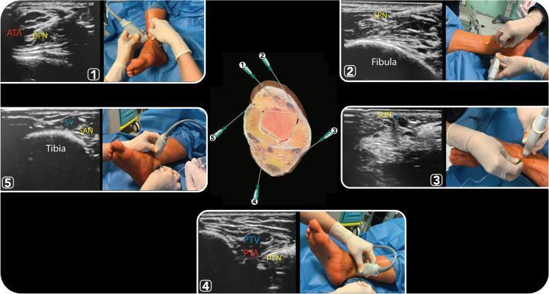
A composite medical educational figure demonstrating ultrasound-guided ankle block techniques for five terminal nerves. A central cross-sectional anatomical diagram of the ankle identifies needle trajectories for the deep peroneal (1), superficial peroneal (2), sural (3), posterior tibial (4), and saphenous (5) nerves. Surrounding this are five numbered panels, each pairing a high-frequency ultrasound scan with a clinical photograph of the transducer and needle placement. (1) Deep Peroneal Nerve (DPN) shown lateral to the anterior tibial artery (ATA) over the tibia. (2) Superficial Peroneal Nerve (SPN) visualized as hyperechoic clusters in the subcutaneous tissue superficial to the fascia near the fibula. (3) Sural Nerve (SUN) identified adjacent to the small saphenous vein (V). (4) Posterior Tibial Nerve (PTN) shown posterior to the posterior tibial artery (PTA) and veins (PTV) at the medial malleolus, exhibiting a classic honeycomb echotexture. (5) Saphenous Nerve (SAN) demonstrated adjacent to the great saphenous vein (SV) over the medial tibia. This resource is designed for anesthesiology training and regional anesthesia protocol development.
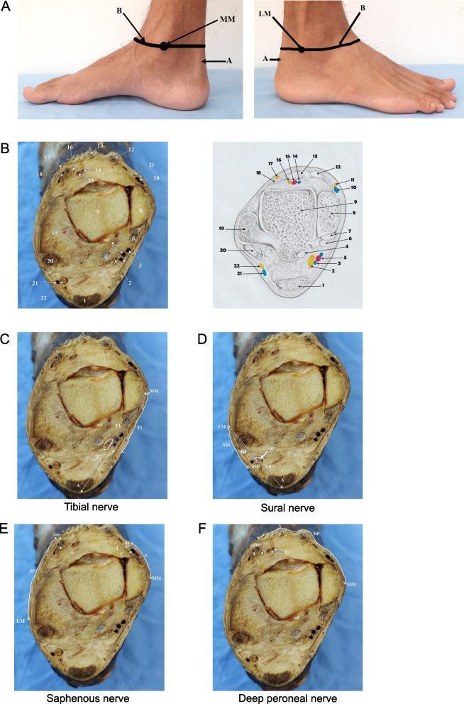
This composite educational resource illustrates the anatomical landmarks and cross-sectional morphology relevant for performing an ankle nerve block. Panel A shows clinical photographs of the medial and lateral ankle with surface markings at the level of the malleoli. Panel B provides a cadaveric cross-section and a corresponding labeled anatomical diagram at the inter-malleolar plane. Key visible structures include the talus (9), Achilles tendon (1), medial (8) and lateral (19) malleoli, and associated neurovascular bundles. Subsequent panels (C-F) map specific regional anesthesia targets: the tibial nerve (C) located posteromedial to the Achilles tendon; the sural nerve (D) located between the lateral malleolus and Achilles; the saphenous and medial dorsal cutaneous nerves (E) along the anterior-medial aspect; and the deep peroneal nerve (F) situated anteriorly between the extensor tendons. Measurements (T1-T3, SR1-SR3, DP) denote distances from palpable bony landmarks to the nerves and depth from the skin surface (dT, dSR, dDP). This content is intended for advanced clinical learners in anesthesiology and orthopedic surgery to understand the spatial relationships required for ultrasound-guided or landmark-based regional anesthesia.
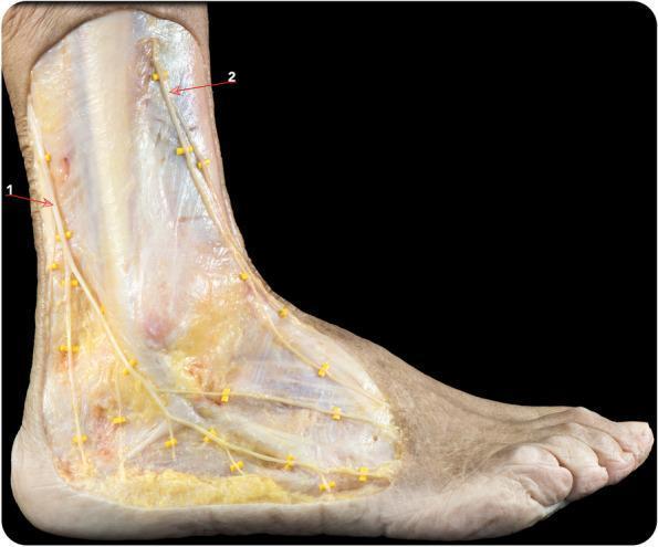
This clinical photograph displays a high-quality anatomical dissection of the lateral and anterolateral aspects of the human distal lower leg, ankle, and foot. The superficial fascia has been removed to expose the neural pathways critical for peripheral nerve blocks. The sural nerve (labeled 1) is shown coursing posterior to the lateral malleolus, demonstrating its characteristic distal arborization into several small branches that provide sensory innervation to the lateral margin of the foot. The superficial peroneal nerve (labeled 2) is visible descending along the anterolateral compartment. It bifurcates into its two primary terminal branches: the medial dorsal cutaneous nerve and the intermediate dorsal cutaneous nerve, which extend toward the dorsum of the foot. The image highlights the superficial nature of these nerves within the subcutaneous tissue, emphasizing their clinical relevance for achieving regional anesthesia through ankle block techniques. Anatomical landmarks such as the lateral malleolus and the extensor tendons are visible under the dissected layers, providing topographical context for nerve localization.
This educational medical graphic illustrates the innervation and regional anesthesia techniques for the hip. The top section features an anatomical diagram of the pelvis and upper thigh in two views: an anterior superficial view showing the Lateral Femoral Cutaneous, Femoral, and Obturator nerves, and a deep view showing the Trochanteric nerve. The bottom section presents three ultrasound images demonstrating specific regional nerve blocks. 1) Obturator Nerve Block: Shows the anterior and posterior branches of the obturator nerve (ObN Ant. Br. and ObN Post Br.). 2) Femoral Nerve Block: Displays the needle tip approaching the femoral nerve, adjacent to the femoral artery, femoral vein, and psoas muscle under the fascia iliaca. 3) PENG (Pericapsular Nerve Group) Block: Highlights landmarks including the Femoral Artery (FA), Pectineus muscle (PE), iliopectineal ligament (LA), and iliacus muscle (M). The diagram serves as a clinical guide for anesthesiology and pain management, comparing traditional nerve blocks with the newer PENG technique for hip analgesia.
Regional Anesthesia of the Foot: Complete Guide for the Emergency Physician
Overview & Indications
- Avoids airway manipulation
- Fewer systemic side effects
- Suitable for ambulatory patients
- Fast onset (~15 minutes)
Anatomy: The Five Nerves of the Foot
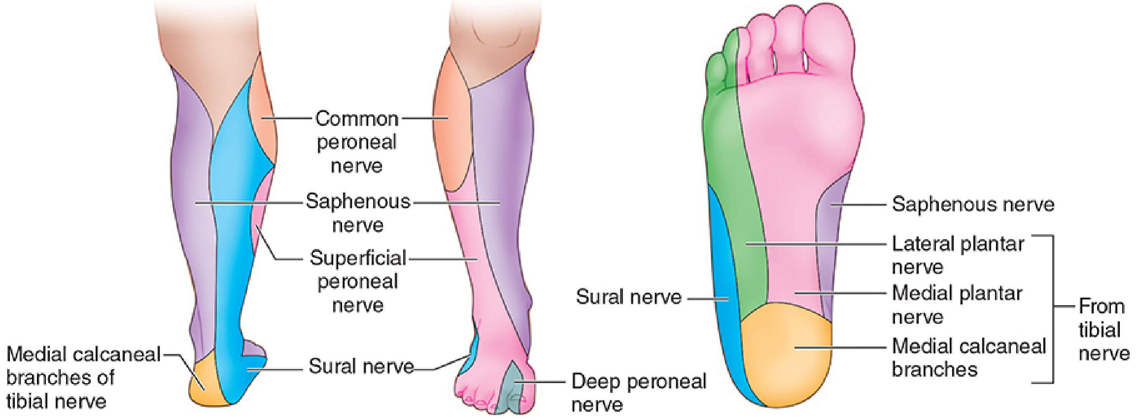
| Nerve | Origin | Landmark | Sensory Territory |
|---|---|---|---|
| Posterior Tibial | Sciatic | Posterior to posterior tibial artery, behind medial malleolus | Heel, sole of foot, plantar toes, medial + lateral plantar |
| Sural | Tibial nerve branch | Between Achilles tendon and lateral malleolus | Lateral foot, lateral proximal sole |
| Deep Peroneal | Common peroneal | Between extensor hallucis longus + extensor digitorum longus tendons; lateral to dorsalis pedis artery | First dorsal web space, short toe extensors |
| Superficial Peroneal | Common peroneal | Superficial, subcutaneous at extensor retinaculum | Dorsum of foot and toes (except 1st web space) |
| Saphenous | Femoral nerve | Anterior to medial malleolus, near saphenous vein | Anteromedial foot strip |
Local Anesthetic Selection
- Lidocaine 1% or 2%: Standard choice, onset 5-10 min, duration ~1-2 hr
- Bupivacaine 0.25-0.5%: Use when longer anesthesia desired
- Ropivacaine 0.5-0.75%: Alternative long-acting agent with lower cardiotoxicity
- Epinephrine: Avoid at the ankle level - risk of ischemic complications to distal circulation. Rosen's notes that for digital blocks of the toe (in healthy patients without vascular disease), lidocaine with epinephrine appears safe per current literature, but the traditional practice of avoiding it is still reasonable.
The Ankle Block: Five-Nerve Technique
1. Posterior Tibial Nerve Block
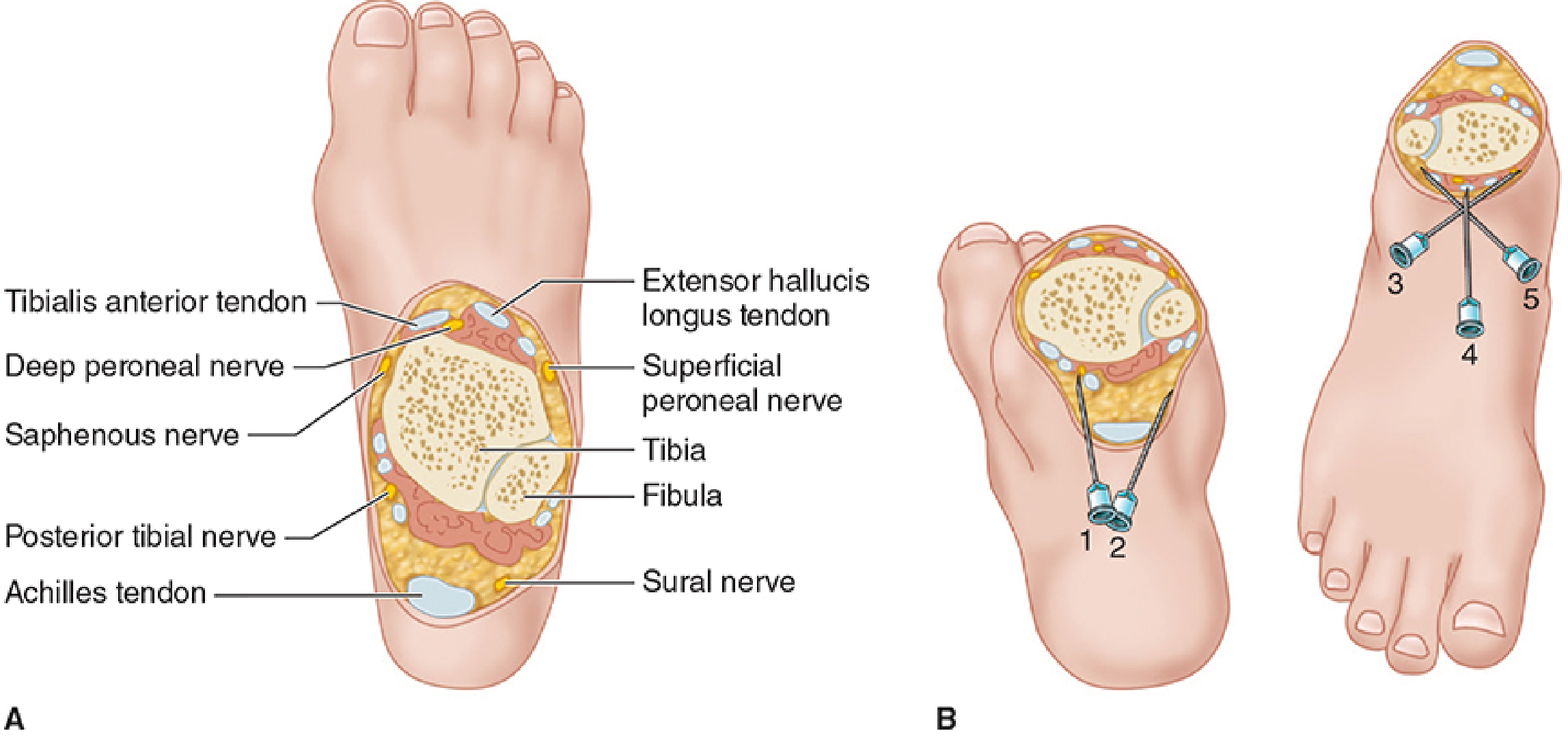
- Insert a 25-gauge needle posterolateral to the artery at the level of the medial malleolus
- If paresthesia obtained: inject 3-5 mL local anesthetic
- If no paresthesia: inject 7-10 mL with fan-shaped technique in the triangle formed by the artery, Achilles tendon, and tibia, slowly withdrawing
- Linear ("hockey stick") 10-MHz probe in transverse (short) axis, posterior and inferior to medial malleolus (or 3-5 cm above)
- Nerve appears hyperechoic, honeycomb-like texture, posterior and deep to the posterior tibial artery
- Color Doppler identifies the artery
- IP or OOP approach; inject 5 mL lateral to nerve
- Identify nerve before it divides into medial and lateral plantar nerves
2. Sural Nerve Block
- Insert 25-gauge needle lateral to the Achilles tendon, direct toward the lateral malleolus
- Subcutaneous injection of 5-10 mL in the groove between lateral malleolus and calcaneus
- US: Identify sural nerve adjacent to the lesser saphenous vein, 1 cm proximal to the lateral malleolus - perivascular approach improves success rate compared to surface landmark alone
3. Deep Peroneal Nerve Block
- Raise a skin wheal just lateral to the arterial pulsation between the two tendons at the intermalleolar line
- Advance 25-gauge needle perpendicular to skin
- Inject 3-5 mL deep to the extensor retinaculum (below fascia)
4. Superficial Peroneal Nerve Block
- Through the same skin wheal used for the deep peroneal nerve block
- Direct needle laterally subcutaneously (not deep), injecting 3-5 mL
- Creates a subcutaneous ridge from the deep peroneal entry point toward the lateral malleolus
- An additional 5-10 mL may be needed for full coverage
- US may visualize the nerve as hyperechoic clusters in subcutaneous tissue superficial to the fascia near the fibula
5. Saphenous Nerve Block
- Through the same skin wheal as deep + superficial peroneal (from the medial direction), or a separate medial injection
- Subcutaneous infiltration of 5 mL of local anesthetic around the saphenous vein anterior to the medial malleolus
- A "wall of anesthesia" between skin and bone suffices
Streamlining: Reducing Needle Insertions
- The deep peroneal, superficial peroneal, and saphenous nerves can all be blocked through a single midline anterior skin wheal at the intermalleolar line - the needle is directed laterally (superficial peroneal), perpendicular/deep (deep peroneal), and medially (saphenous)
- Posterior tibial and sural are blocked posteriorly, requiring 1-2 additional skin punctures
- Minimum practical insertions: 3
Selective Block vs. Complete Block
| Procedure Area | Nerves to Block |
|---|---|
| Plantar forefoot, toes (plantar) | Posterior tibial (dominant), ± sural |
| Dorsum of foot | Superficial peroneal ± deep peroneal |
| 1st web space / great toe dorsum | Deep peroneal |
| Lateral foot | Sural |
| Medial border | Saphenous |
| Entire foot | All five |
Toe / Digital Block
- Position foot with plantar surface accessible
- Insert small needle at the dorsal distal webspace, alongside the edge of the phalanx
- Inject 1-2 mL subcutaneously for dorsal digital nerve
- Redirect volarward, inject 1-2 mL for the plantar digital nerve
- Repeat on the opposite side of the digit
- Total: ~4-6 mL lidocaine 1-2%
Onset, Duration, and Volumes Summary
| Nerve | Volume | Onset |
|---|---|---|
| Posterior tibial | 5 mL (paresthesia) / 7-10 mL (no paresthesia) | 10-20 min |
| Sural | 5-10 mL | 10-15 min |
| Deep peroneal | 3-5 mL | 10-15 min |
| Superficial peroneal | 3-5 mL subcutaneous | 5-10 min |
| Saphenous | 5 mL subcutaneous | 5-10 min |
| Total | ~20-35 mL | ~15 min |
Complications and Contraindications
- Multiple injections cause patient discomfort - premedicate with adequate sedation/analgesia
- Neuropathy (rare): avoid pinning deep nerves (tibial, deep peroneal) against bone; do not inject under high pressure
- Persistent paresthesia: generally self-limited
- Ischemia: do not use epinephrine at the ankle level
- Landmark failure: edema and induration around the ankle make palpation unreliable - use US or consider a more proximal block (popliteal + saphenous blocks)
- Significant ankle edema or infection
- Tourniquet pain expected (e.g., prolonged OR procedure) - ankle block gives no tourniquet analgesia
- Patient needing postoperative continuous catheter analgesia
- Local infection at injection site
- Patient refusal
- Known allergy to local anesthetic class
Ultrasound Guidance: Key Points
- Posterior tibial nerve: Most reliably identified on US; landmark of choice for deep blocks. Probe in short axis posterior to medial malleolus. Honeycomb, hyperechoic structure posterior to the artery.
- Deep peroneal nerve: Difficult to see; rely on identifying the anterior tibial artery with Doppler then inject lateral to it.
- Sural nerve: Identify by the lesser saphenous vein; perivascular injection superior to landmark approach.
- Superficial peroneal & saphenous: Usually blocked by subcutaneous infiltration; US adds little for these.
- US shortens onset time for posterior tibial block and reduces the number of needle passes.
ED Practical Tips (Tintinalli / Rosen's)
- Patient positioning: Elevate foot on folded blanket to allow access posteriorly and anteriorly without repositioning.
- Premedication: Because five injections are needed, 1-2 mg IV midazolam ± fentanyl improves patient tolerance significantly.
- Needle choice: 25-gauge needle throughout; reduces pain on injection.
- Aspiration: Always aspirate before injection, especially near posterior tibial and deep peroneal arteries.
- Wait time: Allow a full 15 minutes before testing block adequacy or beginning the procedure.
- Failed block: If a component fails, supplement with local infiltration at the site, not repeat ankle block injection.
- Toe dislocations: Rosen's recommends digital block + longitudinal traction for interphalangeal joint dislocations; if the great toe is involved, a walking cast with toe plate for 3 weeks post-reduction.
Quick Reference: Nerve Blocks by Clinical Presentation
| ED Presentation | Recommended Block |
|---|---|
| Laceration - plantar forefoot | Posterior tibial nerve block |
| Laceration - dorsum of foot | Superficial ± deep peroneal |
| Subungual hematoma / nail bed injury - toe | Digital (toe) block |
| Foreign body - lateral foot | Sural nerve block |
| Toe fracture | Digital block or partial ankle block |
| Great toe dislocation | Digital block |
| Plantar abscess | Posterior tibial block |
| Full foot surgery / complex laceration | Complete 5-nerve ankle block |
- Morgan & Mikhail's Clinical Anesthesiology 7e, Chapter 46 (pp. 1936-1939)
- Miller's Anesthesia 10e, Chapter 42 (pp. 6277-6283)
- Barash Clinical Anesthesia 9e, Chapter 36 (pp. 3035-3039)
- Rosen's Emergency Medicine, Chapter 42 (pp. 563-566)
- Tintinalli's Emergency Medicine, Chapter 268