
Insect bite like fly management mild swelling and redness
insect fly bite mild swelling redness management treatment 2025
insect bite fly bite skin redness swelling reaction
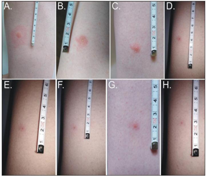
This clinical photographic series (Figures 3A–3H) illustrates the temporal evolution and resolution of a human skin lesion following a bite from the forest fly, Hippobosca equina. The progression is documented over 19 days on the thigh, with a measuring tape providing scale. Initial presentation (19–40 hours) shows painful, urticarial pink papules surrounded by a prominent circular erythematous base approximately 2–3 cm in diameter. By 64 hours (Figure 3C), the lesion transitions to an excoriated erythematous papule with localized dermatitis, reflecting a decrease in surrounding inflammation. The subsequent images (99–260 hours) demonstrate a gradual reduction in size, elevation, and redness. The final stage at 19 days (Figure 3H) reveals a residual hyperpigmented papule, approximately 0.5 cm, marking the resolution of acute inflammation with lingering post-inflammatory hyperpigmentation. This timeline provides a clinical reference for identifying and predicting the natural history of insect-induced dermatological reactions in a human host.
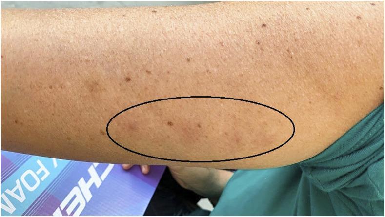
This clinical photograph shows the medial aspect of a human arm presenting with inflammatory skin lesions following insect bites. The patient has a light brown complexion with scattered benign macules (freckles or solar lentigines). Within a black oval annotation, several localized areas of pathology are visible. These findings include erythematous, slightly edematous papules that appear raised relative to the surrounding healthy tissue. The clinical presentation is consistent with a localized hypersensitivity reaction or inflammatory response to triatomine (kissing bug) bites, specifically from Triatoma sanguisuga. This image serves as a diagnostic reference for identifying the dermatological manifestations of Chagas disease vectors in North America. The key educational focus is on the morphology of arthropod-inflicted skin reactions, highlighting the characteristic redness and induration that differentiate the bite sites from general skin irregularities or normal pigmentation.
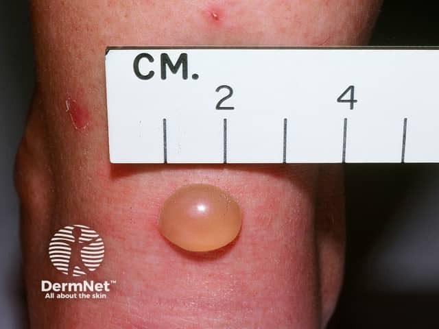
Clinical macroscopic photograph of a solitary cutaneous lesion consistent with an insect bite reaction. Imaging modality: clinical photography with macro close-up, frontal view, ambient lighting; magnification approximately 1.5–2x. Anatomical location: skin of the forearm/wrist region (integumentary system, skin). The lesion is a single, dome-shaped, translucent to skin-colored vesicle/bulla measuring roughly 6–8 mm in diameter, with a smooth, shiny surface and minimal surrounding erythema. The lesion is nonulcerated and without crusting or purulence, suggesting a vesicular blister rather than pustular infection. Clinically, this morphology is typical of a localized insect bite reaction (mosquito, biting fly, or bee) with rapid onset after exposure; differential diagnoses include vesicular contact dermatitis, bullous impetigo, and less likely herpes simplex or varicella zoster vesicles in a solitary focal location. Diagnostic significance lies in recognizing a benign, self-limited cutaneous response requiring symptomatic care and surveillance for secondary infection. Potential clinical use cases include dermatology education, teledermatology triage, image-guided wound assessment, AI-based lesion classification, research on bite reactions, patient counseling, and educational resources for medical students and clinicians.
Fly / Insect Bite Management: Mild Swelling and Redness
Step-by-Step Management
- Move away from the area to avoid further bites.
- Remove any visible stinger or foreign material if present.
- Wash the bite area thoroughly with soap and water to reduce infection risk.
- Apply a cold compress or ice pack (wrapped in a cloth) for 10-20 minutes.
- This is the most effective first step for reducing local swelling and pain.
- Repeat every few hours as needed.
- If the bite is on an arm or leg, elevate the limb - this helps drain edema and reduce swelling faster.
- Hydrocortisone cream (0.5%-1%): Applied 2-3 times daily; reduces local inflammation and itch.
- Calamine lotion: Soothes itching and has a mild astringent effect.
- Baking soda paste: Mix baking soda + water and apply to the bite for itch relief.
- Aloe vera gel: Cooling, anti-inflammatory, and soothing.
- Antihistamines: Cetirizine (10 mg once daily) or loratadine (10 mg once daily) reduce allergic swelling and itch. These are the go-to agents for fly bite reactions per StatPearls on insect bites.
- NSAIDs (ibuprofen 400 mg) or acetaminophen (paracetamol 500-1000 mg): For pain and inflammation, especially with more painful fly bites (horsefly, blackfly).
The "3 Ps" Rule (Indian Journal of Dermatology guideline)
| P | Meaning |
|---|---|
| Protective clothing | Cover skin when in fly-prone areas to prevent further bites |
| Pruritus control | Topical corticosteroids + antihistamines |
| Patience | Most reactions are self-limiting (3-10 days) |
Natural Course
- Mild itching and swelling: usually resolves within 3-7 days
- The bite site may leave a residual hyperpigmented papule for a few weeks after the active reaction subsides (see image above)
When to Seek Medical Attention (Red Flags)
- Swelling spreads rapidly or worsens after 24-48 hours
- Signs of infection: increasing warmth, pus, fever, streaks (lymphangitis)
- Difficulty breathing, throat tightening, or dizziness (anaphylaxis - use epinephrine if available, call emergency services immediately)
- Bite on face/neck with significant swelling
- Swelling involves the eye or lips
- No improvement after 5-7 days of home care
Summary Table
| Intervention | Purpose | Frequency |
|---|---|---|
| Soap & water wash | Prevent infection | Once, immediately |
| Cold compress | Reduce swelling & pain | 10-20 min, every few hours |
| Elevation of limb | Reduce edema | Keep elevated when resting |
| Hydrocortisone cream 1% | Reduce itch & redness | 2-3x daily for 3-5 days |
| Cetirizine 10 mg oral | Reduce allergic reaction | Once daily |
| Ibuprofen 400 mg oral | Pain/inflammation | Every 6-8 hrs with food |