
Imnci guidelines of pneumonia
IMNCI guidelines pneumonia classification treatment 2025
https://nhmodisha.gov.in/wp-content/uploads/2024/02/IMNCI.pd…
IMNCI pneumonia classification chart fast breathing chest indrawing
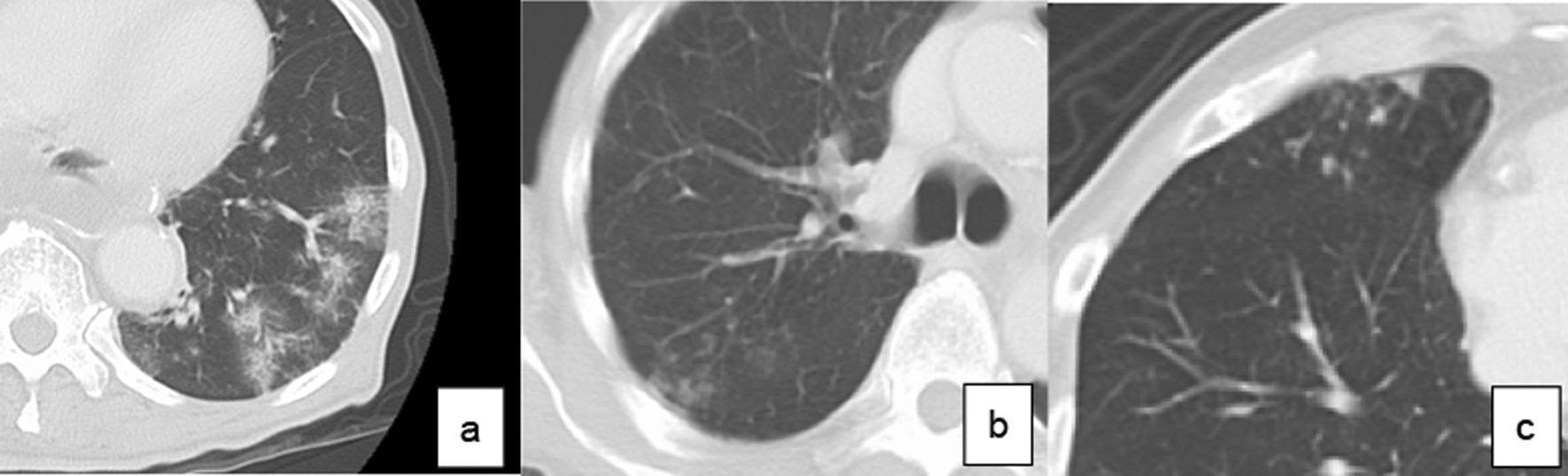
A comparison chart consisting of three axial chest CT scans illustrating radiological classifications of pneumonia. Panel (a) shows the consolidation type, characterized by areas of increased pulmonary density and patchy opacification within the lung parenchyma, consistent with alveolar filling that obscures underlying vascular structures. Panel (b) depicts the reticular type, showing a fine, net-like meshwork of linear opacities reflecting interstitial involvement or thickened interlobular septa. Panel (c) illustrates the nodular type, marked by multiple discrete, rounded opacities or small nodules scattered within the lung field. These images serve as educational reference for identifying long-term postoperative pneumonia (LTPP) patterns, emphasizing the morphological distinctions between alveolar consolidation, interstitial linear patterns, and nodular presentations. The imaging modality is high-resolution computerized tomography (CT), focused on the thoracic cavity and lung parenchyma for diagnostic classification in respiratory medicine.
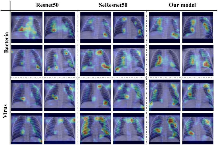
This comparison chart displays a grid of Grad-CAM attention map heatmaps superimposed on pediatric chest X-rays, demonstrating model interpretability for pneumonia classification. The visual is organized into four columns representing three different deep learning architectures: Resnet50, SeResnet50, and a proposed 'Our model'. The rows are categorized by etiology: 'Bacteria' (top two rows) and 'Virus' (bottom two rows). For each model, two representative chest X-ray cases are shown per pathology. The heatmaps use a color gradient (red being highest intensity, blue lowest) to indicate which anatomical regions the AI prioritized for its diagnosis. In bacterial pneumonia cases, 'Our model' shows highly localized, intense 'hot spots' concentrated on pulmonary consolidations compared to the more diffuse attention of Resnet50. For viral cases, the heatmaps illustrate attention distributed across bilateral lung fields, reflecting typical interstitial patterns. This image serves as a diagnostic imaging tool for researchers and medical students to understand how computer vision models identify radiological features of pediatric pneumonia.
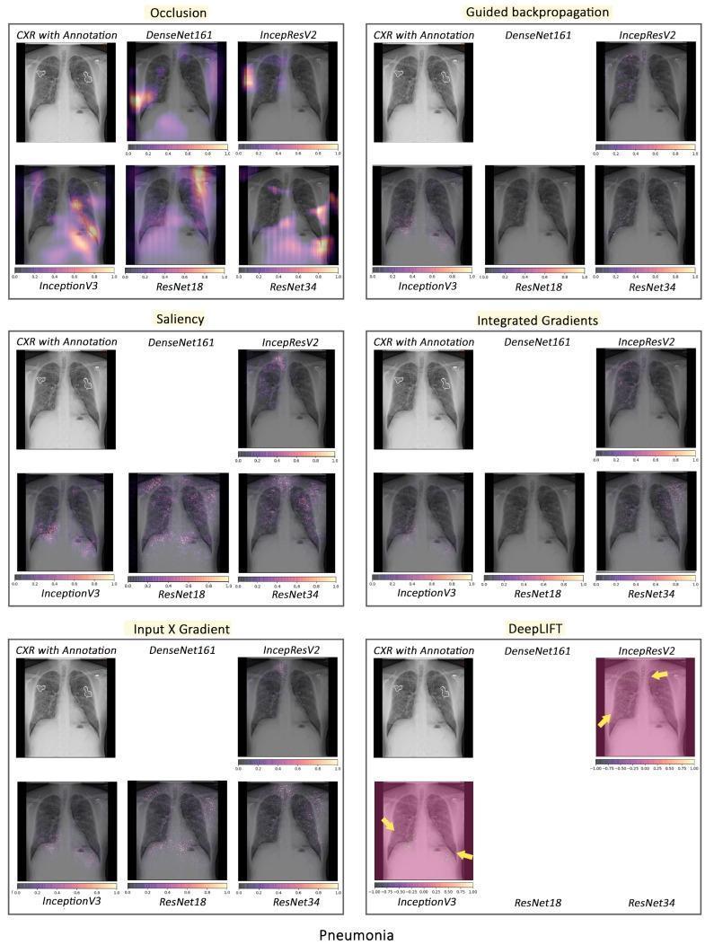
A multi-panel comparison chart illustrating various artificial intelligence (AI) interpretability techniques applied to Chest X-ray (CXR) images for pneumonia detection. The chart evaluates six local interpretability methods: Occlusion, Guided Backpropagation, Saliency, Integrated Gradients, Input X Gradient, and DeepLIFT. Each section includes a baseline 'CXR with Annotation' showing expert-identified pathology zones alongside heatmaps or saliency maps from five neural network architectures: DenseNet161, InceptionResNetV2, InceptionV3, ResNet18, and ResNet34. The heatmaps use color gradients (yellow to purple) to indicate regions of interest that influenced the model's classification. The visualizations demonstrate significant variability in model focus; for instance, the Occlusion method shows concentrated highlighting in the lower lung fields for ResNet models, while InceptionV3 exhibits more diffuse, high-intensity regions across the lungs. Other methods like DeepLIFT and Saliency display more granular, pixel-level importance. This comparison serves to evaluate the clinical validity of AI decision-making by comparing model-highlighted 'important' features against human-annotated radiological signs of pneumonia.
IMNCI Guidelines for Pneumonia
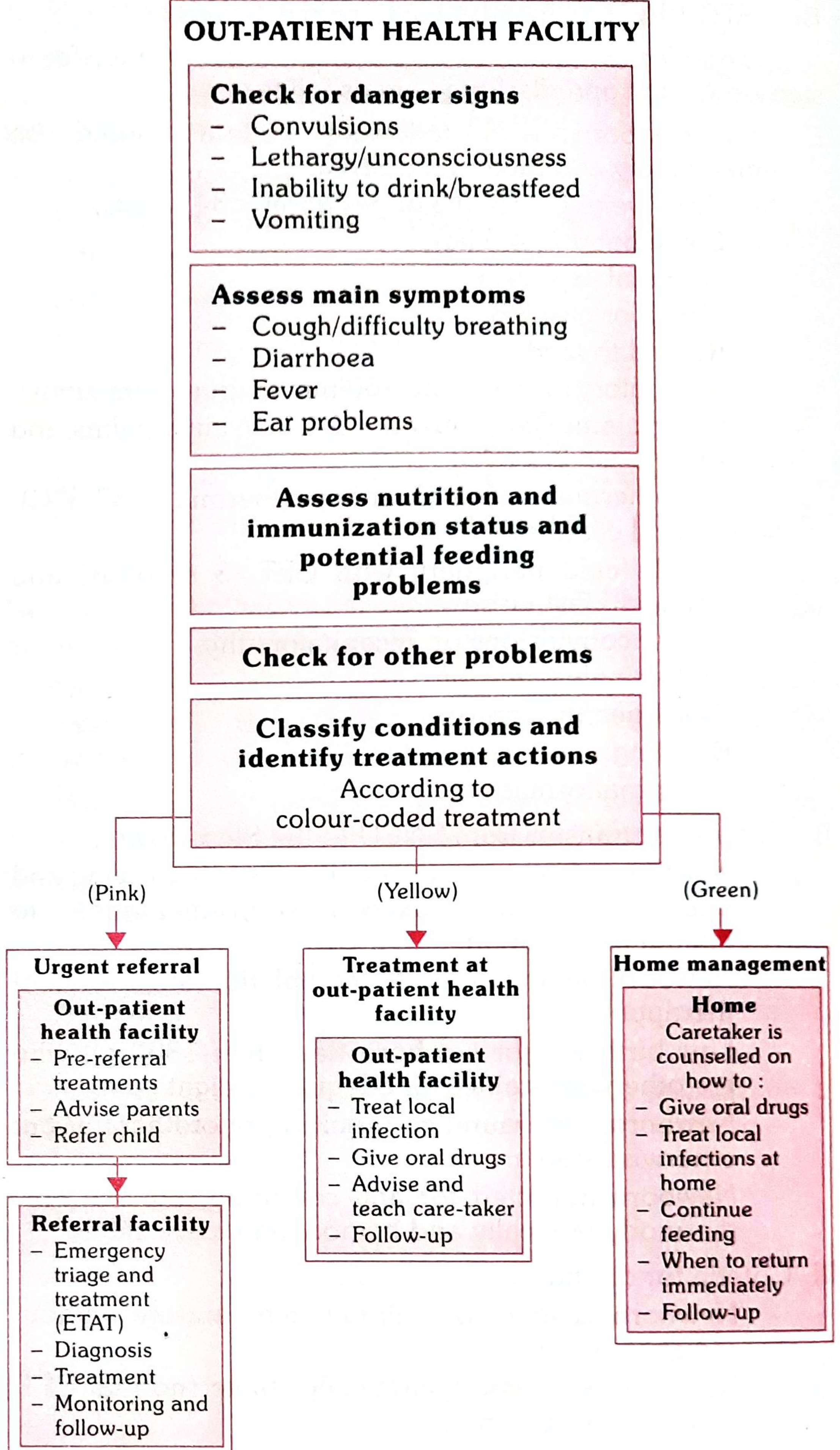
IMNCI divides children into two age groups:
- Young Infants: 0-59 days
- Children: 2 months - 59 months (5 years)
GROUP 1: Young Infants (0-59 days)
Assessment - Possible Serious Bacterial Infection (PSBI)
| Sign | Threshold |
|---|---|
| Not able to feed | Any |
| Convulsions | Any |
| Fast breathing | >60 breaths/min |
| Severe chest in-drawing | Any |
| Axillary temperature | >37.5°C or <35.5°C |
| Abnormal movement | Only when stimulated, or no movement at all |
- Give first dose of oral Amoxicillin + injectable Gentamicin
- Treat to prevent low blood sugar (breastfeed/age-appropriate feed)
- Warm the infant if temperature <35.5°C
- Advise mother how to keep infant warm on the way to hospital
- Refer URGENTLY to hospital
GROUP 2: Children 2 months - 59 months
Step 1 - Assess (ask and look)
- Count respiratory rate for one full minute (when child is calm, not crying)
- Look for chest in-drawing (lower chest wall goes IN when child breathes IN)
| Age | Fast Breathing = |
|---|---|
| 2-11 months | ≥50 breaths/min |
| 12-59 months | ≥40 breaths/min |
Note: Chest in-drawing is significant only when present all the time, in all positions, and not just when crying.
Step 2 - Classify (3 categories)
🔴 PINK - Severe Pneumonia or Very Severe Disease
- Any general danger sign (inability to breastfeed/drink, lethargy/reduced consciousness, convulsions)
- Chest in-drawing
- Stridor in a calm child
- Give first dose of appropriate antibiotic (injectable Chloramphenicol 40 mg/kg IM; if not possible, oral Amoxicillin/Co-trimoxazole)
- Refer URGENTLY to hospital
🟡 YELLOW - Pneumonia
- Fast breathing (≥50/min for 2-11 months; ≥40/min for 12-59 months)
- Chest in-drawing (in updated guidelines, this can classify as pneumonia at outpatient level in non-HIV children)
- Give oral Amoxicillin for 5 days (current/updated protocol)
- Older IMNCI versions used Co-trimoxazole: 2 pediatric tablets twice daily for 2-12 months; 3 tablets twice daily for 12-59 months
- If wheezing, give inhaled bronchodilator for 5 days
- Soothe throat and relieve cough (safe home remedy)
- If HIV-infected child with chest indrawing: give 1st dose Amoxicillin and refer
- If cough >14 days or recurrent wheeze: refer for possible TB or asthma assessment
- Advise mother when to return immediately
- Follow up in 3 days
🟢 GREEN - No Pneumonia: Cough or Cold
- No signs of severe pneumonia or pneumonia
- If wheezing: give inhaled bronchodilator for 5 days
- Soothe throat and relieve cough with safe home remedy
- If cough >14 days or recurrent wheeze: refer for TB/asthma assessment
- Advise mother when to return immediately
- Follow up in 5 days if not improving
Summary Table
| Classification | Key Signs | Color | Action |
|---|---|---|---|
| Severe Pneumonia / Very Severe Disease | Danger signs + chest in-drawing + stridor in calm child | PINK | IM antibiotic + urgent referral |
| Pneumonia | Fast breathing (≥50 or ≥40/min by age) | YELLOW | Oral Amoxicillin x 5 days + follow-up in 3 days |
| No Pneumonia / Cough or Cold | None of the above | GREEN | Home care + follow-up in 5 days |
Key Points to Remember (IMNCI)
- Always count RR for one full minute - the child must be calm and not crying
- Chest in-drawing = lower chest wall moves inward during inspiration; must be present in ALL positions, not just when crying
- Stridor = harsh noise on inspiration; significant only in a calm child
- The color-coded triage: Pink = refer urgently, Yellow = treat at facility, Green = home management
- IMNCI is skill-based and covers preventive, promotive and curative aspects
- Indian adaptation (IMNCI) adds: age group 0-7 days, national guidelines on malaria, anaemia, Vitamin A supplementation, and immunization