
Arteries of heart for 1st years
Reading File
Now let me search the medical library for coronary arteries content aimed at 1st year students (anatomy level).
Finding Sources
Finding Sources
Good - found key sources. Let me read the Color Atlas and Netter's content, which are the most anatomy-focused for 1st years.
Reading File
Reading File
Now let me get the Netter's coronary artery section and fetch the key images:
Reading File
Loading Image
Loading Image
Now let me fetch the key coronary artery diagrams from Netter's:
Loading Image
Loading Image
I now have all the information needed for a complete, well-illustrated 1st-year anatomy answer. Here it is:
Arteries of the Heart (Coronary Arteries)
Overview
The heart has its own dedicated blood supply through the coronary circulation - the coronary arteries are the first branches of the aorta. They arise in the aortic sinuses (behind the cusps of the aortic valve) immediately above the aortic valve, and their main stems run in the coronary sulcus (atrioventricular groove) - which is why they are called "coronary" (from Latin corona = crown). The coronary circulation consists of:
- Coronary arteries → capillary network in the myocardium → coronary veins → coronary sinus → right atrium
There are two main coronary arteries: the Right Coronary Artery (RCA) and the Left Coronary Artery (LCA).
Sternocostal (Anterior) View
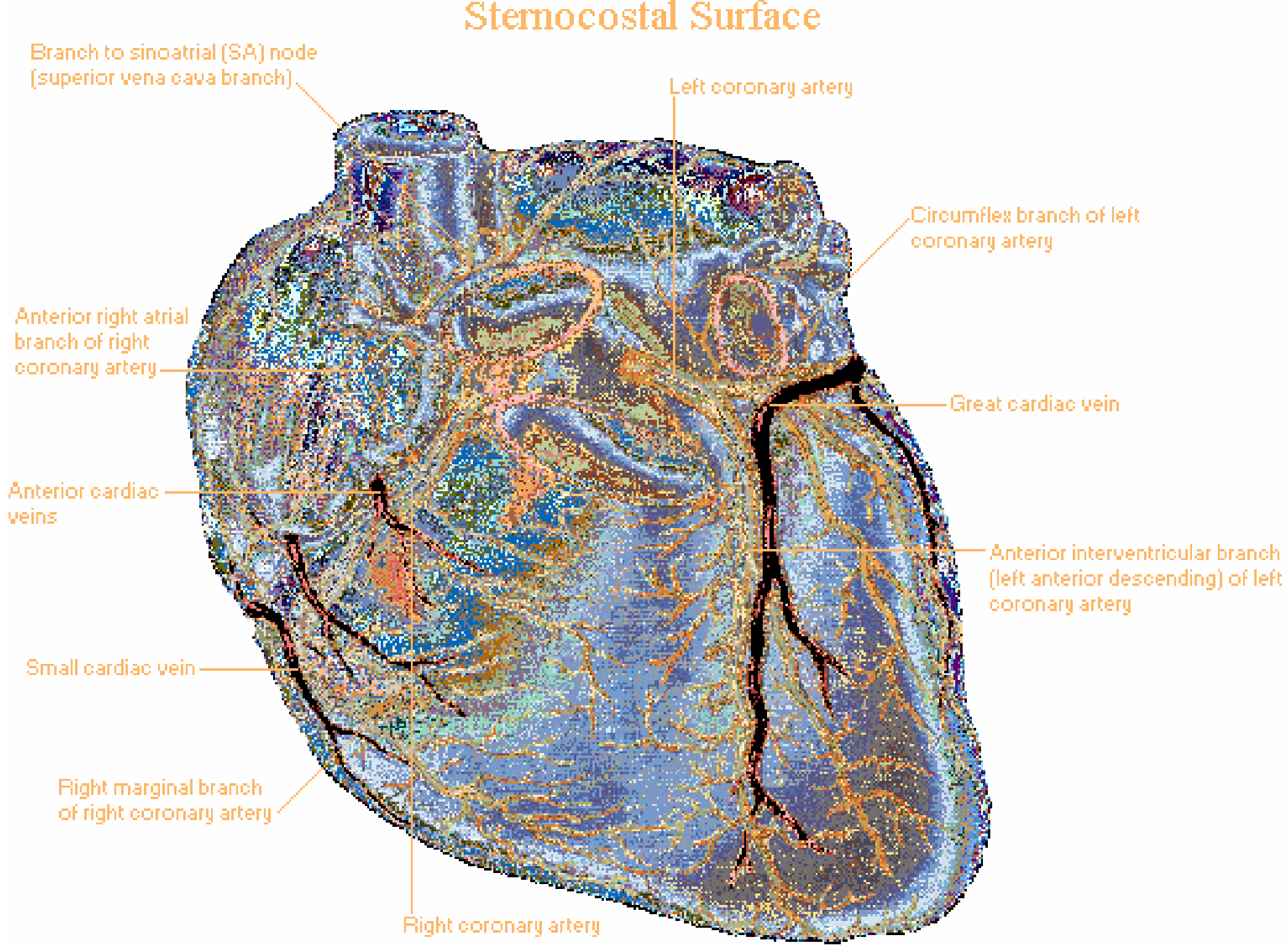
1. Right Coronary Artery (RCA)
- Origin: Right aortic sinus (right semilunar cusp)
- Initial course: Enters the coronary sulcus on the right side, initially hidden under the right auricle
- Course: Runs in the right part of the coronary sulcus, then curves posteriorly to reach the posterior interventricular sulcus
Key branches:
| Branch | Course | Area supplied |
|---|---|---|
| Right atrial branches (including SA nodal branch in ~60% of people) | Branches upward to right atrium | SA node, right atrium |
| Right marginal branch | Runs along the right (acute) margin toward the apex | Right ventricle (anterior surface) |
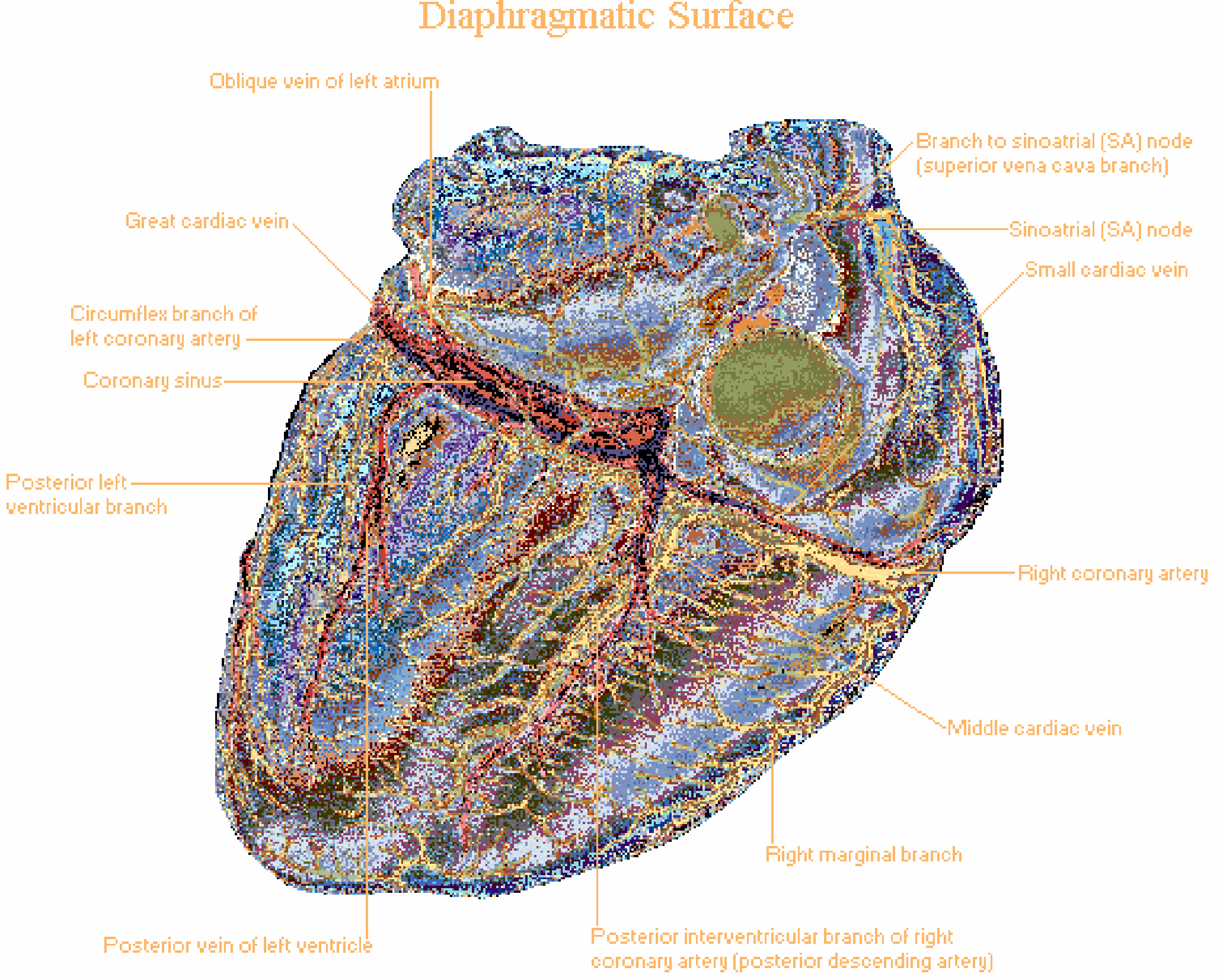
| Posterior interventricular artery (PDA) = Posterior Descending Artery | Runs down posterior interventricular sulcus toward apex | Posterior wall of both ventricles, posterior 1/3 of interventricular septum |
| AV nodal branch | Small branch near the crux cordis | AV node (in ~90% of people, from RCA) |
Overall territory (in balanced/right-dominant circulation):
- Right atrium
- SA node and AV node (conducting system)
- Most of the right ventricle
- Posterior part of the interventricular septum
- Adjacent diaphragmatic surface of the heart
2. Left Coronary Artery (LCA)
- Origin: Left aortic sinus (left semilunar cusp)
- Initial course: Short main stem (left main coronary artery) passes between the pulmonary trunk and left auricle
- Bifurcates into two major branches (occasionally trifurcates when a ramus intermedius is present)
2a. Anterior Interventricular Artery (AIV) = Left Anterior Descending (LAD)
- Runs caudally in the anterior interventricular sulcus toward the apex
- Gives off diagonal branches (to the left ventricle) and septal perforators (to the interventricular septum)
- Supplies: Anterior wall of left ventricle, anterior 2/3 of interventricular septum, apex (the LAD is nicknamed "the widow maker" because its occlusion causes large anterior MI)
2b. Circumflex Artery (Cx)
- Runs posteriorly in the left part of the coronary sulcus
- Gives off obtuse marginal branches to the left ventricle's lateral wall
- In left-dominant circulation: gives rise to the posterior interventricular artery instead of the RCA
- Supplies: Left atrium, lateral and posterior wall of left ventricle
Overall territory of LCA:
- Most of the left ventricle
- Anterior 2/3 of the interventricular septum
- Part of the right ventricle (at the sternocostal surface)
- Left atrium
Diaphragmatic (Posterior) View
Coronary Dominance
The concept of dominance refers to which artery gives rise to the posterior interventricular artery (PDA):
| Type | Frequency | PDA from |
|---|---|---|
| Right dominant | ~70% of people | RCA |
| Balanced | ~10% | Both contribute |
| Left dominant | ~20% | Circumflex branch of LCA |
The dominant artery also supplies the AV node (at the crux cordis).
Quick Summary Table
| Artery | Sulcus it runs in | Main territory |
|---|---|---|
| Right coronary artery | Right coronary sulcus | RA, RV, SA/AV nodes, posterior septum |
| Left anterior descending | Anterior interventricular | LV (anterior), anterior 2/3 septum |
| Circumflex | Left coronary sulcus | LA, LV (lateral/posterior) |
| Posterior interventricular | Posterior interventricular | Posterior LV, RV, posterior 1/3 septum |
| Right marginal | Acute margin | Anterior RV wall |
Clinical Pearl for 1st Years
Coronary arteries are functional end arteries - even though they form small anastomoses, these are not sufficient to develop collateral circulation quickly enough when a vessel is blocked. Occlusion leads to myocardial infarction (heart attack). Over 90% of acute heart attacks arise from fresh coronary thrombosis on a ruptured atherosclerotic plaque.
The location of infarction predicts which artery is blocked:
- Anterior MI - LAD occlusion
- Inferior MI - RCA occlusion (most common)
- Lateral MI - Circumflex occlusion
Sources: Color Atlas of Human Anatomy Vol. 2 (Internal Organs); Netter's Atlas of Human Anatomy
This is a shared conversation. Sign in to Orris to start your own chat.