
Anatomy of frontal lobe
frontal lobe anatomy brain diagram
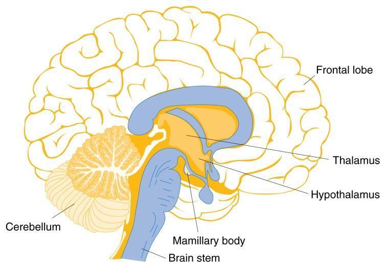
Anatomical Diagram: A mid-sagittal illustration of the human brain, highlighting regions vulnerable to thiamine deficiency and Wernicke-Korsakoff Syndrome. The diagram depicts the frontal lobe with its characteristic gyri and sulci occupying the anterior and superior portions of the cerebrum. Key labeled structures in the central and posterior regions include the thalamus and hypothalamus, located superior to the small, rounded mamillary body. The brain stem is shown extending inferiorly, connecting to the spinal cord. Posterior to the brain stem is the cerebellum, illustrating its distinctive arbor vitae branching pattern. The lateral ventricle is represented as a blue, C-shaped structure above the thalamus. This illustration serves an educational purpose in neurology and nutrition, demonstrating the spatial relationships and functional anatomy of subcortical and brainstem structures often involved in metabolic encephalopathies.
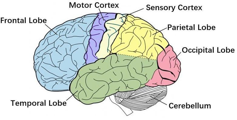
This medical anatomical diagram provides a lateral view of the human brain, utilizing color-coding to delineate primary functional regions and lobes. The frontal lobe is highlighted in light blue at the most anterior position. Progressing posteriorly, the diagram identifies the motor cortex (purple) and the sensory cortex (yellow), which are separated by the central sulcus. The parietal lobe (yellow) follows posteriorly to the sensory cortex, while the occipital lobe is marked in pink at the most posterior pole. The temporal lobe is shaded in green, situated inferior to the frontal and parietal lobes. Below the occipital and temporal regions, the cerebellum is depicted with a distinct striated texture, contrasting with the gyri and sulci patterns of the cerebral cortex. The illustration is designed to teach regional brain anatomy and the spatial relationships essential for understanding corticomuscular coherence (CMC), movement planning, and tactile processing. The labels and color differentiation emphasize the distinct boundaries of these functionally specialized neural structures.
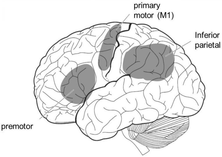
This anatomical diagram provides a lateral view of the human brain, specifically illustrating the cortical regions associated with the mirror neuron system. The line drawing highlights three key functional areas using shaded gray overlays and text labels: the premotor area in the frontal lobe, the primary motor cortex (M1) along the precentral gyrus, and the inferior parietal lobule in the parietal lobe. The diagram clearly depicts the complex surface anatomy of the cerebral cortex, including its gyri and sulci, which define the boundaries of these functional regions. The cerebellum is also visible inferiorly to the occipital and temporal lobes. This illustration is designed to teach the neuroanatomical basis of action-observation processing, demonstrating how the brain bridges visual perception of others' actions with its own motor representation. It is an educational resource suitable for neuroscience and clinical neurology, focusing on sensory-motor integration and the physiological mechanisms of social cognition.
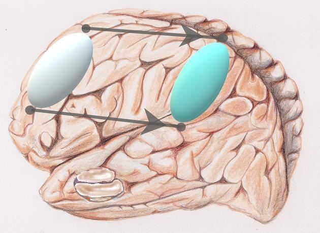
This anatomical diagram provides a lateral view of the human brain, specifically the right cerebral hemisphere, illustrating the concept of neuro-functional reorganization. The background is a detailed medical illustration of the cerebrum, showing prominent gyri and sulci rendered in brownish-red tones. Superimposed on this anatomy are two 3D oval markers. A silver-white oval is positioned over the frontal lobe (specifically the prefrontal cortex area), and a light-blue oval is positioned over the parietal lobe (specifically the inferior parietal region). Two parallel horizontal arrows with circular endpoints connect these regions, pointing from the frontal marker to the parietal marker. This visual represents a 'network translation' or a posterior shift in brain activation patterns often observed during aging. The diagram illustrates how executive functions may shift from frontal resources to parietal regions to maintain cognitive performance as part of a compensatory mechanism in the aging brain. This educational graphic is relevant to neuroanatomy, geriatric psychology, and functional neuroimaging studies.
Anatomy of the Frontal Lobe
Overview and Boundaries
- Posteriorly by the central sulcus (Rolandic fissure), which separates it from the parietal lobe
- Inferolaterally by the Sylvian fissure (lateral sulcus), separating it from the temporal lobe
- Anteriorly it ends at the frontal pole
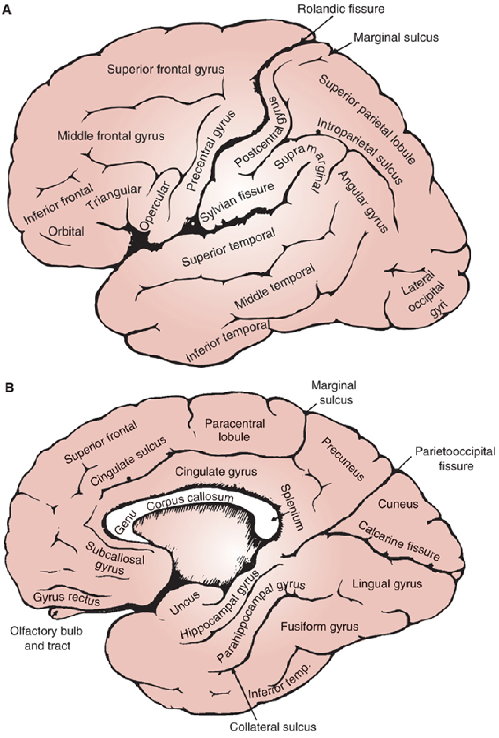
Gyri and Sulci
Lateral Surface
| Gyrus | Location | Key Function |
|---|---|---|
| Superior frontal gyrus | Above the superior frontal sulcus | Higher cognition, working memory |
| Middle frontal gyrus | Between superior and inferior frontal sulci | Prefrontal executive function, frontal eye fields |
| Inferior frontal gyrus | Below the inferior frontal sulcus | Language (dominant hemisphere - Broca's area) |
- Opercular part (pars opercularis) - Brodmann area 44
- Triangular part (pars triangularis) - Brodmann area 45
- Orbital part (pars orbitalis) - Brodmann area 47
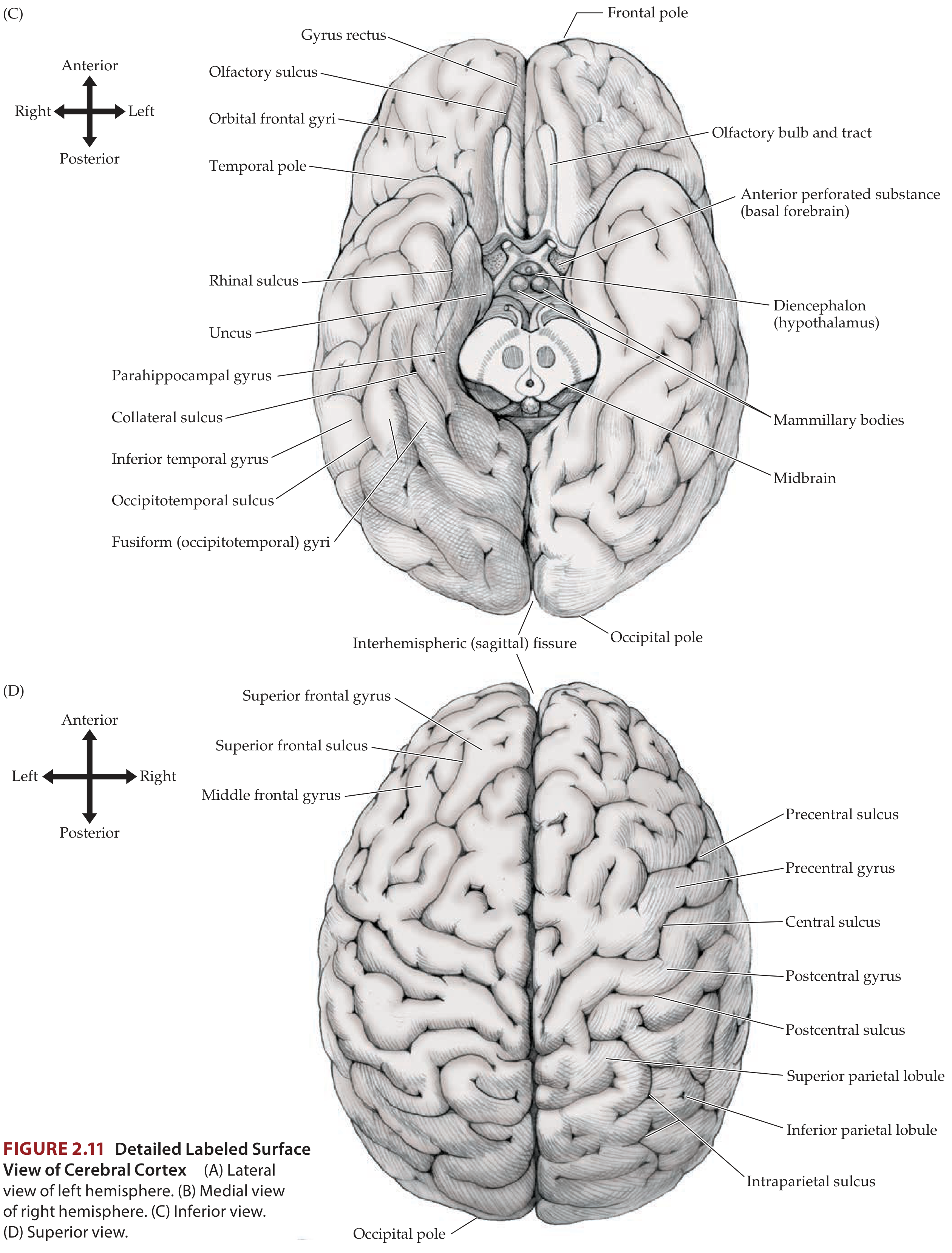
Medial Surface
- Superior frontal gyrus - continues from the lateral surface
- Cingulate gyrus - lies above the corpus callosum, separated from the superior frontal gyrus by the cingulate sulcus
- Subcallosal (paraolfactory) gyrus - at the anteroinferior end near the genu of the corpus callosum
- Paracentral lobule - the medial extension of both the precentral and postcentral gyri; its anterior half corresponds to the motor representation of the lower limb and perineum
Inferior (Orbital) Surface
- Gyrus rectus - a straight gyrus medially, adjacent to the olfactory sulcus
- Olfactory sulcus - carries the olfactory bulb and tract
- Orbital frontal gyri - arranged in an H-shaped pattern around the orbital sulci (medial, anterior, posterior, lateral orbital gyri)
- Olfactory bulb and tract - lie in the olfactory sulcus
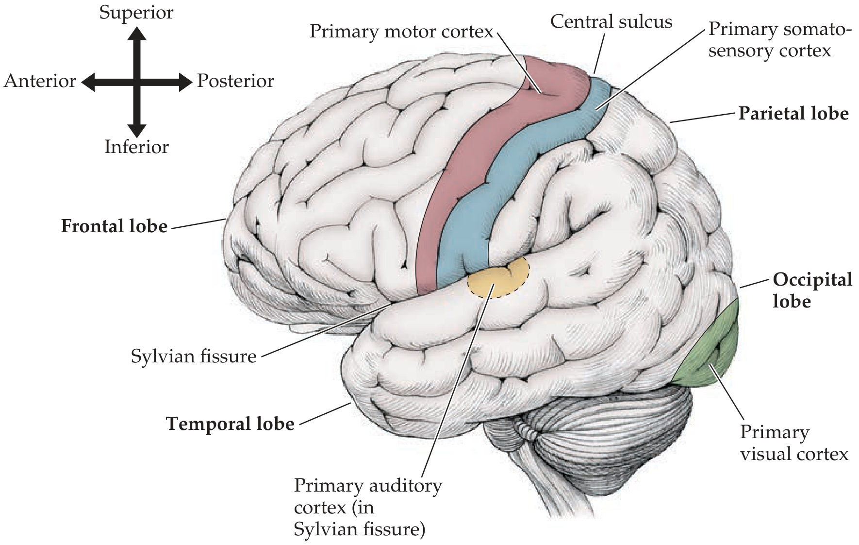
Functional Areas and Brodmann Areas
| Brodmann Area | Name | Location | Function |
|---|---|---|---|
| 4 | Primary motor cortex (M1) | Precentral gyrus | Contralateral voluntary movement (corticospinal tract origin) |
| 6 | Premotor cortex / Supplementary motor area (SMA) | Anterior to precentral gyrus, including mesial frontal | Motor planning, complex motor programs, supplementary eye field |
| 8 | Frontal eye fields (FEF) | Posterior middle frontal gyrus | Voluntary saccadic eye movements (contralateral) |
| 9, 10, 11, 12 | Prefrontal cortex (DLPFC, OFC) | Anterior frontal lobe | Executive function, working memory, judgment, planning |
| 44 | Pars opercularis (Broca's area) | Inferior frontal gyrus | Motor speech (dominant hemisphere) |
| 45 | Pars triangularis (Broca's area) | Inferior frontal gyrus | Expressive language (dominant hemisphere) |
| 47 | Pars orbitalis | Inferior frontal gyrus | Language and orbitofrontal function |
| 24, 32, 33 | Anterior cingulate cortex | Cingulate gyrus (anterior) | Attention, motivation, emotional regulation, error monitoring |
Key Functional Regions in Detail
1. Primary Motor Cortex (Precentral Gyrus, Area 4)
- Located in the precentral gyrus, extending into the posterior bank of the central sulcus
- Contains giant Betz cells in layer V
- Somatotopically organized as a motor homunculus: lower limb represented medially (paracentral lobule), face represented laterally near the Sylvian fissure, with disproportionately large representation for the hand and face
- Projects via the corticospinal (pyramidal) tract to control contralateral voluntary movement
- About 85% of corticospinal fibers decussate at the medullomedullar junction (pyramidal decussation)
2. Premotor and Supplementary Motor Cortex (Area 6)
- Lies immediately anterior to area 4
- Premotor cortex (lateral area 6): complex motor programs, especially learned sequences; sends projections to area 4 and brainstem
- Supplementary motor area (SMA) (mesial area 6): preprogramming of movements, bimanual coordination, speech initiation; lesions cause alien hand syndrome and speech arrest
- The frontal cortex rostral to the precentral gyrus mediates complex motor programs, executed through the subcortical nuclei (basal ganglia, brainstem nuclei) and primary motor cortex
3. Frontal Eye Fields (Area 8)
- Located in the posterior middle frontal gyrus
- Controls voluntary contralateral gaze (saccadic eye movements)
- An acute lesion causes ipsilateral horizontal gaze deviation (eyes deviate toward the lesion); epileptogenic lesions cause contralateral eye deviation
4. Broca's Area (Areas 44+45, Dominant Hemisphere)
- In the posterior part of the inferior frontal gyrus (left side in ~95% of right-handed people)
- Damage causes Broca's (expressive/non-fluent) aphasia: non-fluent speech with intact comprehension
- In the non-dominant hemisphere, the posterior part of the third frontal gyrus is typically smaller than the left
5. Prefrontal Cortex (Areas 9, 10, 46 - DLPFC; Areas 11, 12, 47 - OFC)
- Dorsolateral prefrontal cortex (DLPFC): working memory, executive function, abstract reasoning, antisaccade tasks; lesions produce an abulic (apathetic, slowed) state
- Orbitofrontal cortex (OFC): inhibitory control, social judgment, reward processing; lesions produce disinhibited, impulsive behavior, poor judgment, witzelsucht (inappropriate jocularity)
- Anterior cingulate cortex: motivation, attention, error monitoring; bilateral lesions can cause akinetic mutism
Connections (White Matter)
- Corticospinal and corticobulbar tracts: descend through the corona radiata to the internal capsule (posterior limb and genu respectively)
- Corpus callosum (frontal portion = genu): interhemispheric connections between the two frontal lobes
- Superior longitudinal fasciculus: connects frontal lobe to parietal, temporal, and occipital areas
- Uncinate fasciculus: connects orbitofrontal cortex to anterior temporal lobe
- Cingulum bundle: connects cingulate gyrus throughout the limbic system
- Frontoponto-cerebellar pathway: frontopontine fibers pass through the anterior limb of the internal capsule to the ipsilateral pontine nuclei, then cross to contralateral cerebellar hemisphere
Vascular Supply
| Territory | Artery | Frontal Lobe Region Supplied |
|---|---|---|
| Anterior cerebral artery (ACA) | Medial surface | Medial frontal lobe, paracentral lobule, anterior cingulate, orbital surface (recurrent artery of Heubner) |
| Middle cerebral artery (MCA) | Lateral surface | Precentral gyrus, superior/middle/inferior frontal gyri |
| Anterior communicating artery perforators | Basal forebrain | Subcallosal area, anterior cingulate base |
Clinical Correlations
| Region Damaged | Clinical Sign |
|---|---|
| Primary motor cortex (area 4) | Contralateral spastic hemiparesis (upper motor neuron pattern) |
| Frontal eye fields (area 8) | Ipsilateral gaze deviation (acute stroke); contralateral gaze deviation (seizure) |
| Broca's area (left areas 44/45) | Non-fluent expressive aphasia |
| Supplementary motor area | Alien hand, akinesia, speech arrest |
| Dorsolateral PFC | Abulia, reduced initiative, working memory deficits, perseveration |
| Orbitofrontal cortex | Disinhibition, impulsivity, poor social judgment, confabulation |
| Anterior cingulate (bilateral) | Akinetic mutism |
| Paracentral lobule (ACA territory) | Contralateral leg weakness + urinary incontinence (bilateral) |
Anatomy of parietal lobe
parietal lobe anatomy gyri sulci diagram
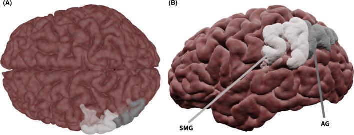
This medical anatomical diagram features a 3D surface-rendered model of the human brain, focusing on the cortical anatomy of the parietal lobe. The brain is presented in two panels: (A) a superior (top-down) view and (B) a lateral (side) view. The rendering utilizes a semi-transparent brown texture that highlights the gyri and sulci of the cerebral cortex. Specific anatomic regions associated with language and cross-modal processing are color-coded for educational identification. The supramarginal gyrus (SMG) is highlighted in opaque white, while the angular gyrus (AG) is highlighted in semi-transparent gray. In the lateral view (B), labels and indicator lines clearly demarcate these two adjacent regions, showing the SMG situated anteriorly to the AG within the inferior parietal lobule. This visualization is designed for neuroanatomical education, specifically to demonstrate the spatial relationship of parietal lobe structures involved in phonological processing and semantic integration, often studied via functional magnetic resonance imaging (fMRI) in clinical contexts like epilepsy or speech-language pathology.
This anatomical diagram provides a lateral view of the human brain, specifically the right cerebral hemisphere, illustrating the concept of neuro-functional reorganization. The background is a detailed medical illustration of the cerebrum, showing prominent gyri and sulci rendered in brownish-red tones. Superimposed on this anatomy are two 3D oval markers. A silver-white oval is positioned over the frontal lobe (specifically the prefrontal cortex area), and a light-blue oval is positioned over the parietal lobe (specifically the inferior parietal region). Two parallel horizontal arrows with circular endpoints connect these regions, pointing from the frontal marker to the parietal marker. This visual represents a 'network translation' or a posterior shift in brain activation patterns often observed during aging. The diagram illustrates how executive functions may shift from frontal resources to parietal regions to maintain cognitive performance as part of a compensatory mechanism in the aging brain. This educational graphic is relevant to neuroanatomy, geriatric psychology, and functional neuroimaging studies.
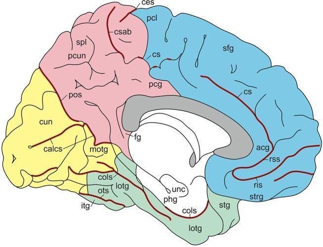
Anatomical diagram of the medial aspect of the human brain, illustrating the major gyri and sulci labeled for neuroanatomical education. The diagram uses color-coding to differentiate cerebral lobes: frontal (blue), parietal (pink), and occipital/temporal (yellow/green). Key landmarks include the cingulate sulcus (cs) and its ascending branch (csab), which separate the superior frontal gyrus (sfg) and paracentral lobule (pcl) from the cingulate gyrus (acg, pcg). The central sulcus (ces) is visible superiorly. In the posterior region, the parieto-occipital sulcus (pos) separates the precuneus (pcun) from the cuneus (cun). The calcarine sulcus (calcs) is shown within the occipital lobe. Inferior structures depicted include the parahippocampal gyrus (phg), uncus (unc), collateral sulcus (cols), and the lateral occipitotemporal gyrus (lotg). This educational resource is designed for advanced neuroanatomy study, providing a standardized map of cortical folding and lobular divisions.
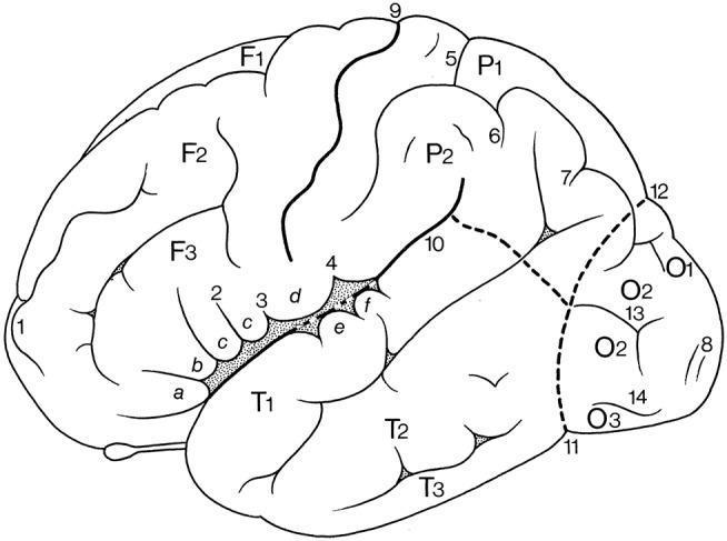
This anatomical diagram illustrates the superolateral surface of the human cerebral hemisphere, highlighting the primary gyri and sulci using a alphanumeric labeling system. The frontal lobe is categorized by gyri F1 (superior), F2 (middle), and F3 (inferior), with F3 further subdivided into orbital (a), triangular (b), and opercular (c) parts. The parietal lobe displays the superior (P1) and inferior (P2) parietal lobules. The temporal lobe contains the superior (T1), middle (T2), and inferior (T3) temporal gyri, while the occipital lobe shows the superior (O1), middle (O2), and inferior (O3) occipital gyri. Major landmark sulci are numbered, including the central sulcus (9), lateral sulcus (10), and parietooccipital sulcus (12). Secondary features such as the subcentral gyrus (d), transverse temporal gyri of Heschl (e, f), and the diagonal sulcus of Eberstaller (2) are also indicated. This schematic provides a foundational reference for neuroanatomical classification and functional localization within the human cerebral cortex.
Anatomy of the Parietal Lobe
Overview and Boundaries
- Anteriorly: Central sulcus (Rolandic fissure) - separates it from the frontal lobe
- Inferiorly (lateral surface): Sylvian fissure (lateral sulcus) - separates it from the temporal lobe
- Posteriorly: An imaginary line drawn from the top of the parietooccipital sulcus (on the medial surface) to the preoccipital notch on the inferior surface - separates it from the occipital lobe
- Medially: The interhemispheric fissure separates the two parietal lobes
Gyri and Sulci
Lateral Surface
- Lies immediately posterior to the central sulcus
- Separated from the rest of the parietal lobe by the postcentral sulcus
- Contains the primary somatosensory cortex (S-I)
- Its most inferior extent, abutting the Sylvian fissure, contains the secondary somatosensory cortex (S-II)
- A transverse sulcus running horizontally across the parietal lobe
- Divides the parietal lobe (posterior to the postcentral sulcus) into:
- Superior parietal lobule (above the IPS)
- Inferior parietal lobule (below the IPS)
- Located above the intraparietal sulcus
- Extends from the postcentral gyrus posteriorly toward the occipital lobe
- Contains Brodmann areas 5 and 7 (somatosensory association cortex)
- Located below the intraparietal sulcus, above the superior temporal sulcus and Sylvian fissure
- Consists of two key gyri:
| Gyrus | Location | Brodmann Area | Key Functions |
|---|---|---|---|
| Supramarginal gyrus | Curves around the posterior end of the Sylvian fissure | 40 | Reading, phonological processing; dominant side: Wernicke's area component; non-dominant: spatial attention |
| Angular gyrus | Curves around the posterior end of the superior temporal sulcus | 39 | Cross-modal integration, reading, calculation, writing; dominant: component of Wernicke's area |
Medial Surface
- Paracentral lobule (posterior half): The medial extension of the postcentral gyrus. The posterior half of this lobule represents the sensory homunculus for the lower limb and perineum. It is bounded anteriorly by the marginal sulcus (a branch of the cingulate sulcus)
- Precuneus: The large medial parietal area posterior to the paracentral lobule, anterior to the parietooccipital sulcus, and above the cingulate isthmus. It is involved in visuospatial processing, episodic memory, and self-referential thought
- Parietooccipital sulcus: A prominent deep sulcus on the medial surface separating the precuneus from the cuneus (occipital lobe)
Functional Areas and Brodmann Areas
| Brodmann Area | Name | Location | Function |
|---|---|---|---|
| 3a | Primary somatosensory cortex | Deepest part of postcentral gyrus (fundus of central sulcus) | Proprioception from muscle spindles |
| 3b | Primary somatosensory cortex | Anterior postcentral gyrus | Cutaneous touch (texture, size) - the "true" primary S1 |
| 1 | Primary somatosensory cortex | Middle postcentral gyrus | Texture discrimination |
| 2 | Primary somatosensory cortex | Posterior postcentral gyrus | Size and shape of objects (stereognosis) |
| 43 | Secondary somatosensory cortex (S-II) | Parietal operculum / upper bank of lateral sulcus | Bilateral somatosensory processing, pain |
| 5 | Somatosensory association cortex | Superior parietal lobule | Integration of somatosensory signals; tactile object recognition |
| 7 | Posterior parietal cortex | Superior parietal lobule (posterior) | Visuospatial processing, integration of visual and somatic inputs |
| 39 | Angular gyrus | Inferior parietal lobule (posterior) | Reading, writing, cross-modal association, calculation |
| 40 | Supramarginal gyrus | Inferior parietal lobule (anterior) | Phonological processing, ideomotor apraxia |
Primary Somatosensory Cortex in Detail
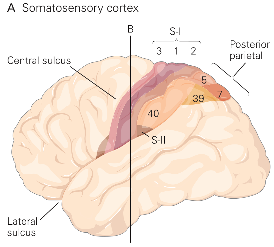
- Areas 3a and 3b lie in the anterior postcentral gyrus (extending into the posterior bank of the central sulcus)
- Area 1 is in the crown of the postcentral gyrus
- Area 2 is in the posterior bank of the postcentral gyrus
- Area 3b - responds to cutaneous mechanoreceptors (touch)
- Area 1 - texture discrimination
- Area 2 - size, shape, complex object features
- Area 3a - deep proprioceptive inputs from muscle spindles
Posterior Parietal Cortex (Areas 5 and 7)
- Primary and secondary somatosensory cortex
- Visual cortex (occipital)
- Auditory cortex (temporal)
- Thalamus (pulvinar)
- Vestibular system
Inferior Parietal Lobule - Language and Higher Cognition
Dominant (Left) Hemisphere IPL
- Angular gyrus (area 39): Reading, writing, arithmetic, semantic processing; lesions cause the Gerstmann syndrome
- Supramarginal gyrus (area 40): Part of Wernicke's area complex (with superior temporal gyrus); phonological processing; lesions contribute to ideomotor apraxia and conduction aphasia
Non-Dominant (Right) Hemisphere IPL
- Spatial attention and directed visuospatial awareness of the contralateral (left) space
- Lesions cause hemispatial neglect, constructional apraxia, and dressing apraxia
Connections (White Matter)
| Fasciculus | Connects | Function |
|---|---|---|
| Arcuate fasciculus / Superior longitudinal fasciculus | Parietal lobe (IPL) ↔ Frontal lobe (Broca's area) | Language, phonological loop |
| Inferior parietal-occipital connections | SPL ↔ Occipital (V5/MT) | Dorsal visual stream ("where/how" pathway) |
| Corpus callosum (posterior body and splenium) | Bilateral parietal and occipital lobes | Interhemispheric sensory and visual transfer |
| Superior thalamic radiation | Thalamus (VPL/VPM) → Postcentral gyrus | Thalamocortical somatosensory relay |
| Parieto-frontal tracts | Posterior parietal → Premotor cortex | Visuomotor coordination, reaching |
Vascular Supply
| Artery | Territory |
|---|---|
| Middle cerebral artery (MCA) - posterior branches | Postcentral gyrus, superior + inferior parietal lobules (supramarginal gyrus, angular gyrus) on the lateral surface |
| Anterior cerebral artery (ACA) - pericallosal branches | Medial parietal lobe: paracentral lobule (posterior portion), precuneus |
| Posterior cerebral artery (PCA) | Most posterior parietal / parietooccipital region |
Clinical Correlations
| Region Damaged | Clinical Syndrome |
|---|---|
| Postcentral gyrus (areas 3a, 3b, 1, 2) - contralateral | Cortical sensory loss: astereognosis, agraphesthesia, impaired two-point discrimination, sensory extinction; primary pain/temperature relatively preserved if postcentral gyrus spared |
| Large acute parietal lesion | Hemisensory loss (mimicking thalamic syndrome), transient contralateral hemiparesis, parietal ataxia |
| Left angular gyrus (area 39) | Gerstmann syndrome: finger agnosia + right-left confusion + acalculia + agraphia; may also cause alexia |
| Left supramarginal gyrus (area 40) | Ideomotor apraxia (bilateral), conduction aphasia |
| Right IPL (area 40) | Hemispatial neglect (left-sided): anosognosia, dressing apraxia, constructional apraxia |
| Right parietal (SPL/IPL) | Constructional apraxia, topographical disorientation |
| Bilateral parietal (areas 5, 7) | Balint syndrome: simultanagnosia + optic ataxia + ocular apraxia |
| Posterior parietal (dominant) | Ideational apraxia |
| Paracentral lobule (ACA, posterior part) | Contralateral leg sensory loss, sensory disturbance in lower limb |