
Tuberculosis pathology
tuberculosis granuloma caseous necrosis histology pathology
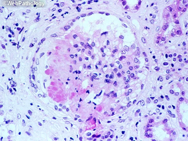
This is a light microscopy image of a formalin-fixed, paraffin-embedded tissue section stained with Hematoxylin and Eosin (H&E). The sample reveals a well-formed granulomatous lesion characterized by a surrounding cuff of lymphocytes and numerous epithelioid histiocytes with elongated, crescent-shaped nuclei. Within the granuloma, multinucleated giant cells (Langhans-type) are visible, and a central amorphous pink necrotic area (caseous necrosis) is evident. The tissue architecture demonstrates organized granulomatous inflammation with a necrotic core, consistent with infectious granulomas. The differential includes mycobacterial disease (tuberculosis) and certain fungal infections (histoplasmosis, coccioidomycosis), though non-necrotizing granulomas can occur in sarcoidosis. The staining highlights chromatin-rich nuclei and cytoplasmic eosinophilia; occasional fibroblasts and mild edema extend into the perigranulomatous region. Clinically, granulomatous tissue with caseation is highly suggestive of TB in the appropriate clinical context and warrants further diagnostic workup (acid-fast bacilli staining, fungal stains, culture, PCR). This image serves as teaching material for histopathology education, differential diagnosis of granulomatous inflammation, and correlation with radiologic and microbiologic findings in suspected infectious etiologies. Additional keywords for indexing: epithelioid macrophages, granuloma, necrosis, caseation, tuberculosis, mycobacteria, fungal infection, lymphohistiocytic rim, tissue biopsy, pathology slide, H&E, histology teaching. Clinical correlation with imaging and sputum analysis improves diagnostic yield.
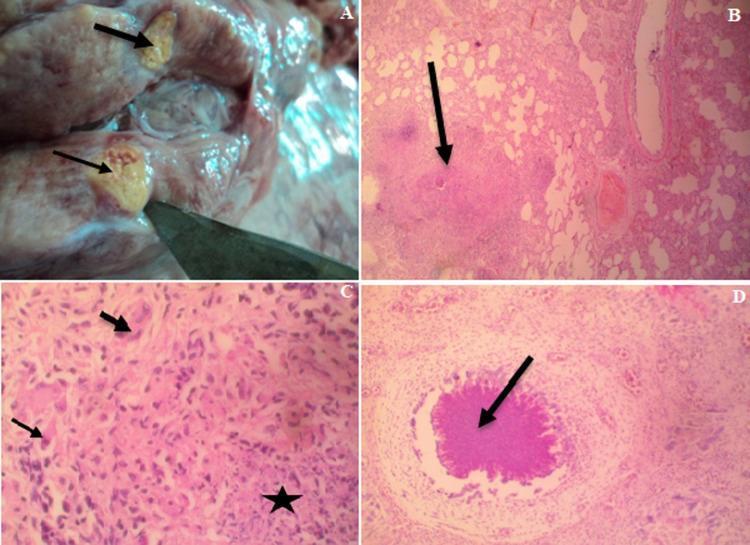
This composite figure illustrates the gross and microscopic pathology of systemic granulomatous inflammation, characteristic of tuberculosis. Panel A shows a gross clinical photograph of a dissected mediastinal lymph node, featuring yellowish, firm-to-hard nodules (arrows) indicating caseation. Panels B, C, and D are hematoxylin and eosin (H&E) stained photomicrographs. Panel B (40x) depicts a large granuloma within lung parenchyma. Panel C (400x) provides a high-magnification view of a lung nodule, identifying a central zone of amorphous caseous necrosis (star) surrounded by a cellular infiltrate of lymphocytes and macrophages, with prominent multinucleated giant cells (arrows) at the periphery. Panel D (100x) shows a splenic granuloma with a distinct central area of caseous necrosis (arrow). Together, these images demonstrate the hallmarks of Mycobacterium-induced infection, including central necrosis, Langhans-type giant cells, and the organized structure of a granuloma across multiple organ systems.
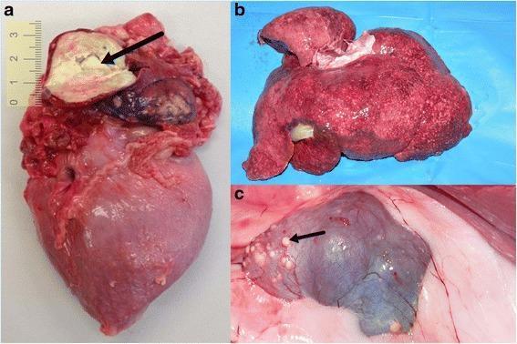
This composite of clinical gross pathology photographs displays multi-organ involvement of granulomatous inflammation consistent with Mycobacterium tuberculosis infection. Panel (a) shows a dissected heart specimen featuring a large, irregularly shaped tuberculoma (approximately 2-3 cm) in the left atrium, characterized by a yellowish-white, semi-solid, caseous necrotic core indicated by a black arrow. Panel (b) illustrates an enlarged, congested liver exhibiting a diffuse miliary pattern, with numerous small, pale foci of granulomatous inflammation scattered throughout the parenchyma. Panel (c) displays a kidney with a distorted, irregular morphology and a dark, cyanotic hue. Multiple small, white subcapsular nodules (granulomas) are visible, with a black arrow highlighting a prominent lesion. These images demonstrate the systemic manifestation of tuberculosis in internal organs, emphasizing the macroscopical features of caseous necrosis and diffuse granuloma formation in the cardiovascular, hepatic, and renal systems. This educational visual is intended for students and professionals in pathology, infectious diseases, and internal medicine to recognize gross signs of disseminated mycobacterial disease.
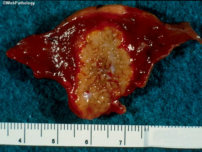
Gross pathology photograph of ex vivo hepatic tissue showing a single, lobulated lesion with a central pale-yellow to tan, crumbly necrotic core surrounded by a hyperemic, reddish-brown peripheral rind. The specimen measures approximately 5.5–6.5 cm in greatest dimension as judged by the metric ruler placed adjacent to the cut surface. The lesion appears well-demarcated from surrounding viable parenchyma, suggesting a focal process such as an infectious granuloma or abscess. The central area exhibits caseous-like necrosis with a cracked, cheese-like consistency; periphery shows congested, friable tissue with mild surface sheen. The overall sample has a smooth to slightly irregular contour. No obvious hemorrhagic stellate patterns noted beyond the margin, though diffuse vascularity is evident on the surface. The cut surface reveals a clear contrast between necrotic core and inflamed rim; there may be microcavitation within the core. These macroscopic features are compatible with granulomatous hepatic disease, notably tuberculosis or fungal infection, though bacterial abscess or parasitic granuloma cannot be excluded without microbiologic and histologic confirmation. This image is useful for education on gross-pathology correlation, differential diagnosis of hepatic necrotizing granulomas, and teaching rounds on infectious liver lesions. Correlate with histology, Ziehl-Neelsen and special stains, culture, and clinical context.
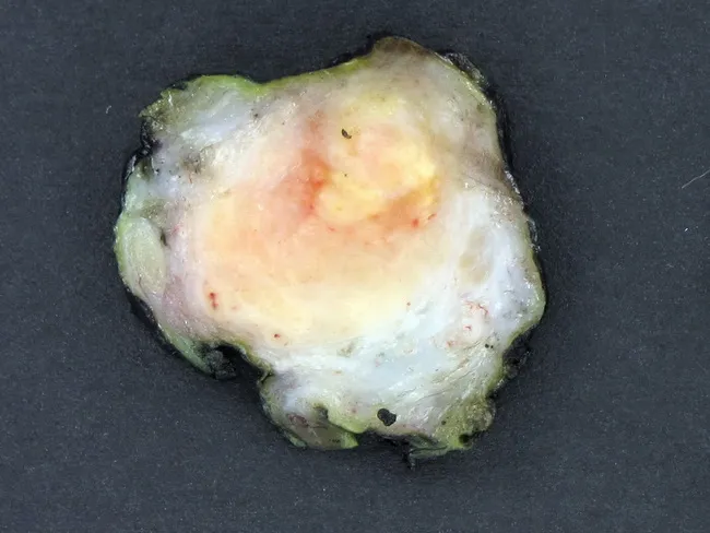
This gross pathology photograph depicts a transverse cross-section of an enlarged epididymis with a centralized, cheese-colored (caseous) necrotic core surrounded by a pale fibrous band. The lesion is consistent with tuberculous epididymo-orchitis, a commonly coexisting genitourinary TB process where granulomatous inflammation frequently involves multiple GU sites such as the prostate or testis and may accompany renal or pulmonary tuberculosis. On the cut surface, the necrotic center contrasts with the surrounding fibrous capsule and residual epididymal parenchyma, reflecting chronic granulomatous disease with fibrosis and scarring. Clinically, patients may report mild scrotal pain, enlargement, low-grade discomfort, infertility, or asymptomatic masses; urinary abnormalities or systemic signs of tuberculosis can be minimal. This image illustrates the macroscopic hallmarks of tuberculosis in the epididymis: caseation necrosis, granuloma formation, and progressive fibrosis. In differential diagnosis, bacterial epididymo-orchitis or fungal granulomatous infections may be considered, but the presence of central caseation strongly supports TB. This specimen is valuable for medical education, pathology training, and research on genitourinary tuberculosis, aiding correlation with histopathology, microbiology, and radiologic findings, and facilitating discussion of diagnostic workup, including urine culture, nucleic acid amplification tests, and anti-tubercular therapy planning. This description enables precise search indexing for TB epididymitis in clinical databases and research.
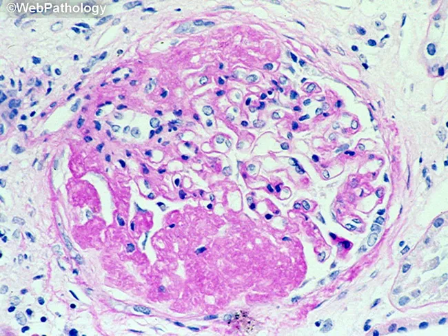
This is a histopathology image obtained with light microscopy from a formalin-fixed paraffin-embedded tissue section stained with Hematoxylin and Eosin. The field demonstrates a granulomatous inflammatory lesion, typically well circumscribed. The central area shows eosinophilic, acellular to granular necrotic material (caseous necrosis), surrounded by a dense mantle of epithelioid histiocytes with elongated, pale-staining nuclei and abundant pale cytoplasm. Multinucleated giant cells of the Langhans type may be present at the periphery. Scattered lymphocytes and occasional plasma cells form a peripheral lymphohistiocytic cuff around the granuloma. The surrounding stroma contains collagen fibers and occasional fibroblasts, with minimal additional inflammatory cells in this field. The morphology is classic for granulomatous inflammation, most commonly associated with infectious etiologies such as Mycobacterium tuberculosis or fungal infections, but can be seen in sarcoidosis or foreign body reactions depending on clinical context. Diagnostic significance: identification of a caseating granuloma raises suspicion for mycobacterial or fungal infection and warrants targeted microbiologic stains (Ziehl-Neelsen for acid-fast bacilli; GMS or PAS for fungi), cultures, and molecular testing. Clinical correlation is essential, including TB exposure history, immune status, and presentation—cough, weight loss, fever, pulmonary nodules or granulomatous lymphadenitis. This image is educational for teaching, differential diagnosis, and research into granulomatous disease pathology.
tuberculosis pathology granuloma
Tuberculosis Pathology
1. The Organism
- Protects against lysosomal enzymes
- Inhibits phagolysosome fusion
- Drives the specific granulomatous immune response
2. Pathogenesis
Phase 1 - First 3 Weeks (Pre-immunity)
- Inhaled bacilli deposit in the distal airspaces of the lower upper lobe or upper lower lobe
- They gain entry into alveolar macrophages via mannose receptor and complement receptors
- Mycobacteria inhibit phagolysosome fusion, surviving and proliferating unchecked within vacuoles
- Bacteria spread via lymphatics and bloodstream - asymptomatic bacteremia seeds multiple organs
- No granuloma forms yet
Phase 2 - After ~3 Weeks (Immune activation)
- Mycobacterial antigens reach draining lymph nodes; dendritic cells and macrophages present antigens to CD4+ T cells
- IL-12 from macrophages drives differentiation into Th1 cells, which secrete IFN-γ
- IFN-γ activates macrophages, which upregulate:
- TNF - recruits monocytes to form granulomas
- iNOS - generates reactive nitrogen intermediates that kill mycobacteria
- Defensins - antimicrobial peptides toxic to mycobacteria
- Activated macrophages differentiate into epithelioid histiocytes - the key cellular component of the granuloma
Key concept: The same Th1 response that confers resistance also mediates hypersensitivity - caseation, cavitation, and tissue destruction are all immunopathological. Tuberculin skin test positivity (PPD/Mantoux) signals both immunity AND hypersensitivity simultaneously. - Robbins & Kumar Basic Pathology
Role of Specific Cytokines
| Cytokine | Source | Effect |
|---|---|---|
| IL-12 | Macrophages | Th1 differentiation |
| IFN-γ | Th1 cells | Macrophage activation (most critical) |
| TNF-α | Activated macrophages | Monocyte recruitment, granuloma maintenance |
| LT-β (lymphotoxin) | Th1 cells | Kills chronically infected macrophages, releases bacteria for destruction |
| NO (iNOS) | Macrophages | Direct mycobactericidal effect |
TNF antagonists used for rheumatoid arthritis/IBD significantly increase TB reactivation risk, confirming TNF's non-redundant role in granuloma maintenance. - Robbins & Kumar Basic Pathology
3. The Granuloma
Structure (from centre outward):
- Central caseous necrosis - cheese-like, acellular, eosinophilic material; results from DTH (delayed-type hypersensitivity) reactions and cytotoxic effects of activated macrophages on oxygen-deprived tissue. Unlike liquefactive or coagulative necrosis, it retains a characteristic "ghost" outline. Mycobacteria are detectable by ZN stain in early caseous material.
- Epithelioid histiocytes - activated macrophages with abundant pale eosinophilic cytoplasm and elongated, "footprint-shaped" nuclei
- Langhans-type giant cells - formed by fusion of epithelioid cells; nuclei arranged in a horseshoe or peripheral ring pattern at the cell rim (distinguish from foreign-body giant cells where nuclei are scattered)
- Peripheral lymphocytic cuff - predominantly CD4+ T cells
- Outer fibrous capsule - forms over time, may calcify
TB Granuloma vs. Sarcoidosis
| Feature | TB granuloma | Sarcoid granuloma |
|---|---|---|
| Caseation | Present (central) | Absent ("naked" granuloma) |
| AFB on ZN stain | Positive (early) | Negative |
| Giant cell type | Langhans | Langhans or foreign body |
| Asteroid bodies | Rare | Common |
4. Histology Images
5. Primary Tuberculosis
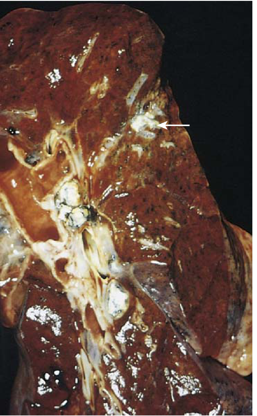
Morphology - The Ghon Complex
- Bacilli implant in the distal airspaces of the lower upper lobe or upper lower lobe, close to the pleura
- A 1-1.5 cm gray-white area of consolidation develops - the Ghon focus
- The center undergoes caseous necrosis
- Bacilli drain to the hilar lymph nodes, which also caseate
- Ghon focus + involved hilar lymph nodes = Ghon complex (also called the Ranke complex when both are calcified)
Outcomes of Primary TB
- Progressive primary TB develops
- No proper granulomatous response (CD4 <200 in HIV)
- Lower/middle lobe consolidation, hilar lymphadenopathy, no cavitation
- Early hematogenous dissemination
6. Secondary (Reactivation) Tuberculosis
- Reactivation of dormant primary focus (decades later) when immunity wanes
- Re-exposure with a large inoculum
Morphology
- Classically localized to the apex of one or both upper lobes (high oxygen tension)
- Prompt tissue response walls off the focus (unlike primary TB)
- Less hilar lymphadenopathy than primary TB
- Initial lesion: <2 cm sharply circumscribed, gray-yellow area with central caseation and peripheral fibrosis
Progression Pathways
- Caseation expands, erodes into a bronchus
- Liquid caseous material is expectorated, leaving a ragged, irregular cavity lined by caseous material
- Poorly walled off by fibrous tissue
- Erosion of vessels → hemoptysis (can be massive if Rasmussen aneurysm forms)
- Cavity is a major source of infectivity - sputum AFB positive
7. Extrapulmonary / Disseminated TB
| Site | Manifestation |
|---|---|
| Cervical lymph nodes | Scrofula - most common extrapulmonary form; usually unifocal |
| Meninges | Tuberculous meningitis - basilar leptomeningitis with granulomas |
| Kidneys | Renal TB - cortical granulomas, cavitation, "putty kidney" calcification |
| Adrenal glands | Can cause Addison disease |
| Spine (vertebrae) | Pott disease - vertebral destruction, kyphosis, paraspinal "cold abscess" extending along tissue planes |
| Fallopian tubes/epididymis | Genital TB - leading cause of TB-related infertility |
| Liver, spleen, bone marrow | Systemic miliary TB - prominent in these organs |
| Intestine | Mucosal ulceration in ileum (trapped in lymphoid aggregates); now mostly from swallowed sputum |
| Pericardium | Constrictive pericarditis from fibrous healing |
8. TB in HIV/AIDS
| CD4 Count | TB Pattern |
|---|---|
| >300/μL | Typical secondary TB - apical cavitary disease |
| <200/μL | Resembles progressive primary TB - lower lobe consolidation, no cavitation, hilar adenopathy |
| Severely immunosuppressed | Disseminated/miliary; extrapulmonary involvement >50%; granulomas absent or poorly formed; sheets of macrophages packed with AFB |
9. Diagnostic Pathology Summary
| Method | Positive Finding | Notes |
|---|---|---|
| H&E stain | Caseating granuloma with Langhans giant cells | Classic finding; non-caseating also occurs |
| Ziehl-Neelsen (ZN) stain | Red acid-fast bacilli (AFB) on blue background | Positive in early/active lesions; negative in fibrocalcific stage |
| Auramine-rhodamine | Fluorescent bacilli | More sensitive than ZN |
| NAAT/PCR | M. tuberculosis DNA | Most sensitive/specific |
| Culture (Lowenstein-Jensen) | Buff/cream-colored colonies (3-6 weeks) | Gold standard |
| Tuberculin/PPD | Induration >10 mm | Tests hypersensitivity, not immunity |
| IGRA (QuantiFERON) | IFN-γ release | Preferred in BCG-vaccinated individuals |
10. Key Pathological Distinctions
- Caseous necrosis is the hallmark of TB. It is "cheese-like," does not liquify immediately, and retains its consistency due to the lipid content of dead mycobacteria.
- Caseation + granuloma = strong presumptive evidence for TB or histoplasmosis/coccidioidomycosis (special stains distinguish them)
- Calcification of a granuloma indicates chronicity/healing but does not mean sterility - viable organisms can persist for decades
- The Ghon complex and its calcified form (Ranke complex) are incidental findings on chest X-ray but represent resolved primary TB