
Case study on kwshirkor and head to examination with nursing care plan
kwashiorkor child malnutrition edema clinical features
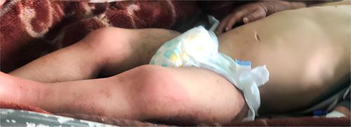
Clinical photograph of a 2-year-old child exhibiting dermatological and systemic manifestations of severe acute malnutrition (SAM). The image displays the torso and lower extremities of a pediatric patient with notable pallor. Significant pitting edema is visible in the lower limbs, particularly involving the legs and feet, giving the extremities a swollen, rounded appearance. The skin texture is markedly dry and xerotic with diffuse scaly areas. These cutaneous changes, including the scaly dermatosis and edema, are classic features of protein-energy malnutrition, specifically kwashiorkor or mixed-type SAM. The image also depicts a distended abdomen, which is frequently associated with underlying nutritional deficiencies, malabsorption syndromes like celiac disease, or hepatomegaly in malnourished populations. This visual serves as an educational reference for identifying the physical signs of severe nutritional failure and secondary skin disorders in a clinical pediatric setting.
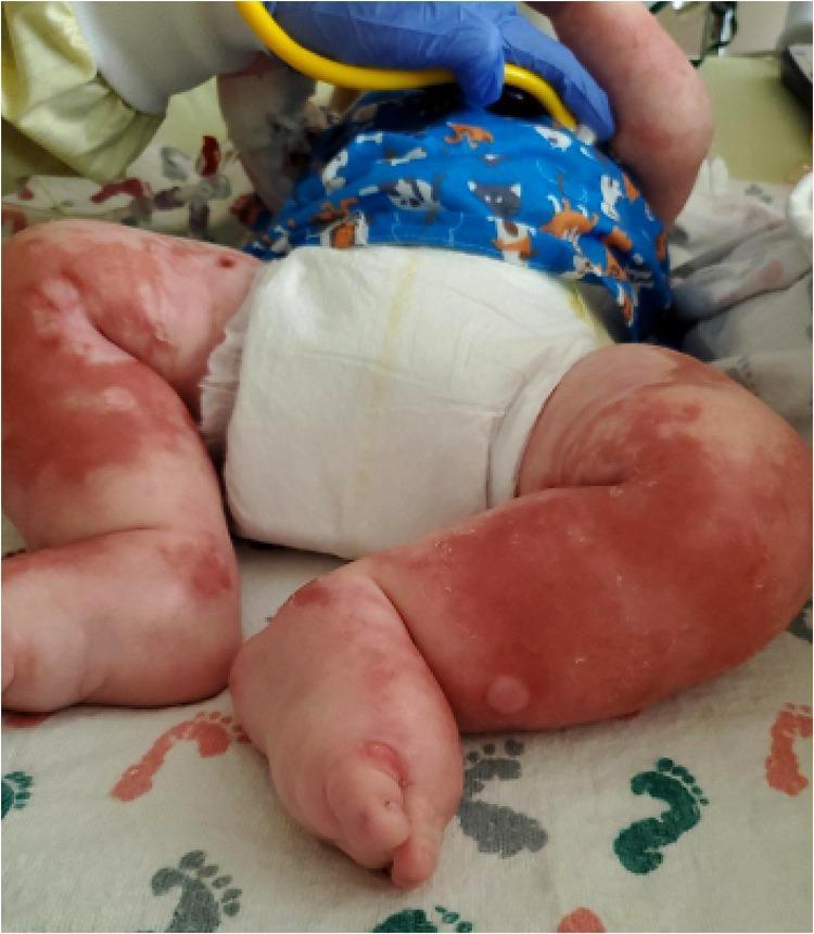
Clinical photograph of an infant's lower extremities exhibiting severe dermatological and systemic manifestations of malnutrition. The image shows diffuse, well-demarcated reddish-brown erythematous plaques with a 'flaky paint' appearance, characteristic of the dermatosis seen in Kwashiorkor. Notable features include widespread scaly plaques and desquamation, as well as several bullous lesions (fluid-filled blisters) on the lateral aspect of the lower leg and dorsal foot. Significant generalized peripheral edema (anasarca) is evident, manifesting as tense, shiny skin and swelling of the thighs, legs, and feet. The distribution is bilateral and involves the flexural surfaces. These visual findings represent the classic cutaneous signs of protein-energy malnutrition, often secondary to malabsorption syndromes such as cystic fibrosis-associated pancreatic insufficiency. The presence of these lesions in a pediatric patient is a critical clinical indicator of severe metabolic and nutritional distress.
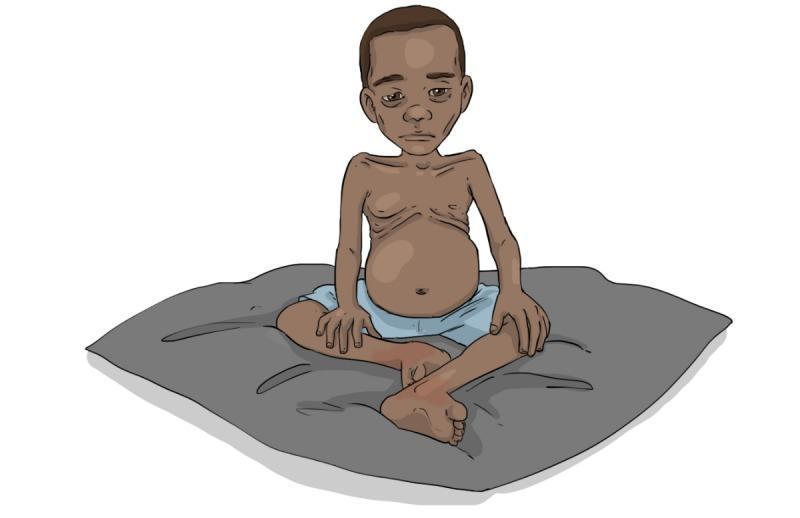
This medical illustration depicts a pediatric patient presenting with severe protein-energy malnutrition (PEM), specifically demonstrating clinical features associated with kwashiorkor or marasmic-kwashiorkor. The child is shown in a seated, cross-legged position. Key visual diagnostic signs include a protuberant, distended abdomen (ascites/hepatomegaly), marked muscle wasting in the upper and lower extremities, and a relative lack of subcutaneous fat, making the rib cage and clavicles visibly prominent. The facial features exhibit a gaunt appearance with sunken eyes and a flat, subdued affect, characteristic of the 'apathetic' demeanor often seen in severely malnourished children. The image serves as an educational tool for identifying the physical manifestations of chronic nutritional deficiency in global health settings, particularly emphasizing the paradox of a bloated abdomen despite generalized emaciation. This illustration is designed for healthcare worker training in malnutrition assessment and triage.
Case Study: Kwashiorkor
Patient Profile
History of Present Illness
Head-to-Toe Physical Examination
General Appearance
- Lethargic, apathetic, miserable-appearing child
- Does not cry; shows no interest in surroundings
- Weight = 70% of expected for age (masked by edema); true lean body mass likely lower
Head & Scalp
- Hair: Sparse, depigmented, reddish-yellow to grey color
- Flag sign: Alternating bands of pale and darker color along individual hair strands (indicating alternating periods of poor and adequate nutrition)
- Hair texture: Dry, lustreless, straightened (formerly curly), "crackled" hair noted
- Scalp: Easy pluckability - hair comes out with minimal traction
- No lymphadenopathy in the cervical region
Face
- Puffiness and periorbital edema
- Flat, subdued, "moon face" appearance
- Sunken eyes
- Pale conjunctivae (anemia)
- No active corneal lesions; however, vitamin A deficiency should be excluded
- Dry lips; angular stomatitis possible (riboflavin/B-complex deficiency)
Eyes
- Conjunctival pallor (anemia)
- Bitot's spots should be screened for (Vitamin A deficiency co-exists)
- No obvious corneal ulceration at this time
Mouth & Throat
- Angular cheilitis noted
- Pale oral mucous membranes
- Dry tongue
- No thrush at this time
Neck
- No significant lymphadenopathy
- Neck supple, no thyroid enlargement
Chest & Respiratory
- Bilateral breath sounds present and equal
- Mild subcostal recession
- No crackles; screening required for TB or pneumonia (susceptibility increased)
- Respiratory rate: 32/min (mild tachypnea)
- Chest wall relatively thin; mild rib prominence visible
Cardiovascular
- Heart rate: 98 bpm, regular
- Heart sounds: normal S1 and S2, no murmurs
- Capillary refill: 2.5 seconds (borderline)
- Blood pressure: 78/50 mmHg (low for age)
Abdomen
- Distended, pot-belly appearance - characteristic finding
- Hepatomegaly palpable 4 cm below right costal margin; smooth surface, non-tender
- Fatty liver (steatosis): due to reduced synthesis of apolipoprotein carrier protein component of lipoproteins, causing triglyceride accumulation
- Mild ascites on percussion (shifting dullness)
- Bowel sounds present, mild hyperactivity
- No splenomegaly
Skin (entire body)
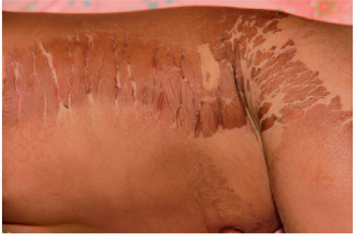
- "Flaky paint" dermatosis - hyperpigmented patches with alternating zones of desquamation and hypopigmentation
- Areas first affected: pressure areas - groin, buttocks, flexures, elbows, knees
- Lesions resemble "crazy paving," "cracked paint," or "mosaic skin"
- In severe areas: peeling leaves pale, ulcerated hypopigmented areas with hyperpigmented borders
- Nails: Soft, thin, brittle
- Skin turgor is poor despite edema
Extremities / Musculoskeletal
- Bilateral pitting edema of lower limbs (2+ pitting), extending to the dorsum of feet
- Mild peripheral muscle wasting (masked by edema)
- Relative sparing of subcutaneous fat compared to marasmus
- Mid-upper arm circumference (MUAC): 10.8 cm (severe malnutrition: <11.5 cm)
Neurological
- Markedly apathetic, listless
- No spontaneous play
- Decreased response to stimulation
- Developmental regression: unable to sit unsupported (was walking at 14 months)
- Cranial nerves grossly intact
- No focal neurological deficits
Clinical Images
Laboratory Investigations
| Investigation | Result | Normal | Significance |
|---|---|---|---|
| Serum albumin | 1.4 g/dL | 3.5-5.0 g/dL | Severe hypoalbuminemia - drives edema |
| Total protein | 3.2 g/dL | 6.0-8.0 g/dL | Reduced |
| Hemoglobin | 7.2 g/dL | 11-14 g/dL | Anemia |
| Serum potassium | 2.8 mEq/L | 3.5-5.0 mEq/L | Hypokalemia |
| Serum sodium | 128 mEq/L | 135-145 mEq/L | Hyponatremia |
| Blood glucose | 62 mg/dL | 70-100 mg/dL | Hypoglycemia risk |
| Serum zinc | Low | Zinc deficiency (contributes to skin changes) | |
| LFTs | AST/ALT mildly elevated | Fatty liver changes | |
| Stool exam | Ova/cysts of Giardia | Co-existing infection | |
| Weight for age | 70% expected | SAM (severe acute malnutrition) | |
| MUAC | 10.8 cm | >13.5 cm = normal | Severe malnutrition |
Diagnosis
- Hypoalbuminemia with generalized edema
- Fatty hepatomegaly
- Iron deficiency anemia
- Electrolyte imbalance (hypokalemia, hyponatremia)
- Giardiasis (co-infection)
- Vitamin deficiencies (A, B-complex, zinc)
Nursing Care Plan
Nursing Diagnosis 1: Imbalanced Nutrition - Less Than Body Requirements
| Nursing Interventions | Rationale |
|---|---|
| Follow WHO F-75 therapeutic milk (75 kcal/100 mL) in acute/stabilization phase (Days 1-7) | Prevents refeeding syndrome; gentle resuscitation of metabolic processes |
| Transition to F-100 (100 kcal/100 mL) in rehabilitation phase (Week 2 onward) | Higher protein/calorie density to promote catch-up growth |
| Feed small frequent amounts every 2-3 hours; do not force feed | Prevents overloading a compromised gut; promotes tolerance |
| Monitor and record daily weight, MUAC, edema grading | Tracks treatment response; edema should resolve before weight gain expected |
| Supplement with therapeutic zinc (2 mg/kg/day), Vitamin A, and B-complex | Co-deficiencies worsen outcomes; zinc essential for skin healing |
| Provide Ready-to-Use Therapeutic Food (RUTF) like Plumpy'Nut once stabilized | High energy-protein paste for outpatient nutritional rehabilitation |
| Educate mother on age-appropriate protein-rich weaning foods (eggs, lentils, milk) | Sustainable dietary improvement post-discharge |
Nursing Diagnosis 2: Excess Fluid Volume (Edema)
| Nursing Interventions | Rationale |
|---|---|
| Avoid IV fluids unless in shock - use oral/NG route when possible | IV fluids can worsen fluid overload in hypoalbuminemic state |
| Monitor edema grade daily (0-3+ scale) and document changes | Edema resolves as albumin improves; tracking guides treatment |
| Restrict sodium in early feeds (F-75 is low in sodium) | Limits fluid retention; WHO therapeutic diets are formulated appropriately |
| Position child with legs elevated when at rest | Promotes venous return and reduces dependent edema |
| Monitor respiratory rate and oxygen saturation | Detect early pleural effusion or pulmonary edema |
| Weigh daily (expected weight may initially decrease as edema resolves) | Edema loss often mistaken for failure to gain weight |
| Avoid diuretics unless severe symptomatic overload | Diuretics can worsen electrolyte imbalance (esp. potassium) |
Nursing Diagnosis 3: Impaired Skin Integrity
| Nursing Interventions | Rationale |
|---|---|
| Keep skin clean and dry; gentle sponge bath twice daily | Prevents secondary bacterial/fungal infection in broken skin |
| Apply barrier cream (zinc oxide or petroleum jelly) to affected areas | Protects fragile skin; reduces friction damage |
| Avoid adhesive tapes and tight bandages on edematous areas | Edematous skin tears easily; tape avulsion can create wounds |
| Use soft cotton clothing; no synthetic fabrics | Reduces friction and pressure on damaged skin |
| Inspect skin at every nursing assessment; document and photograph lesions | Early detection of secondary infection |
| Administer zinc supplementation as prescribed | Zinc is critical for epidermal repair and immune function |
| Maintain oral hygiene; apply lip balm for angular stomatitis | Prevents worsening of mucosal lesions; promotes comfort |
Nursing Diagnosis 4: Risk for Infection
| Nursing Interventions | Rationale |
|---|---|
| Practice strict hand hygiene before all patient contact | Most important infection prevention measure in immunocompromised host |
| Ensure broad-spectrum antibiotic therapy as prescribed (e.g., amoxicillin per WHO guidelines) | WHO recommends empirical antibiotics for all hospitalized SAM children |
| Administer Vitamin A supplementation (100,000 IU for age 6-12m; 200,000 IU for >12 months) | Vitamin A is essential for immune mucosa; single megadose corrects deficiency |
| Ensure measles vaccination is given before discharge if not immunized | Measles is rapidly fatal in malnourished children |
| Screen for TB and treat parasitic co-infections (e.g., albendazole for helminthiasis) | Co-infections worsen nutritional state; giardiasis confirmed in this child |
| Isolate from other infectious patients if possible | Compromised immune defenses cannot fight nosocomial pathogens |
| Monitor temperature 4-hourly; report any fever or hypothermia promptly | Malnourished children may not mount a fever; hypothermia is a danger sign |
Nursing Diagnosis 5: Impaired Growth and Development
| Nursing Interventions | Rationale |
|---|---|
| Provide structured play stimulation for at least 30 minutes daily | Nutritional recovery alone is insufficient; play therapy improves brain development |
| Encourage mother to hold, talk to, and interact with the child frequently | Emotional stimulation during recovery accelerates neurological improvement |
| Assess developmental milestones at admission and weekly during recovery | Tracks neurological recovery; identifies residual delays |
| Provide a safe, calm ward environment | Reduces distress in apathetic child; facilitates recovery |
| Refer to developmental pediatrician if milestones do not improve after nutritional rehabilitation | Long-term protein deficiency can cause permanent cognitive impairment |
Nursing Diagnosis 6: Deficient Knowledge (Caregiver)
| Nursing Interventions | Rationale |
|---|---|
| Assess mother's literacy, cultural food beliefs, and economic constraints | Education must be contextually appropriate and realistic |
| Teach protein-rich local foods: eggs, lentils, beans, groundnuts, fish, milk | Addresses cause of kwashiorkor; empowers sustainable dietary change |
| Demonstrate preparation of energy-dense weaning porridge (cereal + legume + fat) | Practical skills are more effective than verbal instructions alone |
| Discuss importance of continued breastfeeding alongside complementary foods | Breastfeeding provides immune factors and nutrition beyond 6 months |
| Educate on growth monitoring: regular weight checks and road-to-health card use | Early detection of growth faltering prevents recurrence |
| Provide written/pictorial take-home feeding guide (as appropriate for literacy) | Reinforces teaching after discharge |
| Link family to community nutrition program / government food support scheme | Addresses underlying food insecurity - the root cause |
Medical Management Summary
| Phase | Duration | Formula/Food | Key Actions |
|---|---|---|---|
| Stabilization | Days 1-7 | WHO F-75 therapeutic milk (75 kcal/100 mL) | Treat hypoglycemia, hypothermia, dehydration, infections; correct electrolytes |
| Rehabilitation | Weeks 2-6 | WHO F-100 / RUTF | Catch-up growth feeding; zinc, Vitamin A, folic acid, multivitamins |
| Follow-up | 3-6 months | Age-appropriate home diet | Nutritional education, immunizations, growth monitoring |
- Amoxicillin (empirical antibiotic) - all SAM children
- Vitamin A (200,000 IU single dose for >12 months)
- Folic acid (5 mg Day 1, then 1 mg/day)
- Zinc (2 mg/kg/day for 2 weeks)
- Albendazole (400 mg single dose, after Day 7 of feeding)
Prognosis
- Robbins & Kumar Basic Pathology - Kwashiorkor, Chapter 7
- Robbins, Cotran & Kumar Pathologic Basis of Disease - Chapter 9
- Lippincott's Biochemistry, 8th Ed - Protein-Energy Malnutrition, Chapter 27
- Park's Textbook of Preventive and Social Medicine - PEM
- Andrews' Diseases of the Skin, 13th Ed - Kwashiorkor dermatology
- Harper's Illustrated Biochemistry, 32nd Ed - Chapter on kwashiorkor
Pediatric nursing viva question
Which topic(s) do you want viva questions on? · What level are the viva questions for? · What format do you prefer?
🏥 Pediatric Nursing Viva Questions - B.Sc Nursing
UNIT 1: GROWTH & DEVELOPMENT
Q1. What is the difference between growth and development?
- Growth = increase in physical size (height, weight, head circumference) - quantitative change
- Development = increase in functional capacity and skill acquisition - qualitative change
- Growth can be measured; development is assessed through milestones
- Growth is more rapid in infancy and puberty; development is continuous throughout life
Q2. State the developmental milestones in the first 2 years of life.
| Age | Gross Motor | Language/Social/Visual |
|---|---|---|
| 1 month | Raises head in prone | Visually follows to midline, alerts to sound |
| 2 months | Lifts chest off table | Social smile, recognizes parent |
| 4 months | Rolls over | Laughs, orients to voice |
| 6 months | Sits unsupported | Babbles |
| 9 months | Pulls to stand, cruises | Says "mama/dada" indiscriminately |
| 12 months | Walks alone | 2 words other than mama/dada |
| 15 months | Creeps upstairs, walks backward | Uses 4-6 words |
| 18 months | Runs | 7-10 words, knows 5 body parts |
| 24 months | Walks up/down stairs independently | 50-word vocab, two-word sentences |
Viva tip: "Walks alone at 12 months, runs at 18 months, walks up stairs at 24 months" - examiners commonly ask this.
Q3. MCQ: At what age does a child first sit unsupported?
- a) 3 months
- b) 6 months ✓
- c) 9 months
- d) 12 months
Q4. What are the important growth parameters in a child? What are the normal values?
| Parameter | Normal at Birth | 6 months | 1 year | 5 years |
|---|---|---|---|---|
| Weight | 2.5-3.5 kg | ~6.5-7 kg | ~9-10 kg (3×birth) | ~18-20 kg |
| Length/Height | ~50 cm | ~65 cm | ~75 cm | ~110 cm |
| Head circumference | ~34 cm | ~43 cm | ~46-47 cm | ~50-51 cm |
| Chest circumference | ~32 cm | Equals head at 1 year | > head after 1 year |
- Birth weight doubles by 5-6 months, triples by 1 year, quadruples by 2 years
- Head circumference equals chest circumference at 1 year
- After 1 year, chest > head
Q5. What are the Tanner stages of puberty?
- Girls: Breast development and pubic hair growth (Stage I = prepubertal; Stage V = adult)
- Boys: Testicular enlargement begins first (Stage II onward), then pubic hair, penile growth, voice change
- Menarche usually occurs at Tanner Stage III-IV
- Puberty in girls: 8-13 years; Boys: 9-14 years
UNIT 2: NEONATAL NURSING
Q6. What is the APGAR score? How is it calculated?
| Parameter | 0 | 1 | 2 |
|---|---|---|---|
| Appearance (color) | Blue/pale all over | Blue extremities, pink body | Completely pink |
| Pulse (heart rate) | Absent | <100 bpm | ≥100 bpm |
| Grimace (reflex) | No response | Grimace only | Cry/cough/sneeze |
| Activity (muscle tone) | Limp | Some flexion | Active motion |
| Respiration | Absent | Slow/irregular | Good, crying |
- 7-10: Normal
- 4-6: Moderate depression - needs stimulation/oxygen
- 0-3: Severe depression - needs immediate resuscitation
Important: A low Apgar score does NOT predict long-term neurological outcome. Resuscitation should NOT be delayed to calculate the Apgar score.
Q7. MCQ: When is the APGAR score repeated if initial score is <7?
- a) Every 10 minutes for 30 minutes
- b) Every 5 minutes up to 20 minutes ✓
- c) Only once at 10 minutes
- d) At 1 hour of life
Q8. What is the New Ballard Score? What does it assess?
- The New Ballard Score assesses gestational age of a newborn
- It has two components:
- Neuromuscular maturity (posture, square window, arm recoil, popliteal angle, scarf sign, heel-to-ear)
- Physical maturity (skin, lanugo, plantar surface, breast, eye/ear, genitals)
- Scores are summed and plotted on a maturity rating scale
- More accurate when performed within 12-20 hours of birth
Q9. How are newborns classified by gestational age and birth weight?
- Preterm: <37 weeks
- Term: 37-42 weeks
- Post-term: >42 weeks
- LBW (Low birth weight): <2500 g
- VLBW: <1500 g
- ELBW: <1000 g
- SGA (Small for gestational age): <10th percentile
- AGA (Appropriate for gestational age): 10th-90th percentile
- LGA (Large for gestational age): >90th percentile
Q10. What are the danger signs in a neonate that require immediate referral?
- Not feeding at all or feeding very poorly
- Convulsions
- Fast breathing (≥60/min)
- Severe chest indrawing
- Grunting
- Hypothermia (axillary temp <35.5°C) or fever (>37.5°C)
- Yellow palms and soles (jaundice)
- Severe skin pustules or umbilical redness extending to skin
- Lethargic or unconscious
Q11. What are the causes and management of neonatal jaundice?
- Appears Day 2-3, disappears by Day 7-10 (term), Day 14 (preterm)
- Due to high red cell breakdown + immature liver conjugation
- Hemolytic: ABO incompatibility, Rh incompatibility, G6PD deficiency
- Sepsis, metabolic disorders, hypothyroidism
- Biliary atresia (conjugated hyperbilirubinemia - pale stools, dark urine)
- Phototherapy: First-line for unconjugated hyperbilirubinemia
- Exchange transfusion: For severe cases / risk of kernicterus
- Treat underlying cause
UNIT 3: NUTRITIONAL DISORDERS
Q12. What are the differences between Kwashiorkor and Marasmus?
| Feature | Kwashiorkor | Marasmus |
|---|---|---|
| Primary deficiency | Protein (with relatively adequate calories) | Both protein AND calories |
| Weight for age | 60-80% of normal | <60% of normal |
| Edema | Present (hallmark) | Absent |
| Muscle/fat wasting | Relatively spared (masked by edema) | Markedly wasted |
| Serum albumin | Markedly low | Low-normal |
| Fatty liver | Present | Absent |
| Skin changes | "Flaky paint" dermatosis | Dry, wrinkled skin |
| Hair changes | Flag sign, depigmented | Thinning but less striking |
| Age group | 1-5 years (post-weaning) | <1 year |
| Appearance | "Moon face," pot belly, edema | Wizened/"old man" face |
Q13. MCQ: Which feature is the HALLMARK of Kwashiorkor that distinguishes it from Marasmus?
- a) Hair depigmentation
- b) Fatty liver
- c) Pitting edema ✓
- d) Growth retardation
Q14. What is the WHO 10-step management of Severe Acute Malnutrition (SAM)?
- Treat/prevent hypoglycemia
- Treat/prevent hypothermia
- Treat/prevent dehydration (use ReSoMal - Rehydration Solution for Malnutrition)
- Correct electrolyte imbalance (potassium, magnesium)
- Treat/prevent infection (empirical antibiotics)
- Correct micronutrient deficiencies
- Begin cautious feeding with F-75 (75 kcal/100 mL)
- Achieve catch-up growth (transition to F-100 or RUTF)
- Stimulate emotional and sensory development
- Prepare for follow-up after recovery
Q15. What is Vitamin D deficiency rickets? List its clinical features.
- Skull: Craniotabes (softening of skull bones), frontal bossing, delayed fontanelle closure, delayed dentition
- Chest: Rickety rosary (enlarged costochondral junctions), Harrison's sulcus (groove along lower chest margin), pigeon chest
- Limbs: Bowing of legs (genu varum), knock-knees (genu valgum), thickening at wrists and ankles
- Spine: Kyphoscoliosis
- General: Growth retardation, hypotonia, delayed walking, irritability, seizures (hypocalcemia)
Q16. What are the normal vitamin A prophylaxis doses for a child?
| Age | Dose | Route |
|---|---|---|
| 9 months (with measles) | 1,00,000 IU | Oral |
| 16-18 months | 2,00,000 IU | Oral |
| Every 6 months until 5 years | 2,00,000 IU | Oral |
UNIT 4: RESPIRATORY DISORDERS
Q17. What is the classification of Acute Respiratory Infections (ARI) in children per IMNCI?
| Classification | Signs | Action |
|---|---|---|
| Very severe disease | Central cyanosis, unable to drink, convulsions, altered consciousness, severe chest indrawing | Urgent referral + oxygen + antibiotics |
| Severe pneumonia | Chest indrawing without above danger signs | Refer + amoxicillin |
| Pneumonia | Fast breathing only (≥60/min in <2m; ≥50/min in 2-12m; ≥40/min in 1-5y) | Oral amoxicillin + home care |
| No pneumonia (cough/cold) | No fast breathing, no chest indrawing | Home care, soothe throat |
Q18. What is epiglottitis? How is it different from croup?
| Feature | Croup (Laryngotracheobronchitis) | Epiglottitis |
|---|---|---|
| Age | 6 months - 3 years | 2-7 years |
| Cause | Parainfluenza virus (most common) | Haemophilus influenzae type b |
| Onset | Gradual | Sudden, rapidly progressive |
| Cough | Barking, seal-like | Muffled, minimal |
| Drooling | No | Yes (cannot swallow) |
| Position | Prefers lying down | Tripod position, leans forward |
| Fever | Low-grade | High (>39°C), toxic appearance |
| Stridor | Inspiratory, worse at night | Inspiratory, continuous |
| X-ray | Steeple sign (subglottic narrowing) | Thumbprint sign (epiglottis) |
| Treatment | Cool mist, nebulized epinephrine, steroids | Intubation, IV cefotaxime, no tongue depressor |
Nursing alert: Never use a tongue depressor or attempt throat examination in suspected epiglottitis - can cause complete airway obstruction.
Q19. MCQ: What is the X-ray finding characteristic of croup?
- a) Thumbprint sign
- b) Steeple sign ✓
- c) Sail sign
- d) Ground glass appearance
UNIT 5: CARDIOVASCULAR - CONGENITAL HEART DISEASE
Q20. Classify congenital heart defects. Give one example of each.
- Ventricular Septal Defect (VSD) - most common CHD
- Atrial Septal Defect (ASD)
- Patent Ductus Arteriosus (PDA)
- Pulmonary Stenosis (obstructive)
- Tetralogy of Fallot (TOF) - most common cyanotic CHD
- Transposition of Great Arteries (TGA)
- Truncus Arteriosus
- Total Anomalous Pulmonary Venous Connection (TAPVC)
- Tricuspid Atresia
- Pulmonary stenosis
- Right ventricular hypertrophy
- Override of aorta (overriding aorta)
- Ventricular septal defect
- E = Tet spells (hypercyanotic episodes)
Q21. What is a "Tet spell"? What is the nursing management?
- Crying, feeding, defecation, fever, early morning
- Place child in knee-chest position (squatting position) - increases systemic vascular resistance
- Administer 100% oxygen by face mask
- Calm the child; minimize stimulation
- Administer morphine (0.1 mg/kg IV/SC) - reduces respiratory drive and hyperpnea
- IV fluids (fluid bolus) to increase preload
- Propranolol (0.1-0.2 mg/kg IV) - reduces infundibular spasm
- Sodium bicarbonate for metabolic acidosis
Q22. MCQ: The most common congenital heart defect is:
- a) Atrial Septal Defect
- b) Ventricular Septal Defect ✓
- c) Tetralogy of Fallot
- d) Patent Ductus Arteriosus
UNIT 6: GASTROINTESTINAL DISORDERS
Q23. What is the IMNCI classification and management of dehydration in a child?
| Classification | Signs | Treatment |
|---|---|---|
| Severe dehydration | 2 or more of: sunken eyes, very slow skin pinch, drinks poorly/unable to drink, lethargic/unconscious | Plan C: IV Ringer's lactate 100 mL/kg (Infants: 30 mL/kg in 1hr + 70 mL/kg in 5hrs; Older children: 30+70 in 3hrs) |
| Some dehydration | 2 or more of: restless, sunken eyes, drinks thirstily, slow skin pinch | Plan B: ORS 75 mL/kg over 4 hours |
| No dehydration | Not enough signs for above | Plan A: Home fluids, continue feeding, return if worse |
- Sodium 75 mEq/L, Chloride 65 mEq/L, Glucose 75 mEq/L, Potassium 20 mEq/L, Citrate 10 mEq/L
- Osmolarity: 245 mOsm/L (low osmolarity)
Q24. What is intussusception? What are its features?
- Intussusception = telescoping of one segment of bowel into an adjacent segment
- Most common in 5-9 months of age (peak 6-18 months)
- Most common type: Ileocolic
- Intermittent, colicky abdominal pain (child draws legs up, screams, then is comfortable between episodes)
- Vomiting (initially reflex, later bilious)
- Red currant jelly stools (blood + mucus per rectum - late sign)
UNIT 7: NEUROLOGICAL DISORDERS
Q25. What are the types and causes of seizures in children?
- Simple: Single, <15 min, generalized, no recurrence in 24 hrs, normal child
- Complex: >15 min, focal, recurs in 24 hrs, or child has neurological abnormality
- Age: 6 months - 6 years; peak 18 months
- Risk of epilepsy: <3% after simple febrile seizure
| Age | Common Causes |
|---|---|
| Neonates (0-28 days) | Hypoxic-ischemic encephalopathy, hypoglycemia, hypocalcemia, meningitis, IVH |
| Infants | Febrile seizures, meningitis, metabolic, infantile spasms (West syndrome) |
| Children | Febrile, epilepsy, meningitis, head injury, toxins |
- Do NOT restrain; protect from injury (side rails, padded)
- Turn to lateral/recovery position (prevents aspiration)
- Time the seizure
- Ensure airway patency - suction if needed
- Oxygen by mask
- IV access; give diazepam (0.2-0.3 mg/kg IV) or midazolam (buccal/nasal)
- Monitor vitals; check glucose (treat hypoglycemia)
- Do NOT put anything in mouth
Q26. MCQ: What is the first-line drug for status epilepticus in children?
- a) Phenytoin
- b) Phenobarbitone
- c) Diazepam (benzodiazepine) ✓
- d) Carbamazepine
UNIT 8: IMMUNIZATION
Q27. State the National Immunization Schedule for children in India (NIS 2023).
| Age | Vaccines |
|---|---|
| At birth | BCG, OPV-0 (zero dose), Hepatitis B (birth dose) |
| 6 weeks | OPV-1, Pentavalent-1 (DPT+HepB+Hib), IPV-1, Rotavirus-1, PCV-1 |
| 10 weeks | OPV-2, Pentavalent-2, IPV-2, Rotavirus-2, PCV-2 |
| 14 weeks | OPV-3, Pentavalent-3, IPV-3, Rotavirus-3, PCV-3 |
| 9-12 months | MR (Measles-Rubella), JE-1 (endemic areas), Vitamin A (1st dose) |
| 16-24 months | DPT booster-1, OPV booster, MR-2, JE-2, Vitamin A (2nd dose) |
| 5-6 years | DPT booster-2 |
| 10 years | TT/Td |
| 16 years | TT/Td |
Q28. What are contraindications to vaccination?
- Live vaccines (MMR, OPV, BCG, Varicella) are contraindicated in severely immunocompromised children (HIV with low CD4, on chemotherapy)
- Anaphylactic reaction to previous dose or vaccine component
- Mild fever / mild illness
- Current antibiotic therapy
- Premature birth (vaccinate at chronological age)
- Malnutrition (in fact, priority group)
- Breastfeeding
UNIT 9: COMMON PEDIATRIC EMERGENCIES
Q29. What are the PALS (Pediatric Advanced Life Support) age-based definitions?
| Category | Age |
|---|---|
| Neonate | 0-28 days |
| Infant | 1-12 months |
| Toddler | 1-3 years |
| Preschool | 3-6 years |
| School age | 6-12 years |
| Adolescent | 12-18 years |
| Age | Normal HR |
|---|---|
| Neonate | 120-160/min |
| Infant | 100-160/min |
| Toddler | 90-150/min |
| School age | 70-120/min |
Q30. What is the nurse's role in Child Abuse recognition?
- Injury inconsistent with developmental stage (bruises in a non-ambulatory infant)
- Multiple injuries at different stages of healing
- Delay in seeking medical care
- Unexplained fractures (especially spiral fractures, posterior rib fractures)
- "Shaken baby syndrome" signs: retinal hemorrhages, SDH without external injury
- Burn marks in unusual patterns (cigarette burns, immersion burns)
- Behavioral signs: fearfulness, regression, inappropriate sexual knowledge
- Document findings objectively - use child's exact words, describe wounds accurately with measurements
- Report to child protection services (mandatory reporter obligation)
- Do NOT confront the caregiver alone; involve social worker
- Ensure child's safety; do NOT discharge to unsafe environment
- Maintain confidentiality and non-judgmental attitude
UNIT 10: HIGH-YIELD VIVA SHORT ANSWERS
Q31. What is Kawasaki disease? What are its diagnostic criteria?
- C - Conjunctival injection (bilateral, non-exudative)
- R - Rash (polymorphous)
- A - Adenopathy (cervical lymph node ≥1.5 cm)
- S - Strawberry tongue / oral changes (red cracked lips, pharyngeal injection)
- H - Hand/foot changes (erythema, edema of hands/feet; periungual desquamation in Week 2-3)
Q32. What is Reye's syndrome? How is it related to aspirin?
- Reye's syndrome = acute non-inflammatory encephalopathy + hepatic failure in children
- Associated with aspirin use during viral illness (varicella, influenza)
- Presents with vomiting, altered consciousness, liver dysfunction after a viral illness
- Aspirin is CONTRAINDICATED in children <12 years for viral illnesses (except Kawasaki disease under specialist supervision)
Q33. What are the nursing priorities in a child with meningitis?
- Maintain airway, breathing, circulation
- Seizure precautions - padded side rails, emergency medications at bedside
- Isolation precautions (droplet for bacterial meningitis until 24 hours of antibiotic therapy)
- Position: HOB elevated 30°, neutral neck position (avoid jugular compression)
- Administer IV antibiotics promptly (ceftriaxone + ampicillin empirically)
- Dexamethasone before or with first antibiotic dose (reduces risk of neurological sequelae - esp. deafness in H. influenzae)
- Monitor for raised ICP: avoid lumbar puncture if papilledema/focal neuro signs
- Monitor neuro vitals hourly: GCS, pupil reactivity, fontanelle tension (infants)
- Strict fluid restriction (SIADH common complication)
- Monitor hearing after recovery (sensorineural hearing loss is a complication)
Q34. MCQ: Which of the following is the most common cause of bacterial meningitis in infants 1-3 months?
- a) Streptococcus pneumoniae
- b) Neisseria meningitidis
- c) Streptococcus agalactiae (Group B Strep) / E. coli ✓
- d) Haemophilus influenzae
Q35. What is Intussusception vs. Hirschsprung disease - key differences?
| Feature | Intussusception | Hirschsprung Disease |
|---|---|---|
| Age | 6-18 months | Newborn/infant |
| Presentation | Acute, episodic pain, red currant jelly stools | Chronic constipation, failure to pass meconium in first 24 hrs |
| Cause | Telescoping of bowel | Absence of ganglion cells in distal colon |
| Diagnosis | Ultrasound / air enema | Rectal biopsy (gold standard) |
| Treatment | Air enema / surgery | Surgical pull-through (Swenson's) |
Key Exam Tips for Pediatric Nursing Viva (B.Sc Level):
- Always know normal values by age (HR, RR, BP, developmental milestones, weight)
- Know the IMNCI algorithm (classifications and treatment plans)
- Know immunization schedule in detail - examiners frequently ask exact doses and ages
- For any pediatric emergency, structure your answer as: Assess → Airway/Breathing/Circulation → Specific drug → Monitoring → Parent education
- The 5 "never do" items: Never leave a seizing child alone; never use tongue depressor in epiglottitis; never give aspirin to febrile children; never force fluid in severe edema; never discharge a suspected abuse case to unsafe home
Kwasirkor treatment drugs dose route frequency & action
WHO severe acute malnutrition kwashiorkor drug treatment doses routes 2023
https://www.unicef.org/rwanda/media/141/file/2018-SAM-Protoc…
Kwashiorkor - Treatment Drugs: Dose, Route, Frequency & Action
PHASE 1: STABILIZATION (Days 1-7)
1. ANTIBIOTICS (Empirical - Given to ALL SAM/Kwashiorkor children)
First-line (Uncomplicated, no apparent infection signs):
| Drug | Dose | Route | Frequency | Duration | Action |
|---|---|---|---|---|---|
| Amoxicillin | 25 mg/kg/dose | Oral | Every 12 hours (BD) | 5 days | Broad-spectrum penicillin; kills gram +ve and gram -ve bacteria; treats subclinical gut infections common in SAM |
First-line (Complicated SAM - with medical complications, anorexia, lethargy):
| Drug | Dose | Route | Frequency | Duration | Action |
|---|---|---|---|---|---|
| Ampicillin | 50 mg/kg/dose | IV or IM | Every 6 hours (QID) | 2 days, then switch to oral amoxicillin | Broad-spectrum beta-lactam; covers gram +ve cocci and gram -ve rods; first-line parenteral option |
| → Then Amoxicillin | 25 mg/kg/dose | Oral | Every 12 hours (BD) | 5 days | As above |
| Gentamicin | 5 mg/kg | IV or IM | Once daily (OD) | 7 days | Aminoglycoside; bactericidal against gram -ve bacilli (E. coli, Klebsiella, Pseudomonas); synergistic with ampicillin |
Nursing alert: Monitor renal function and urine output with gentamicin. Malnourished children have altered kidney function - toxicity risk is higher. Space gentamicin and cefotaxime by at least 1 hour (they inactivate each other).
Second-line (Failure to improve in 48 hrs / serious complications):
| Drug | Dose | Route | Frequency | Duration | Action |
|---|---|---|---|---|---|
| Ceftriaxone | 80 mg/kg/day (50 mg/kg if <2 kg) | IV | Once daily (OD) over 30 min | 7 days | 3rd-gen cephalosporin; broad-spectrum; excellent for meningitis, pneumonia, septicemia |
| Cefotaxime | 100 mg/kg/day | IV | Divided in 3 doses (TDS) | 7 days | 3rd-gen cephalosporin; covers gram -ve including E. coli and Klebsiella |
| Ciprofloxacin | 20-30 mg/kg/day | PO or IV | Divided in 2 doses (BD) | 5-7 days | Fluoroquinolone; broad-spectrum; used for resistant gram -ve infections |
| Metronidazole | 10 mg/kg/day (reduced from standard 30 mg/kg - due to hepatic impairment in SAM) | Oral | Once or twice daily | Max 7 days | Nitroimidazole; anaerobic organisms and protozoa (Giardia, E. histolytica); fatty liver in SAM reduces drug metabolism |
2. HYPOGLYCEMIA TREATMENT
| Drug | Dose | Route | Frequency | Action |
|---|---|---|---|---|
| Dextrose 10% | 5 mL/kg | IV bolus | Stat (immediate) if BGL <3 mmol/L | Rapidly raises blood glucose; prevents seizures and brain damage; malnourished children cannot gluconeogenize adequately |
| Sugar water (50 mL of 10% glucose) | 50 mL | Oral/NGT | Immediately if conscious | Same as above - oral route preferred if child is conscious |
3. HYPOTHERMIA TREATMENT
| Drug/Intervention | Action |
|---|---|
| No specific drug | Warm the child: Kangaroo method (skin-to-skin with mother), warm blankets, keep room warm (25-30°C), feed frequently (glucose), treat infection |
PHASE 1 & 2: MICRONUTRIENT SUPPLEMENTATION
4. VITAMIN A
| Age | Dose | Route | Frequency | Timing | Action |
|---|---|---|---|---|---|
| <6 months | 50,000 IU | Oral | Single dose | Day 1 | Fat-soluble vitamin; essential for epithelial integrity, immune function, vision; deficiency causes xerophthalmia, night blindness, increased susceptibility to infections |
| 6-12 months | 1,00,000 IU | Oral | Single dose | Day 1 | Same as above |
| >12 months | 2,00,000 IU | Oral | Single dose | Day 1 | Same as above |
| If eye signs present (xerophthalmia) | Same dose | Oral | Repeat on Day 2 and Day 14 | 3 doses total | Aggressive repletion to prevent corneal ulceration and blindness |
Note: Children receiving RUTF (Ready-to-Use Therapeutic Food) that meets WHO specifications do NOT need additional vitamin A supplementation as it is already incorporated. Routine supplementation given before discharge if not already received.
5. FOLIC ACID
| Drug | Dose | Route | Frequency | Duration | Action |
|---|---|---|---|---|---|
| Folic acid | 5 mg (loading dose) | Oral | Day 1 ONLY - single dose | 1 day | B-vitamin; cofactor for DNA synthesis and red cell production; loading dose corrects acute deficiency |
| Folic acid | 1 mg/day | Oral | Once daily (OD) | Weeks 2 onward | Maintenance; supports ongoing erythropoiesis and cell division during catch-up growth |
6. ZINC
| Drug | Dose | Route | Frequency | Duration | Action |
|---|---|---|---|---|---|
| Zinc sulfate | 2 mg/kg/day | Oral | Once daily (OD) | 2 weeks | Essential trace element; required for >300 enzyme functions; promotes wound healing, skin repair (critical for flaky paint dermatosis), immune function, protein synthesis, gut mucosal repair |
Important: Zinc supplementation is NOT needed if the child is receiving WHO-compliant therapeutic foods (F-75, F-100, RUTF) - adequate zinc is already incorporated. Only supplement if therapeutic foods are unavailable.
7. POTASSIUM
| Drug | Dose | Route | Frequency | Action |
|---|---|---|---|---|
| Potassium chloride | 3-4 mEq/kg/day | Oral | In divided doses with feeds | Corrects hypokalemia (a hallmark electrolyte disorder in kwashiorkor); essential for cardiac conduction, muscle function, and cell membrane potential |
F-75 therapeutic milk is already supplemented with extra potassium. Additional oral KCl added to feeds.
8. MAGNESIUM
| Drug | Dose | Route | Frequency | Action |
|---|---|---|---|---|
| Magnesium sulfate | 0.3-0.6 mEq/kg/day (or 50% MgSO4: 0.3 mL/kg/day) | IM or added to feeds | Daily | Corrects hypomagnesemia; co-deficiency in SAM; essential for ATP production, enzyme activation, cardiac rhythm; low Mg worsens hypokalemia |
9. MULTIVITAMIN/MINERAL SUPPLEMENT (CMV - Combined Mineral Vitamin mix)
| Supplement | Content | Route | Frequency | Action |
|---|---|---|---|---|
| WHO-CMV mineral mix | Potassium, magnesium, zinc, copper, selenium, iodine | Added to therapeutic foods | Daily with feeds | Corrects multiple co-deficiencies common in SAM; supports metabolism and recovery |
PHASE 2: REHABILITATION (Week 2 onward)
10. IRON
| Drug | Dose | Route | Frequency | Timing | Action |
|---|---|---|---|---|---|
| Ferrous sulfate / Ferrous fumarate | 3 mg/kg/day of elemental iron | Oral | Once or twice daily | Start only in Week 2 (after stabilization, NOT in Week 1) | Corrects iron-deficiency anemia; iron given in Week 1 can worsen oxidative damage and bacterial infection (free iron fuels pathogens) |
Critical: NEVER give iron in the first week (stabilization phase). Starting iron too early in SAM worsens outcomes by promoting oxidative stress and bacterial growth (pathogens use iron).
11. DEWORMING (Antihelminthic)
| Drug | Dose | Route | Frequency | Timing | Action |
|---|---|---|---|---|---|
| Albendazole | 200 mg (<2 years) / 400 mg (≥2 years) | Oral | Single dose | After Day 7 (rehabilitation phase) | Broad-spectrum antihelminthic; treats roundworm, hookworm, whipworm, Giardia; reduces gut parasitic load that worsens malnutrition |
| Mebendazole | 500 mg | Oral | Single dose | After Day 7 | Alternative antihelminthic; similar spectrum |
THERAPEUTIC FOODS (NOT drugs, but core treatment)
| Product | Energy | Protein | Route | Phase | Purpose |
|---|---|---|---|---|---|
| F-75 | 75 kcal/100 mL | 0.9 g/100 mL | Oral/NGT | Phase 1 (Days 1-7) | Low-protein, low-energy milk; gentle metabolic support; prevents refeeding syndrome; stabilizes hypoglycemia |
| F-100 | 100 kcal/100 mL | 2.9 g/100 mL | Oral/NGT | Phase 2 (Week 2+) | Higher energy/protein for catch-up growth |
| RUTF (Plumpy'Nut) | ~500 kcal/sachet | 13.6 g/sachet | Oral | Rehabilitation/outpatient | Ready-to-use peanut paste; all micronutrients incorporated; used for community management |
| ReSoMal | Special ORS | Low Na, high K | Oral/NGT | Phase 1 (dehydration) | Rehydration solution for SAM - lower sodium (45 mEq/L vs standard 90 mEq/L) to prevent fluid overload in edematous kwashiorkor |
QUICK REFERENCE SUMMARY TABLE
| Drug | Dose | Route | Frequency | Duration | Phase |
|---|---|---|---|---|---|
| Amoxicillin (uncomplicated) | 25 mg/kg/dose | Oral | BD (12 hourly) | 5 days | Phase 1 |
| Ampicillin (complicated) | 50 mg/kg/dose | IV/IM | QID (6 hourly) | 2 days | Phase 1 |
| Gentamicin | 5 mg/kg | IV/IM | OD | 7 days | Phase 1 |
| Ceftriaxone (2nd line) | 80 mg/kg/day | IV (30 min) | OD | 7 days | Phase 1 |
| Metronidazole | 10 mg/kg/day | Oral | OD or BD | Max 7 days | Phase 1 |
| Vitamin A (<6m) | 50,000 IU | Oral | Single dose | Day 1 | Phase 1 |
| Vitamin A (6-12m) | 1,00,000 IU | Oral | Single dose | Day 1 | Phase 1 |
| Vitamin A (>12m) | 2,00,000 IU | Oral | Single dose | Day 1 | Phase 1 |
| Folic acid (loading) | 5 mg | Oral | Single dose | Day 1 only | Phase 1 |
| Folic acid (maintenance) | 1 mg/day | Oral | OD | Week 2+ | Phase 2 |
| Zinc sulfate | 2 mg/kg/day | Oral | OD | 2 weeks | Phase 1-2 |
| Potassium chloride | 3-4 mEq/kg/day | Oral | In divided doses | Ongoing | Phase 1-2 |
| Magnesium sulfate | 0.3 mL/kg/day (50%) | IM/oral | OD | Phase 1 | Phase 1 |
| Iron (elemental) | 3 mg/kg/day | Oral | OD/BD | Week 2 onward | Phase 2 ONLY |
| Albendazole (≥2 yr) | 400 mg | Oral | Single dose | After Day 7 | Phase 2 |
| Dextrose 10% | 5 mL/kg | IV bolus | Stat for hypoglycemia | Once | Emergency |
Key Nursing Points
- Gentamicin: Monitor renal function, urine output; space from cefotaxime by ≥1 hour
- Iron: NEVER in Week 1 - worsens oxidative damage and promotes infection
- Metronidazole: Reduce dose to 10 mg/kg/day in SAM (liver impairment); max 7 days only
- Vitamin A: Single megadose on Day 1; repeat only if eye signs (xerophthalmia)
- Folic acid: 5 mg loading on Day 1, then 1 mg/day from Week 2
- Feed every 2-3 hours around the clock to prevent hypoglycemia
- ReSoMal (NOT standard ORS) for dehydration - lower Na to prevent worsening edema
- Zinc in RUTF/therapeutic foods - do NOT double-dose
Vitals - anthropometric measurements
Vital Signs & Anthropometric Measurements
PART A: VITAL SIGNS
1. TEMPERATURE (T)
| Site | Normal Range | Notes |
|---|---|---|
| Oral | 36.5 - 37.5°C (97.7 - 99.5°F) | Most common in adults; not used in children <5 yrs or unconscious patients |
| Axillary | 36.0 - 37.0°C (96.8 - 98.6°F) | 0.5°C lower than oral; preferred in infants and children; safe |
| Rectal | 37.0 - 38.0°C (98.6 - 100.4°F) | 0.5°C higher than oral; most accurate core temp; used in neonates |
| Tympanic (ear) | 36.8 - 37.9°C | Infrared; quick; good for children; affected by ear wax |
| Temporal artery | ~37.0°C | Non-invasive; forehead scan |
- Hypothermia: <35°C (danger sign in neonates and SAM children)
- Low-grade fever: 37.5 - 38.0°C
- Fever: >38.0°C (rectal) / >37.5°C (axillary)
- Hyperpyrexia: >41°C (medical emergency)
- Wash hands; explain to patient/parent
- Dry the axilla (moisture gives false low reading)
- Place thermometer in the center of the axilla, arm held close to body
- Hold for 3-5 minutes (glass) or until beep (digital)
- Read, record, and document
2. PULSE (P)
Normal Pulse Rates by Age (Sabiston Textbook of Surgery)
| Age Group | Normal Pulse Rate (bpm) | Upper Limit |
|---|---|---|
| Premature neonate | 120 - 180 | <180 |
| Newborn (0-3 months) | 100 - 160 | <180 |
| Infant (3-12 months) | 100 - 160 | <160 |
| Toddler (1-3 years) | 90 - 150 | <150 |
| Preschool (3-6 years) | 80 - 140 | <140 |
| School age (6-12 years) | 70 - 120 | <120 |
| Adolescent (≥13 years) | 60 - 100 | <100 |
| Adult | 60 - 100 |
- Tachycardia: >100 bpm (adult); varies by age in children
- Bradycardia: <60 bpm (adult)
| Site | Used For |
|---|---|
| Radial artery (wrist) | Routine adult pulse |
| Brachial artery | BP measurement, infants |
| Carotid artery | Cardiac arrest check (adults) |
| Femoral artery | Cardiac arrest check (children) |
| Brachial/femoral | Cardiac arrest (infants) |
| Apical (chest, stethoscope) | Infants, arrhythmia, cardiac conditions |
| Dorsalis pedis | Peripheral circulation assessment |
| Temporal artery | Children, pulse in head |
- Rate: Beats per minute (count for 60 seconds for irregular; 30 sec × 2 for regular)
- Rhythm: Regular or irregular
- Volume: Full/bounding, weak/thready, normal
- Character: Collapsing (aortic regurgitation), pulsus paradoxus (cardiac tamponade), alternating
3. RESPIRATION (R)
Normal Respiratory Rates by Age
| Age Group | Normal RR (breaths/min) | Tachypnea Threshold |
|---|---|---|
| Premature neonate | 40 - 60 | ≥60 |
| Newborn (0-2 months) | 40 - 60 | ≥60 |
| Infant (2-12 months) | 30 - 50 | ≥50 |
| Toddler (1-5 years) | 20 - 40 | ≥40 |
| School age (6-12 years) | 18 - 25 | ≥25 |
| Adolescent | 15 - 20 | >20 |
| Adult | 12 - 20 | >20 |
IMNCI tachypnea cut-offs: ≥60/min (<2 months), ≥50/min (2-12 months), ≥40/min (1-5 years)
- Tachypnea: Fast breathing - pneumonia, fever, acidosis, anxiety
- Bradypnea: Slow breathing - opioids, CNS depression
- Cheyne-Stokes: Cycles of increasing/decreasing depth then apnea - raised ICP, heart failure
- Kussmaul's: Deep, rapid, sighing - diabetic ketoacidosis (respiratory compensation for metabolic acidosis)
- Biot's: Irregular with periods of apnea - meningitis, brainstem injury
- Apnea: No breathing >20 seconds - danger sign in neonates
4. BLOOD PRESSURE (BP)
Normal Blood Pressure by Age (Sabiston Textbook of Surgery - Table 42.1)
| Age Group | Normal Systolic BP | Normal Diastolic BP | Minimum Acceptable SBP |
|---|---|---|---|
| Premature/Neonate | 60 - 80 mmHg | 40 - 50 | >60 |
| Infant (0-12 months) | 70 - 100 mmHg | 50 - 65 | >60-70 |
| Toddler (1-3 years) | 80 - 110 mmHg | 50 - 70 | >70 |
| Preschool (3-6 years) | 85 - 110 mmHg | 55 - 70 | >75 |
| School age (6-12 years) | 90 - 120 mmHg | 60 - 75 | >80 |
| Adolescent | 100 - 130 mmHg | 65 - 80 | >90 |
| Adult | 100 - 120 mmHg | 60 - 80 | >90 |
SBP (min) = 70 + (2 × age in years)
- Hypertension (adult): >130/80 mmHg (ACC/AHA 2017)
- Hypotension: SBP <90 mmHg (adult) / below formula value in children
- Pulse pressure = Systolic - Diastolic (normal: 30-40 mmHg); narrow in cardiac tamponade; wide in aortic regurgitation
5. OXYGEN SATURATION (SpO2) - 5th Vital Sign
| Normal Value | Concerning | Critical |
|---|---|---|
| 95 - 100% | 90 - 94% (requires monitoring) | <90% (hypoxia - requires O2 therapy) |
- Measured by pulse oximetry on finger, toe, or earlobe
- Reflects peripheral oxygen saturation (not direct arterial PaO2)
- Falsely low: nail polish, poor perfusion, hypothermia, probe misplacement
- Falsely high: carbon monoxide poisoning (CO binds hemoglobin like O2)
6. PAIN - 6th Vital Sign (Subjective)
| Scale | Used For |
|---|---|
| Numeric Rating Scale (NRS) 0-10 | Adults, children >8 years |
| Wong-Baker FACES Scale | Children 3-8 years |
| FLACC Scale (Face, Legs, Activity, Cry, Consolability) | Infants and non-verbal children |
| CRIES Scale | Neonatal pain |
| VAS (Visual Analogue Scale) | Adults |
PART B: ANTHROPOMETRIC MEASUREMENTS
1. WEIGHT
| Age | Normal Weight | Key Milestones |
|---|---|---|
| At birth | 2.5 - 3.5 kg (average 3 kg) | Low birth weight = <2.5 kg |
| 5-6 months | ~6-7 kg | Doubles birth weight |
| 12 months | ~9-10 kg | Triples birth weight |
| 2 years | ~12 kg | Quadruples birth weight |
| 5 years | ~18-20 kg | |
| Adult male | ~65-70 kg | |
| Adult female | ~55-60 kg |
Weight (kg) = Age (years) × 2 + 8 Example: 3-year-old = 3×2+8 = 14 kg (estimate)
- Infants: Naked on infant weighing scale (beam balance/digital pan scale)
- Children >2 years: Standing on digital/platform scale; light clothing, no shoes
- Weigh at same time of day (morning, before feeds)
- Document in grams (infants) or kilograms
| Grade | Weight-for-Age (% of expected) | Classification |
|---|---|---|
| Normal | >90% | Normal |
| Grade I | 75 - 90% | Mild malnutrition |
| Grade II | 60 - 75% | Moderate malnutrition |
| Grade III | <60% | Severe malnutrition (Marasmus range) |
Kwashiorkor: 60-80% (edema masks true weight loss)
2. HEIGHT / LENGTH
| Age | Method | Normal Values |
|---|---|---|
| <2 years | Recumbent length (lying on infantometer) | Birth: ~50 cm |
| >2 years | Standing height (stadiometer) | 1 year: ~75 cm |
| 2 years: ~87 cm | ||
| 4 years: ~100 cm (doubles birth length) | ||
| Adult male: ~170 cm |
- Infants: One person holds head, other extends legs, measure from crown to heel
- Children: Stand erect, heels together, back and buttocks touching wall, head in Frankfurt plane (line from lower border of eye socket to top of ear canal is horizontal)
- No shoes, hair ornaments removed
3. HEAD CIRCUMFERENCE (HC)
| Age | Normal Head Circumference | Key Facts |
|---|---|---|
| At birth | 33 - 35 cm (average 34 cm) | |
| 6 months | ~43 cm | Grows ~1 cm/month in first 6 months |
| 1 year | 46 - 47 cm | Grows ~0.5 cm/month (6-12 months) |
| 2 years | ~49 cm | |
| Adult | ~55 cm |
- At 6 months: head circumference = chest circumference (both ~43 cm)
- After 6 months: chest becomes larger than head
- At 1 year: head circumference = chest circumference again (~46 cm)
- Microcephaly: HC <2 SD below mean for age/sex - causes: TORCH infections, genetic
- Macrocephaly: HC >2 SD above mean - causes: hydrocephalus, megalencephaly
4. CHEST CIRCUMFERENCE (CC)
| Age | Normal | Relation to HC |
|---|---|---|
| At birth | 32 - 33 cm (average 32 cm) | HC > CC by ~2 cm at birth |
| 6 months | ~43 cm | HC = CC at 6 months |
| >6 months | Exceeds HC | CC > HC after 6 months |
Exam memory aid: "Head bigger at birth; chest bigger after 6 months"
5. MID-UPPER ARM CIRCUMFERENCE (MUAC)
| MUAC | Classification | Action |
|---|---|---|
| ≥13.5 cm | Well-nourished / Normal | No action needed |
| 12.5 - 13.5 cm | Moderate Acute Malnutrition (MAM) | Supplementary feeding program |
| <12.5 cm | Severe Acute Malnutrition (SAM) | Hospital admission / therapeutic feeding |
| <11.5 cm | SAM with very high mortality risk | Urgent inpatient care |
- Green zone: ≥13.5 cm - normal
- Yellow zone: 12.5-13.5 cm - MAM
- Red zone: <12.5 cm - SAM
- Locate midpoint between tip of acromion (shoulder) and tip of olecranon (elbow)
- Mark midpoint with a pen
- Place non-stretchable tape around the arm at the midpoint
- Tape should be snug but not tight (1 finger should pass under)
- Read in mm at the zero mark
6. SKINFOLD THICKNESS
| Site | Technique | What It Measures |
|---|---|---|
| Triceps skinfold (TSF) | Back of upper arm, midway between shoulder and elbow | Most common; reflects peripheral fat stores |
| Biceps skinfold | Front of upper arm | Subcutaneous fat |
| Subscapular skinfold | Below tip of left scapula | Central fat stores |
| Suprailiac skinfold | Just above iliac crest | Abdominal fat |
- Adult male: 12.5 mm
- Adult female: 16.5 mm
- Values <5th percentile = depleted fat stores
MAMC (cm) = MUAC (cm) - [π × TSF (cm)] Reflects muscle mass/protein stores
7. BODY MASS INDEX (BMI)
BMI = Weight (kg) ÷ Height² (m²)
| BMI | Classification |
|---|---|
| <18.5 kg/m² | Underweight |
| 18.5 - 24.9 | Normal |
| 25.0 - 29.9 | Overweight |
| 30.0 - 34.9 | Obese Class I |
| 35.0 - 39.9 | Obese Class II |
| ≥40.0 | Obese Class III (Morbid obesity) |
Important: BMI alone does NOT diagnose malnutrition - a patient can be malnourished at any BMI range. (Yamada's Gastroenterology)
8. WAIST CIRCUMFERENCE & WAIST-HIP RATIO
| Measurement | Site | Normal (Male) | Normal (Female) |
|---|---|---|---|
| Waist circumference | At umbilical level | <94 cm | <80 cm |
| Waist-Hip Ratio (WHR) | Waist ÷ Hip circumference | <0.9 | <0.85 |
- Reflects central/abdominal obesity
- Predicts cardiovascular risk and metabolic syndrome better than BMI alone
QUICK REFERENCE SUMMARY
Vital Signs Normal Values
| Vital | Neonate | Infant | Toddler | School Age | Adult |
|---|---|---|---|---|---|
| Temp (°C) | 36.5-37.5 | 36.5-37.5 | 36.5-37.5 | 36.5-37.5 | 36.5-37.5 |
| Pulse (bpm) | 120-160 | 100-160 | 90-150 | 70-120 | 60-100 |
| RR (breaths/min) | 40-60 | 30-50 | 20-40 | 18-25 | 12-20 |
| SBP (mmHg) | 60-80 | 70-100 | 80-110 | 90-120 | 100-120 |
| SpO2 | ≥95% | ≥95% | ≥95% | ≥95% | 95-100% |
Anthropometric Quick Reference
| Measurement | Birth | 6 months | 1 year | 2 years |
|---|---|---|---|---|
| Weight | 3 kg | 6.5 kg (×2) | 9-10 kg (×3) | 12 kg (×4) |
| Length/Height | 50 cm | 65 cm | 75 cm | 87 cm |
| Head circumference | 34 cm | 43 cm | 46-47 cm | 49 cm |
| Chest circumference | 32 cm | 43 cm (= HC) | 46-47 cm | >HC |
| MUAC | - | - | Normal ≥13.5 cm | Normal ≥13.5 cm |
- Sabiston Textbook of Surgery - Table 42.1 Normal Vital Signs by Age Group
- Yamada's Textbook of Gastroenterology 7e - Anthropometry chapter
- Park's Textbook of Preventive & Social Medicine - MUAC classification
- UNICEF/WHO SAM Protocol - MUAC cut-offs for malnutrition
According to the patient written in
What would you like me to do based on the Kwashiorkor patient (Arjun K., 2.5 yrs) from the case study?
Patient Assessment Record
Vital Signs & Anthropometric Measurements
SECTION A: VITAL SIGNS
| Vital Sign | Patient's Value | Normal for 2.5 yrs (Toddler) | Status | Clinical Interpretation |
|---|---|---|---|---|
| Temperature | 36.1°C (Axillary) | 36.5 - 37.5°C | 🔴 LOW | Hypothermia - danger sign in SAM; impaired thermoregulation due to loss of subcutaneous fat, poor metabolic rate, and reduced muscle mass; requires immediate warming |
| Pulse Rate | 98 bpm | 90 - 150 bpm | 🟢 Normal | Within normal range; however, monitor closely - hypokalemia (K+ 2.8 mEq/L) can precipitate arrhythmias; tachycardia may develop with infection or fluid shifts |
| Respiratory Rate | 32 breaths/min | 20 - 40 breaths/min | 🟡 Borderline | Upper end of normal; mild tachypnea may indicate early respiratory compromise, metabolic acidosis, or early pneumonia - reassess frequently |
| Blood Pressure | 78/50 mmHg | SBP: 80 - 110 mmHg | 🔴 LOW | Hypotension - SBP below the minimum [70+(2×2.5) = 75 mmHg formula] acceptable, but just above 75; indicates circulatory compromise; severe hypoalbuminemia reduces oncotic pressure and effective circulating volume |
| SpO2 | 96% | ≥95% | 🟢 Normal | Adequate oxygenation at room air; monitor if respiratory rate worsens |
| Pain Score (FLACC) | 3/10 | 0 = No pain | 🟡 Mild | Child shows mild discomfort (facial grimacing during examination, slight leg movement); related to abdominal distension and skin lesions |
Vital Signs - Nursing Notes:
Hypothermia alert: Temperature 36.1°C is a WHO danger sign in SAM. Initiate warming immediately:
- Kangaroo care (skin-to-skin with mother)
- Warm blanket/clothing
- Keep room temperature at 25-30°C
- Feed immediately (glucose source)
- Treat underlying infection
Hypotension alert: BP 78/50 mmHg - Do NOT give IV fluids aggressively (risk of fluid overload in edematous kwashiorkor). Use F-75 therapeutic feeds. Monitor closely.
Hypokalemia risk: With serum K+ = 2.8 mEq/L, monitor for cardiac arrhythmias. Apply cardiac monitor. Correct potassium via feeds (not IV bolus).
SECTION B: ANTHROPOMETRIC MEASUREMENTS
| Measurement | Patient's Value | Normal for 2.5 yrs (Male) | % of Normal / Z-score | Status | Interpretation |
|---|---|---|---|---|---|
| Weight | 9.2 kg | ~13 kg (expected) | 70.7% of expected | 🔴 Severely Low | Grade III malnutrition (Gomez classification) - True weight loss is masked by edema; actual lean body mass loss is greater than apparent |
| Height/Length | 84 cm | ~90-92 cm (expected) | ~91% of expected | 🟡 Mildly Low | Mild stunting - indicates chronic undernutrition; height-for-age Z-score approximately -1.5 SD |
| Head Circumference | 46 cm | 48-49 cm | Below normal | 🟡 Below normal | Slightly smaller than expected for age; brain growth affected by chronic protein deficiency; monitor for cognitive delay |
| Chest Circumference | 45 cm | ~50 cm | Below normal | 🔴 Low | Decreased chest muscle mass; CC should be > HC at this age (2.5 yrs), but both are reduced |
| MUAC | 10.8 cm | ≥13.5 cm = Normal | <11.5 cm | 🔴 SAM | Confirms Severe Acute Malnutrition - MUAC <11.5 cm = very high mortality risk; requires urgent inpatient therapeutic feeding |
| BMI | 13.0 kg/m² | 15.0-17.0 kg/m² (approx.) | Below 5th percentile | 🔴 Severely Underweight | Significant wasting; however, BMI underestimates protein depletion in edematous kwashiorkor |
| Edema | 2+ Bilateral pitting edema (feet and legs) | Absent | Present | 🔴 Abnormal | Hallmark of Kwashiorkor - caused by hypoalbuminemia (1.4 g/dL); edema adds ~1-2 kg to actual body weight, further masking true weight loss |
BMI Calculation for Arjun:
BMI = Weight (kg) ÷ Height² (m²) BMI = 9.2 ÷ (0.84)² BMI = 9.2 ÷ 0.706 BMI = 13.0 kg/m² → Below 5th percentile for age
Gomez Classification (Weight-for-Age):
| Calculation | Value |
|---|---|
| Actual weight | 9.2 kg |
| Expected weight for age (2.5 yrs) | ~13 kg |
| % of expected = (9.2 ÷ 13) × 100 | = 70.7% |
| Gomez Grade | Grade III (Severe Malnutrition) - <75% |
Note: True weight loss is even greater because 1-2 kg of edema fluid is included in the 9.2 kg recorded weight.
MUAC Interpretation:
| MUAC Tape Color | Value | Classification |
|---|---|---|
| 🔴 Red Zone | 10.8 cm | Severe Acute Malnutrition (SAM) |
| Expected for age | ≥13.5 cm | Normal |
| Difference | -2.7 cm below minimum | High mortality risk |
SECTION C: INTEGRATED ASSESSMENT SUMMARY TABLE
| Parameter | Arjun's Value | Normal (2.5 yr Male) | Finding |
|---|---|---|---|
| Temperature | 36.1°C (axillary) | 36.5-37.5°C | ⚠️ Hypothermia |
| Pulse | 98 bpm | 90-150 bpm | ✅ Normal |
| Respiration | 32/min | 20-40/min | ⚠️ Borderline high |
| Blood Pressure | 78/50 mmHg | 80-110 / 55-70 | ⚠️ Hypotension |
| SpO2 | 96% | ≥95% | ✅ Normal |
| Pain (FLACC) | 3/10 | 0 | ⚠️ Mild discomfort |
| Weight | 9.2 kg | ~13 kg | 🔴 70.7% - Grade III |
| Height | 84 cm | ~91 cm | ⚠️ Mild stunting |
| Head Circumference | 46 cm | 48-49 cm | ⚠️ Below normal |
| Chest Circumference | 45 cm | ~50 cm | 🔴 Reduced |
| MUAC | 10.8 cm | ≥13.5 cm | 🔴 SAM (Red zone) |
| BMI | 13.0 kg/m² | ~15-17 kg/m² | 🔴 <5th percentile |
| Bilateral pitting edema | 2+ | Absent | 🔴 Kwashiorkor sign |
SECTION D: PRIORITY NURSING ACTIONS BASED ON FINDINGS
| Finding | Priority Action |
|---|---|
| Hypothermia (36.1°C) | Warm child immediately; kangaroo care; warm feeds every 2-3 hrs |
| Hypotension (78/50) | Monitor hourly; avoid aggressive IV fluids; start F-75 feeds via NGT if not feeding |
| MUAC 10.8 cm (SAM) | Admit to inpatient nutritional rehabilitation unit; start WHO SAM protocol |
| Weight 70.7% (Grade III) | Begin F-75 therapeutic milk; record weight daily; expect edema to fall before weight rises |
| Edema 2+ | Monitor edema grade daily; restrict sodium; ReSoMal if dehydrated |
| RR 32/min (borderline) | Chest auscultation; screen for pneumonia; oxygen ready |
| FLACC pain 3/10 | Reposition; topical zinc oxide on skin lesions; gentle handling |
Health education
Health Education for the Patient with Kwashiorkor
(For Arjun K., 2.5 years - and his Family)
Target audience: Mother (primary caregiver), father, and family members Setting: Bedside, ward, and discharge counselling Method: Demonstration, pictorial charts, verbal explanation, return demonstration Language: Simple, local language; non-judgmental approach
HEALTH EDUCATION PLAN - OVERVIEW
| Topic | Method | Who to Teach | When |
|---|---|---|---|
| 1. Understanding kwashiorkor | Verbal + pictures | Mother & father | Day 1 (admission) |
| 2. Nutrition & protein-rich foods | Demonstration + food models | Mother | Daily during admission |
| 3. Breastfeeding & weaning | Counselling + demonstration | Mother | Day 2 |
| 4. Feeding schedule | Written chart | Mother | Day 2-3 |
| 5. Hygiene & food safety | Demonstration | Mother & family | Day 3 |
| 6. Growth monitoring | Practical demonstration | Mother | Before discharge |
| 7. Immunization | Verbal + schedule card | Both parents | Before discharge |
| 8. Danger signs - when to return | Verbal + take-home sheet | Both parents | Discharge day |
| 9. Family planning | Counselling & referral | Both parents | Discharge day |
| 10. Follow-up & community support | Written referral | Both parents | Discharge day |
1. UNDERSTANDING KWASHIORKOR
What to Teach:
-
What is kwashiorkor?
- "Your child's body did not get enough protein (body-building food) in his diet."
- "When a child is fed only rice, corn gruel, or watery foods after weaning and does not get milk, eggs, dal, or meat - the body cannot build muscle and keep fluid in the right place."
- "This caused the swelling in his legs, the dull behavior, the skin changes, and the big belly."
-
Cause in Arjun's case:
- Abrupt stopping of breastfeeding when new sibling was born
- Diet of only rice gruel - no protein source
- Repeated diarrhea worsened protein loss
-
This is NOT a curse or spiritual illness - it is a medical condition caused by lack of protein and treatable with proper food and medicines.
Key Message:
"Kwashiorkor is caused by lack of protein food - it is PREVENTABLE and TREATABLE."
2. NUTRITION EDUCATION - PROTEIN-RICH FOODS
The Three Food Groups - Teach with a "Food Plate":
| Group | Purpose | Examples |
|---|---|---|
| Body-building foods (PROTEIN) | Build muscles, repair cells, prevent kwashiorkor | Eggs, milk, curd, dal (lentils), rajma, chana, groundnuts, fish, chicken, paneer, soyabean |
| Energy foods (CARBOHYDRATES + FATS) | Provide fuel and calories | Rice, wheat, roti, potato, oil, ghee, jaggery |
| Protective foods (VITAMINS & MINERALS) | Immunity, growth, eye health | Green leafy vegetables, carrots, tomatoes, fruits, amla |
Practical Teaching Points:
- Eggs: 1 egg/day = 6 g protein; boil or scramble; given from 6 months
- Dal/Lentils: Red dal, moong dal, masoor - cook soft; 1 katori = 7-8 g protein
- Groundnuts/peanuts: Cheap, high protein and energy; give as chutney/boiled
- Milk/curd: 1 cup = 8 g protein; essential for children
- Soyabean: Highest plant protein; use as flour in porridge
- Fish: Affordable in coastal/river areas; excellent protein source
Mix cereal (rice/wheat) + pulse (dal) + fat (oil/ghee) in 3:1:0.5 ratio This is called "khichdi" - a complete meal for children from 6 months+ Add mashed banana or sweet potato for energy
- Small amounts frequently are better than large amounts once or twice
- Never skip a meal
- Do not dilute food with excess water
- "When your child has fever or diarrhea - continue feeding, do not stop food."
- "Feed at least 1 extra meal per day during and after illness for 2 weeks (catch-up feeding)."
3. BREASTFEEDING & WEANING EDUCATION
Key Teaching Points:
- Breast milk is the BEST food for infants up to 6 months - exclusive breastfeeding
- Breastfeeding should continue alongside complementary foods until 2 years and beyond
- Never stop breastfeeding abruptly - especially when a new baby is born
- Breast milk provides antibodies that protect against infections
- Breastfeeding was stopped suddenly at 10 months
- No adequate protein substitute was given
- This is a common cause of kwashiorkor in younger siblings
| Age | What to Give | How Much | How Often |
|---|---|---|---|
| 0-6 months | Breast milk ONLY | On demand | 8-12 times/day |
| 6 months | Start semi-solid food (mashed rice + dal + vegetable) + breast milk | 2-3 tbsp | 2-3 times/day |
| 7-8 months | Thicker porridge, mashed family foods, egg yolk | ½ cup | 3 times/day |
| 9-11 months | Chopped/finger foods, dal, vegetables, egg | ¾ cup | 3-4 times/day |
| 12-23 months | Family foods + breast milk | 1 full cup | 4-5 times/day |
| >2 years | Full family foods + extra protein snacks | 5 times/day |
- Start weaning at exactly 6 months - not before, not too late
- Introduce one new food at a time every 3-4 days
- Never add salt or sugar to food for infants <1 year
- Always add a little oil or ghee to increase calorie density
- Continue breastfeeding alongside solid foods
4. FEEDING SCHEDULE FOR ARJUN (POST-DISCHARGE)
| Time | Food | Quantity |
|---|---|---|
| 7:00 AM | Egg (boiled/scrambled) + 1 small roti | 1 egg + ½ roti |
| 9:30 AM | Mashed banana or papaya | ½ fruit |
| 12:00 PM | Khichdi (rice + moong dal + ghee) + mashed vegetable | 1 cup |
| 3:00 PM | Milk / curd / groundnut chutney | ½ cup milk or 2 tbsp chutney |
| 6:00 PM | Dal + rice / roti | 1 cup |
| 8:30 PM | Warm milk or daliya (wheat porridge) | ½ cup |
Post-discharge RUTF (Plumpy'Nut): If prescribed, give 1-3 sachets/day as instructed. Do NOT share with siblings or add water to it.
5. HYGIENE & FOOD SAFETY EDUCATION
Personal Hygiene:
- Handwashing with soap and water:
- Before preparing food
- Before feeding the child
- After using the toilet
- After cleaning the child after defecation
- 20 seconds minimum; use soap (not just water)
Food Safety:
- Cook food freshly - do not store cooked food for >2 hours at room temperature
- Use clean, covered utensils
- Use safe/boiled water for drinking and cooking
- Wash fruits and vegetables before giving to child
- Do not give street food or roadside snacks to young children
Environmental Hygiene:
- Use toilets; practice open defecation-free (ODF) behavior
- Dispose of garbage away from the home
- Cover food to prevent flies (flies carry diarrheal infections)
- Keep the child's play area clean
Skin Care for Arjun (special instruction):
- Bathe gently with lukewarm water and soft cloth - no scrubbing
- Apply prescribed barrier cream (zinc oxide) on skin lesions
- Keep skin clean and dry - especially in skin folds (groin, armpits, neck)
- Dress in loose, soft cotton clothing - avoid tight or synthetic fabrics
- Do NOT peel off skin flakes forcibly - allow natural shedding
6. GROWTH MONITORING
Teach Mother to Use the Road-to-Health (Growth) Card:
- Weigh the child every month at the nearest Anganwadi / PHC
- Plot weight on the growth chart
- The curve should always go upward - flat or downward curve = danger
- Red line on chart = action needed immediately
- Keep the growth card safe - bring it to every health visit
Simple signs of good growth to look for at home:
- Child is active and playful
- Eating well and gaining weight
- No swelling in legs or face
- Skin improving, no new sores
- Hair becoming darker and thicker
- Alert and interested in surroundings
When to return IMMEDIATELY (Danger Signs):
⚠️ Return to hospital/clinic without delay if:
- Swelling returns in legs or face
- Child stops feeding or refuses food
- Diarrhea or vomiting starts
- Fever or convulsions
- Child becomes very weak or unconscious
- Skin sores worsen or new ulcers appear
- Child loses weight despite feeding
7. IMMUNIZATION EDUCATION
Teach:
-
Explain the immunization schedule (National Immunization Program)
-
Arjun needs:
- Measles-Rubella (MR) vaccine if not given (measles is devastating in malnourished children)
- All due vaccines completed before discharge
- Carry immunization card to every visit
-
"Vaccines protect your child from dangerous diseases like measles, which can kill a malnourished child."
-
"Vaccines are FREE at Anganwadi, PHC, and government hospitals."
-
"Even if your child has mild fever or cold - still give the vaccine. Only delay if very sick."
8. FAMILY PLANNING EDUCATION
Why Teach This:
- Kwashiorkor often occurs when a new baby is born and the older child is abruptly weaned
- Close birth spacing (< 2 years) means each child competes for limited food and maternal care
- Arjun's condition was directly triggered by birth of his sibling
Key Messages:
- Space pregnancies at least 2-3 years apart - this protects both the mother and child
- Family planning services are available FREE at PHC/CHC
- Options include: Condoms, OCP, IUD (copper T), Depo-Provera injection, sterilization
- "A healthy child needs time and attention - fewer children allow better care for each child."
9. SOCIOECONOMIC & COMMUNITY SUPPORT
Refer Family to:
- ICDS (Integrated Child Development Services) / Anganwadi centre: Supplementary nutrition, pre-school education, growth monitoring - FREE
- Mid-Day Meal scheme for older siblings in school
- PDS (Public Distribution System): Subsidized rice, wheat, dal through ration card
- PM Poshan / POSHAN Abhiyaan: Government nutrition program for children and mothers
- National Food Security Act (NFSA): Eligible families receive 5 kg grain/person/month
- NGO / nutrition rehabilitation centre (NRC): If inpatient rehabilitation needed
10. DISCHARGE HEALTH EDUCATION SUMMARY CARD
✅ DO's:
- ✅ Give protein-rich food at every meal (egg / dal / milk / fish)
- ✅ Feed 5 times a day - small, frequent, energy-dense meals
- ✅ Continue breastfeeding until 2 years
- ✅ Wash hands with soap before feeding
- ✅ Weigh child every month at Anganwadi
- ✅ Complete all vaccines
- ✅ Give RUTF sachets as instructed
- ✅ Give medicines (zinc, folic acid, albendazole) as prescribed
- ✅ Keep follow-up appointment in 2 weeks
❌ DON'Ts:
- ❌ Do NOT give only rice gruel / rice water as main food
- ❌ Do NOT stop breastfeeding suddenly
- ❌ Do NOT skip meals during illness - feed extra instead
- ❌ Do NOT give aspirin, anti-vomiting drugs, antihistamines without doctor advice
- ❌ Do NOT miss follow-up visits
- ❌ Do NOT share RUTF with other children
EVALUATION OF HEALTH EDUCATION
| Evaluation Question | Expected Response |
|---|---|
| Name 3 protein-rich foods | Egg, dal, milk / groundnut / fish |
| Demonstrate handwashing steps | 6-step WHO technique, ≥20 sec with soap |
| Show how to prepare khichdi | Correct rice + dal + ghee combination |
| Identify 2 danger signs for return | Swelling, refusal to feed, fever, convulsions |
| State feeding frequency | At least 5 times a day |
| When to weigh the child | Every month at Anganwadi |
Lab investigation to diarrhea
Laboratory Investigations for Diarrhea
CLASSIFICATION OF DIARRHEA (determines which investigations to order)
| Type | Duration | Common Causes |
|---|---|---|
| Acute watery diarrhea | <14 days | Rotavirus, ETEC, Vibrio cholerae, Cryptosporidium |
| Acute bloody diarrhea (Dysentery) | <14 days | Shigella, EIEC, Campylobacter, Entamoeba histolytica |
| Persistent diarrhea | ≥14 days | Giardia, Cryptosporidium, ETEC, malabsorption |
| Chronic diarrhea | >4 weeks | IBD, celiac, lactose intolerance, parasites |
STOOL INVESTIGATIONS (PRIMARY - Most Important)
1. Stool Routine Examination (Macroscopic + Microscopic)
| Parameter | What is Examined | Normal | Abnormal Finding | Significance |
|---|---|---|---|---|
| Colour | Visual inspection | Yellow/brown | - Green: Rapid transit, bile salts - Black/tarry: Upper GI bleed - Red/bloody: Lower GI bleed / dysentery - Rice-water: Cholera - Pale/clay: Biliary obstruction | Indicates site and nature of pathology |
| Consistency | Visual | Formed | Watery, semi-formed, loose | Watery = secretory/viral; mucoid = inflammatory |
| Mucus | Visual | Absent | Present | Indicates colonic inflammation (Shigella, Entamoeba, IBD) |
| Blood | Visual | Absent | Present (macroscopic) | Dysentery - Shigella, Entamoeba, EIEC |
| Odour | Smell | Normal | - Foul/offensive: Fat malabsorption (Giardia, celiac) - Sour/acidic: Carbohydrate malabsorption, rotavirus | Diagnostic clue |
| WBC (Pus cells) | Microscopy (saline preparation) | 0-2/HPF | >5 WBC/HPF = inflammatory diarrhea | Suggests invasive bacteria (Shigella, Salmonella, Campylobacter, EIEC) |
| RBC | Microscopy | Absent | Present | Invasive pathogens - Shigella, Entamoeba, EIEC; differentiates dysentery from watery diarrhea |
| Ova & Cysts (O&P) | Microscopy (iodine + saline prep) | Absent | - Giardia cysts/trophozoites - Entamoeba histolytica trophozoites/cysts - Ascaris ova - Hookworm ova - Trichuris ova - Cryptosporidium oocysts | Identifies parasitic cause; Arjun has Giardia confirmed |
| Fat globules | Sudan III stain | Absent | Present (steatorrhoea) | Fat malabsorption - Giardia, pancreatitis, celiac, kwashiorkor |
| Reducing substances | Clinitest on fresh liquid stool | Absent | Positive (>0.5%) | Carbohydrate malabsorption; lactose intolerance; post-diarrheal enteropathy |
| pH | pH paper | 7-8 (alkaline) | <6 (acidic) | Carbohydrate fermentation; lactose intolerance; viral gastroenteritis |
2. Stool Culture & Sensitivity (C/S)
| Organism Sought | Culture Medium | Expected Result in | Significance |
|---|---|---|---|
| Shigella spp. | MacConkey, XLD, SS agar | 24-48 hours | Most common cause of bloody diarrhea in children |
| Salmonella spp. | MacConkey, XLD, Blood agar | 24-48 hours | Typhoid, non-typhoidal salmonellosis |
| Vibrio cholerae | TCBS agar (thiosulfate citrate bile salt) | 24 hours | Cholera - rice-water stools |
| Campylobacter | Campy-BAP agar, 42°C, microaerophilic | 48-72 hours | Bloody diarrhea with cramps; poultry source |
| E. coli (ETEC, EIEC, EHEC) | MacConkey + sorbitol (for EHEC O157:H7) | 24-48 hours | Traveller's diarrhea (ETEC); HUS (EHEC) |
| Clostridium difficile | CCFA selective agar | 48-72 hours | Post-antibiotic diarrhea; pseudomembranous colitis |
3. Stool Antigen Tests (Rapid Diagnostic Tests)
| Test | Organism | Method | Time | Significance |
|---|---|---|---|---|
| Rotavirus antigen | Rotavirus | ELISA / Rapid lateral flow | 30 min | Most common cause of acute watery diarrhea in children <5 yrs; no antibiotic needed |
| Giardia antigen (GSA-65) | Giardia lamblia | ELISA / Rapid strip | 30 min | More sensitive than microscopy; confirms giardiasis (relevant for Arjun) |
| Cryptosporidium antigen | Cryptosporidium parvum | ELISA | 2-4 hours | Common in immunocompromised and malnourished children |
| H. pylori stool antigen | H. pylori | ELISA | 2-4 hours | Chronic diarrhea, gastritis |
| C. difficile toxin A/B | C. diff | EIA / PCR | 2-6 hours | Antibiotic-associated diarrhea |
| Norovirus antigen | Norovirus | ELISA | 4-6 hours | Epidemic vomiting + diarrhea; foodborne |
4. Stool Special Tests
| Test | Indication | What it Detects |
|---|---|---|
| Modified Ziehl-Neelsen (acid-fast) stain | Persistent diarrhea, HIV, malnutrition | Cryptosporidium oocysts (appear as pink-red circles on blue background) |
| Gram stain | Suspected bacterial dysentery | Gram-negative rods (Shigella, Salmonella, Campylobacter - comma-shaped) |
| Dark-field microscopy | Suspected cholera | Characteristic "shooting star" motility of V. cholerae |
| Fluorescent antibody test | Giardia, Cryptosporidium | Immunofluorescent detection; most sensitive |
| PCR (stool) | Outbreak investigation, atypical pathogens | DNA identification of specific pathogens including ETEC, norovirus, rotavirus |
| Stool osmolality & osmotic gap | Distinguish osmotic vs. secretory diarrhea | Osmotic gap >125 mOsm/kg = osmotic diarrhea (stops with fasting); <50 = secretory |
BLOOD INVESTIGATIONS
5. Complete Blood Count (CBC) with Differential
| Parameter | Normal (Child 2-5 yrs) | Expected in Diarrhea | Significance |
|---|---|---|---|
| Hemoglobin | 11-13 g/dL | Low (7.2 g/dL in Arjun) | Anemia from malnutrition + chronic infection; iron deficiency |
| Total WBC count | 5,000-15,000/mm³ | - Elevated (>15,000): Bacterial infection - Normal/Low: Viral, parasitic - Very high (>30,000): Severe bacterial sepsis | Differentiates bacterial from viral etiology |
| Neutrophils | 40-60% | - Increased in bacterial - Increased bands (left shift) in sepsis | Left shift = systemic bacterial infection |
| Lymphocytes | 30-45% | Elevated in viral diarrhea | Viral etiology (rotavirus, norovirus) |
| Eosinophils | 1-4% | Elevated (>5%) = eosinophilia | Parasitic infestation (Giardia, Ascaris, hookworm) - relevant for Arjun |
| Platelets | 1,50,000-4,00,000 | Low in EHEC (HUS) / sepsis | Thrombocytopenia in hemolytic-uremic syndrome (E. coli O157:H7) |
| Hematocrit (PCV) | 35-42% | Elevated in dehydration | Hemoconcentration; indicates degree of fluid loss |
6. Serum Electrolytes
| Electrolyte | Normal Range | Finding in Diarrhea | Significance |
|---|---|---|---|
| Sodium (Na⁺) | 135-145 mEq/L | Arjun: 128 mEq/L (Low) | - Hyponatremia (<130): Hypotonic dehydration; sodium loss in watery stools; common in infants - Hypernatremia (>150): Hypertonic dehydration; water loss > salt loss; fever + inadequate ORS intake |
| Potassium (K⁺) | 3.5-5.0 mEq/L | Arjun: 2.8 mEq/L (Low) | Hypokalemia - potassium lost in profuse watery diarrhea; risk of cardiac arrhythmia, muscle weakness, ileus |
| Chloride (Cl⁻) | 98-106 mEq/L | Low in diarrhea | Lost with sodium in diarrheal fluid |
| Bicarbonate (HCO₃⁻) | 22-26 mEq/L | Low (<20) | Metabolic acidosis - most important complication of severe diarrhea; diarrheal fluid is alkaline (rich in bicarbonate); loss causes acidosis |
| Blood pH (ABG) | 7.35-7.45 | <7.35 | Metabolic acidosis - compensated by deep sighing (Kussmaul) breathing |
7. Serum Biochemistry
| Test | Normal | In Diarrhea | Significance |
|---|---|---|---|
| Blood Urea Nitrogen (BUN) | 7-20 mg/dL | Elevated | Pre-renal azotemia from dehydration; reduced renal perfusion |
| Serum Creatinine | 0.3-0.7 mg/dL (child) | Elevated in severe dehydration | Assesses renal function; elevated = acute kidney injury from dehydration |
| Blood glucose | 70-100 mg/dL | Low (<60 mg/dL) in SAM with diarrhea | Hypoglycemia - malnourished children have no glycogen stores; life-threatening |
| Serum albumin | 3.5-5.0 g/dL | Low (1.4 g/dL in Arjun) | Hypoalbuminemia - protein malnutrition; worsened by protein loss in chronic diarrhea |
| Serum protein (total) | 6.0-8.0 g/dL | Low | Protein-losing enteropathy from chronic diarrhea |
| Liver function tests | Normal | May be altered | Hepatic injury from sepsis, amoebic abscess; fatty liver in kwashiorkor |
| Serum calcium | 8.5-10.5 mg/dL | May be low | Hypocalcemia in persistent diarrhea + malabsorption; risk of tetany/seizures |
| C-Reactive Protein (CRP) | <6 mg/L | Elevated in bacterial infection | Differentiates bacterial (high CRP) from viral (low/normal CRP) diarrhea |
| Procalcitonin (PCT) | <0.1 ng/mL | >0.5: bacterial; >2.0: systemic sepsis | More specific than CRP for bacterial infection |
8. Blood Culture
| Culture | Organism Sought | Time | Significance |
|---|---|---|---|
| Blood culture × 2 sets | Salmonella typhi/paratyphi, Shigella, E. coli, Staphylococcus | 5-7 days (Salmonella) | Gold standard for enteric fever; positive in Week 1 of typhoid in 80% |
| Widal test | Antibodies against S. typhi (O & H antigens) | 30 min (agglutination) | Positive after Week 2 of illness; titre ≥1:160 significant; false positives common |
9. Serum Osmolality
| Test | Normal | Significance |
|---|---|---|
| Serum osmolality | 275-295 mOsm/kg | Elevated in hypernatremic dehydration; low in hyponatremic dehydration; guides type of rehydration fluid |
URINE INVESTIGATIONS
10. Urine Routine & Microscopy
| Parameter | Normal | In Diarrhea with Dehydration | Significance |
|---|---|---|---|
| Urine specific gravity | 1.010-1.025 | >1.030 | Concentrated urine = significant dehydration; kidney conserving water |
| Urine output | >1 mL/kg/hr | <0.5 mL/kg/hr = oliguria | Decreased output = severe dehydration / pre-renal AKI |
| Urine colour | Pale yellow | Dark yellow/amber | Dehydration |
| Urine ketones | Absent | Present | Starvation, vomiting, poor oral intake |
| Urine creatinine | Used with serum Cr for FENa calculation | Distinguishes pre-renal from intrinsic renal failure |
IMAGING & SPECIAL INVESTIGATIONS
11. Abdominal X-ray (Erect + Supine)
| Finding | Significance |
|---|---|
| Air-fluid levels | Intestinal obstruction / ileus from hypokalemia |
| Free gas under diaphragm | Perforation (typhoid, amoeba) |
| Thumb-printing of colon | Ischaemic colitis / inflammatory bowel |
| Dilated loops | Toxic megacolon (rare in children) |
12. Abdominal Ultrasound
| Indication | What it Shows |
|---|---|
| Suspected amoebic liver abscess | Hypoechoic lesion in right lobe of liver |
| Intussusception | "Doughnut sign" / "target sign" |
| Ascites (Arjun has mild ascites) | Free fluid in abdomen |
| Bowel wall thickening | Inflammatory diarrhea, Crohn's disease |
13. Colonoscopy / Sigmoidoscopy (Rarely in Children)
- Indicated for chronic/persistent diarrhea unresponsive to treatment
- Diagnoses: Inflammatory bowel disease (IBD), pseudomembranous colitis, amoebic colitis
SUMMARY TABLE - INVESTIGATIONS FOR ARJUN (Kwashiorkor + Diarrhea)
| Investigation | Arjun's Likely Result | Significance |
|---|---|---|
| Stool R/E | Giardia cysts, fat globules, WBC 2-4/HPF | Confirms giardiasis; fat malabsorption |
| Stool C/S | Growth of Giardia (antigen positive) | Guides antibiotic (metronidazole) |
| Stool Giardia antigen | Positive | Confirms Giardia lamblia infestation |
| CBC | Hb 7.2, WBC 14,000, eosinophilia | Anemia + parasitic eosinophilia |
| Serum Na⁺ | 128 mEq/L | Hyponatremia |
| Serum K⁺ | 2.8 mEq/L | Hypokalemia - cardiac risk |
| Blood glucose | 62 mg/dL | Hypoglycemia risk |
| Serum albumin | 1.4 g/dL | Severe hypoalbuminemia |
| BUN/Creatinine | Mildly elevated | Mild pre-renal azotemia |
| Urine specific gravity | >1.025 | Mild dehydration |
| Abdominal USG | Mild ascites, hepatomegaly | Consistent with kwashiorkor |
NORMAL VALUES - QUICK REFERENCE FOR DIARRHEA LAB REPORTS
| Test | Normal Value | Critical Value |
|---|---|---|
| Serum Na⁺ | 135-145 mEq/L | <125 or >155 mEq/L |
| Serum K⁺ | 3.5-5.0 mEq/L | <2.5 or >6.5 mEq/L |
| Serum HCO₃⁻ | 22-26 mEq/L | <15 mEq/L = severe acidosis |
| Blood glucose | 70-100 mg/dL | <45 mg/dL = hypoglycemia |
| Serum albumin | 3.5-5.0 g/dL | <2.0 g/dL = severe hypoalbuminemia |
| BUN | 7-20 mg/dL | >40 mg/dL = pre-renal AKI |
| Creatinine (child) | 0.3-0.7 mg/dL | >2.0 = significant renal impairment |
| Hematocrit (PCV) | 35-42% | >50% = severe hemoconcentration |
| Stool WBC | 0-2/HPF | >5/HPF = inflammatory/bacterial |
| Stool pH | 7-8 | <6 = carbohydrate malabsorption |
Nursing role in lab investigations:
- Collect stool sample correctly (fresh, clean container, examine within 1-2 hrs)
- Label all samples accurately with name, age, date, time
- Maintain cold chain for samples if delay in transport
- Record and report critical values immediately to the physician
- Explain procedures to the mother in simple language
- Monitor for results and initiate treatment promptly (e.g., correct electrolytes, treat Giardia with metronidazole)