
Case discussion on bronchiectasis
bronchiectasis
bronchiectasis management
bronchiectasis HRCT chest CT scan signet ring
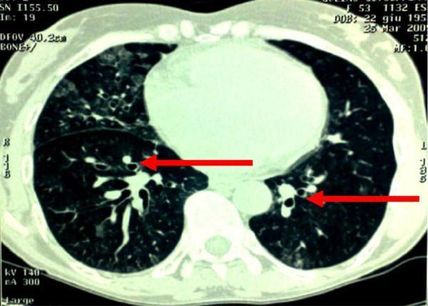
This diagnostic image is an axial slice from a Computed Tomography (CT) scan of the chest, displayed in a lung window. The primary finding is widespread cylindrical bronchiectasis, indicated by red arrows pointing to dilated, thick-walled bronchi. In cross-section, these dilated airways demonstrate the characteristic 'signet ring' sign, where the diameter of the bronchus is significantly larger than its accompanying pulmonary artery. The distribution of the bronchiectasis is bilateral and diffuse, involving multiple lobes. The background lung parenchyma exhibits heterogeneous areas of increased opacity and textural irregularity consistent with chronic inflammatory or post-infectious changes, though no acute pulmonary consolidation is visible. This imaging illustrates the typical morphological presentation of secondary bronchiectasis, often associated with chronic respiratory conditions or recurrent aspiration. The scan demonstrates the diagnostic utility of HRCT in evaluating airway dilation and bronchial wall thickening.
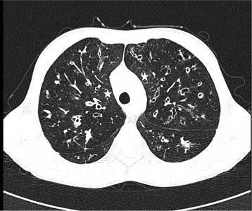
This diagnostic image is a thin-section high-resolution computed tomography (HRCT) axial reconstruction of the chest from a 15-year-old male with cystic fibrosis (CF). The image demonstrates severe bilateral bronchiectasis, characterized by significant bronchial dilatation. Multiple instances of the 'signet ring sign' are visible, where the internal diameter of the bronchus is larger than its accompanying pulmonary artery. There is a notable lack of normal bronchial tapering, with dilated airways extending into the lung periphery and visible within 1 cm of the pleural surface. The parenchyma shows mosaic attenuation; areas of decreased CT attenuation consistent with air trapping are observed in the central parenchyma, while regions marked with asterisks represent areas of relatively normal lung attenuation. The findings illustrate advanced chronic suppurative lung disease and small airway involvement common in pediatric CF patients with impaired pulmonary function (FEV1 45%).
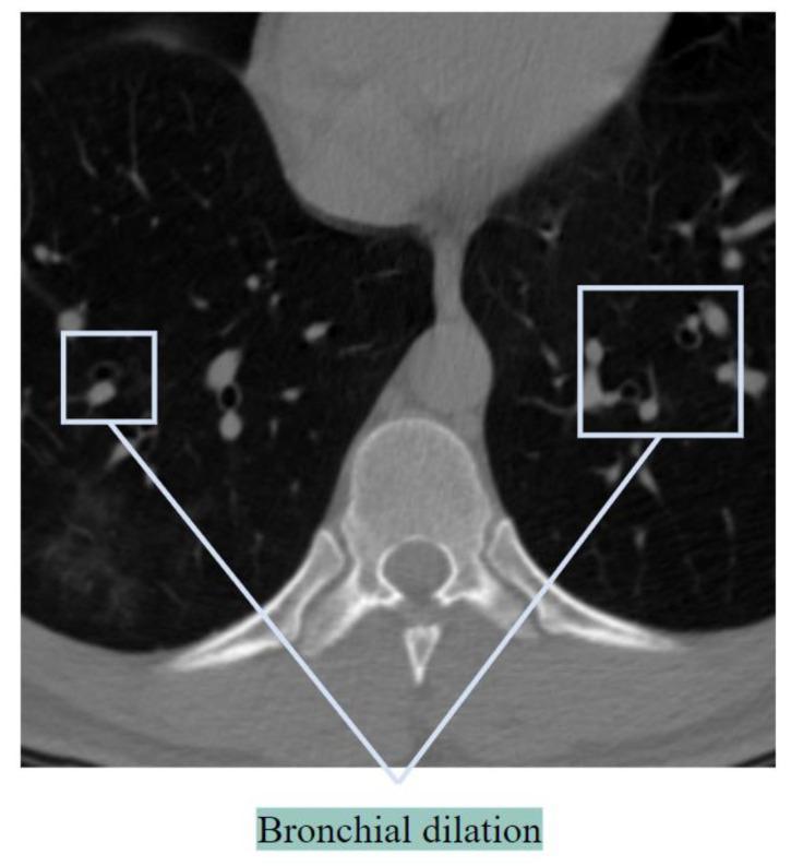
This diagnostic axial CT scan of the chest focuses on the lower lung parenchyma and mediastinal structures at the level of a vertebral body. The image primarily demonstrates the 'signet ring sign,' a hallmark radiological finding of bronchiectasis. Specifically, highlighted regions show pathological bronchial dilation where the internal diameter of the air-filled bronchus is significantly larger than its accompanying pulmonary artery. In a healthy lung, the bronchus-to-artery (BA) ratio is typically 1:1 or less; here, the enlarged, thin-walled bronchus (the ring) adjacent to the smaller, opaque circular artery (the signet) indicates airway disease. The surrounding lung parenchyma appears largely hypodense and air-filled, though multiple discrete BA pairs across both lung fields exhibit similar morphological changes. This visual is used to teach diagnostic criteria for chronic obstructive or inflammatory lung conditions, such as bronchiectasis, by illustrating the lack of normal airway tapering and disproportionate bronchial enlargement.
Bronchiectasis - Case Discussion
Definition
Etiology & Predisposing Conditions
| Category | Examples |
|---|---|
| Post-infectious | TB (major cause in endemic areas), Staphylococcus aureus, Klebsiella, pertussis, SARS-CoV-2 |
| Bronchial obstruction | Tumors, foreign bodies, mucus impaction, atopic asthma |
| Congenital/Hereditary | Cystic fibrosis, primary ciliary dyskinesia (immotile cilia syndrome), Kartagener syndrome |
| Immunodeficiency | Hypogammaglobulinemia (recurrent bacterial infections) |
| Parenchymal fibrosis | Traction bronchiectasis from fibrotic retraction |
Pathogenesis
- Obstruction impairs secretion clearance → bacterial colonization
- Infection causes necrotizing inflammation → bronchial wall damage
- Cytokines, bacterial toxins, and proteases perpetuate the cycle
- Irreversible dilation and peribronchial fibrosis result
- Robbins & Kumar Basic Pathology, p.455
Types (Pathological Classification)
- Cylindrical - uniform, regular airway dilation (least severe)
- Varicose - non-uniform, serpiginous ("beaded") dilation
- Cystic/Saccular - most severe; string or cluster of cysts, often with air-fluid levels
Clinical Features
| Feature | Description |
|---|---|
| Cough | Chronic, productive - the most prominent symptom |
| Sputum | ≥30 mL/day of mucopurulent, sometimes foul-smelling sputum |
| Hemoptysis | Can range from blood-streaked sputum to massive bleed |
| Dyspnea | Especially in extensive disease |
| Rhinosinusitis | Common associated finding |
| Systemic | Fever, weight loss during exacerbations |
Complications
- Obstructive ventilatory defect with hypoxemia and hypercapnia
- Cor pulmonale (pulmonary hypertension from chronic hypoxia)
- Brain abscess (hematogenous spread)
- Secondary amyloidosis (now rare with modern treatment)
- Massive hemoptysis
Radiology
Chest X-Ray Signs
- Tramlines - parallel line opacities (bronchial wall thickening seen in profile)
- Ring opacities - bronchi seen end-on
- Mucus plugs - tubular or ovoid opacities
- Overinflation (in CF-associated bronchiectasis)
- Atelectasis in affected segments
HRCT (Gold Standard)
-
Signet ring sign - bronchus diameter > adjacent pulmonary artery (most recognized sign)
-
Lack of normal bronchial tapering (the cardinal sign)
-
Bronchi visible within 1 cm of costal pleura (normally they are not)
-
Varicose pattern - beaded configuration
-
Cystic pattern - string/cluster of cysts, may have air-fluid levels
-
Mucus-filled dilated bronchi ("glove-finger" opacities)
-
Grainger & Allison's Diagnostic Radiology, p.155-156
Management
Airway Clearance (cornerstone of therapy)
- Chest physiotherapy and postural drainage - essential in all patients
- Active cycle of breathing techniques (ACBT)
- Inhaled beta-2 agonists - improve mucociliary clearance and reverse bronchoconstriction
Antibiotics
- Targeted at identified pathogens during exacerbations
- Long-term macrolide therapy (e.g., azithromycin) - reduces exacerbation frequency and improves lung function in recurrent disease
- Inhaled tobramycin for Pseudomonas-colonized patients: a 2025 meta-analysis (PMID: 40339664) confirmed benefit in non-CF bronchiectasis
Mucoactive Agents
- A 2026 systematic review (PMID: 42342264) assessed mucoactive agents (hypertonic saline, mannitol, dornase alfa) - evidence for their routine use remains selective
DPP-1 Inhibitors (Emerging)
- Brensocatib (DPP-1/dipeptidyl peptidase-1 inhibitor) reduces neutrophilic airway inflammation
- A 2025 GRADE meta-analysis (PMID: 41299471) confirmed efficacy and safety in bronchiectasis - represents a newer disease-modifying approach
Surgical
- Lobectomy/segmentectomy for localized disease refractory to medical therapy or causing massive hemoptysis
- Lung transplantation in end-stage CF-related bronchiectasis
Antitussives
- Not recommended in bronchiectasis - cough is the primary airway clearance mechanism
Key Points for Exam
| Point | Detail |
|---|---|
| Diagnostic gold standard | HRCT chest |
| Most common morphology | Cylindrical (least severe) |
| Most severe morphology | Cystic/saccular |
| Most common lower lobe pathogen | Pseudomonas aeruginosa |
| Why antitussives are contraindicated | Cough clears secretions - suppressing it worsens disease |
| CF-related bronchiectasis location | Upper lobes (unlike most other causes - lower lobes) |
| Pathological hallmark | Irreversible dilation beyond 4x normal diameter |
Pulmonary hypertension case study
pulmonary arterial hypertension treatment
pulmonary hypertension plexiform lesion histology right heart catheterization
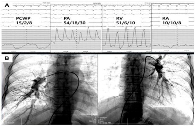
This composite educational image illustrates diagnostic modalities for Chronic Thromboembolic Pulmonary Hypertension (CTEPH). Part A displays Right Heart Catheterization (RHC) hemodynamic waveforms and pressure readings. The pulmonary artery (PA) pressure (54/18/30 mmHg) and right ventricular (RV) pressure (51/6/10 mmHg) show significant systolic elevation, while the pulmonary capillary wedge pressure (PCWP 15/2/8 mmHg) remains within the normal range, confirming a precapillary pulmonary hypertension profile. Part B presents Digital Subtraction Angiography (DSA) of the bilateral pulmonary vasculature. A diagnostic catheter is seen traversing the right heart into the pulmonary arteries. The angiography reveals characteristic CTEPH findings, including irregular arterial branching, segmental stenoses, and 'pruned' distal vasculature, particularly evident on the left side. These visual features signify organized thrombi and microvasculopathy. This material is designed for advanced medical education in cardiology and pulmonology to demonstrate the integration of invasive hemodynamics and vascular imaging in the confirmatory diagnosis of Group 4 pulmonary hypertension.
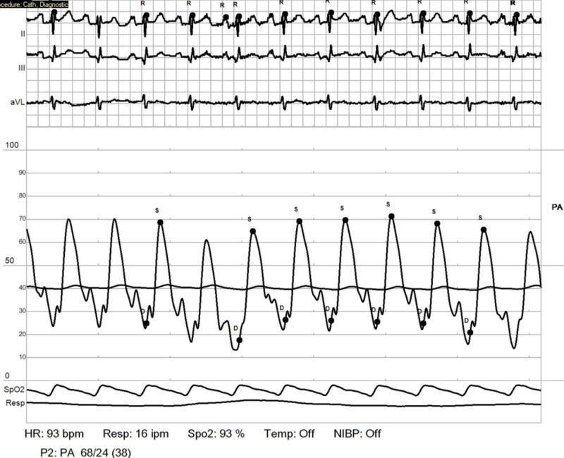
A multi-parameter medical monitor display from a right heart catheterization (RHC) procedure. The primary visual focus is a pulmonary artery (PA) pressure waveform tracing, which demonstrates significantly elevated pressures. The PA waveform shows cyclical systolic peaks (labeled 'S') and diastolic troughs (labeled 'D'), with a numerical measurement of 68/24 (38) mmHg, indicating pulmonary hypertension. Positioned above the PA tracing are three electrocardiogram (ECG) leads (II, III, and aVL) showing a sinus rhythm at a heart rate (HR) of 93 beats per minute. Below the pressure tracing are continuous waveforms for pulse oximetry (SpO2) and respiration (Resp). Numerical data at the bottom of the screen confirm a heart rate of 93 bpm, a respiratory rate of 16 inspirations per minute, and an oxygen saturation level of 93%. This clinical image illustrates hemodynamic monitoring in a patient with systemic sclerosis-associated pulmonary arterial hypertension (SSc-PAH) following medical management.
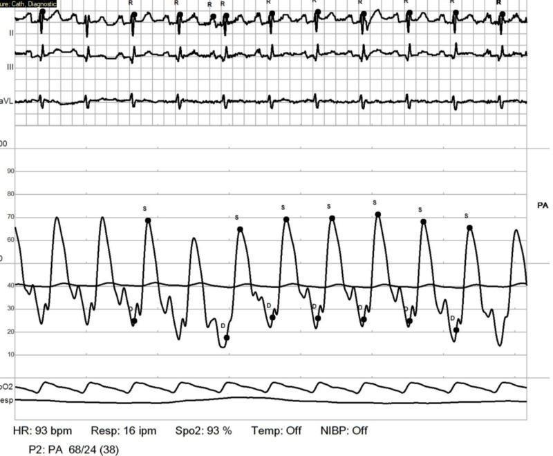
A diagnostic hemodynamic tracing from a right heart catheterization (RHC) procedure. The image displays synchronized multi-parameter monitoring data, including a three-lead electrocardiogram (ECG) at the top showing leads II, III, and aVL with a heart rate of 93 bpm. The central section features a pressure-volume waveform representing pulmonary artery (PA) pressures. Systolic peaks (S) are visualized reaching approximately 68-70 mmHg, and diastolic troughs (D) are noted at approximately 24 mmHg, indicating significant pulmonary hypertension. A concurrent flatter waveform represents the pulmonary capillary wedge pressure (PCWP), with a mean value indicated around 38 mmHg. Bottom panels display digital telemetry data including a plethysmographic oxygen saturation (SpO2) of 93% and a respiratory rate of 16 inspirations per minute (ipm). This tracing is used in clinical cardiology and pulmonology to evaluate pulmonary vascular resistance and hemodynamic response in patients with systemic sclerosis-associated pulmonary arterial hypertension.
pulmonary hypertension pathology plexiform lesion vascular remodeling
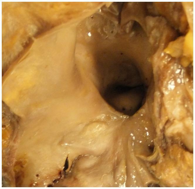
This clinical photograph displays a gross pathological specimen of an open pulmonary artery. The luminal surface of the vessel exhibits significant morphological alterations, characterized by multiple raised, yellowish intimal plaques consistent with atheroma. These features are a classic hallmark of chronic pulmonary hypertension, which in this clinical context is associated with Eisenmenger syndrome—a condition where long-standing left-to-right congenital heart shunts cause pulmonary vascular remodeling. The presence of atherosclerotic-like changes in the typically low-pressure pulmonary circuit indicates severe, prolonged elevation in pulmonary arterial pressure. The image serves as an educational tool for identifying macroscopic signs of vascular pathology in the setting of advanced congenital heart disease and pulmonary hypertensive vascular disease.
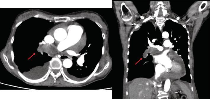
This diagnostic image consists of two panels showing a thoracic contrast-enhanced computed tomography (CT) scan in axial (left) and coronal (right) views. A red arrow in both views points to a prominent, low-attenuation space-occupying lesion located within the right pulmonary artery, extending toward the hilum. This finding is characteristic of a central pulmonary thrombus or embolism, appearing as a filling defect that obstructs the contrast-filled vessel lumen. Additional clinical findings include a large right-sided pleural effusion, visible as a crescentic fluid collection in the posterior-dependent portion of the right hemithorax on the axial view. The heart appears enlarged, consistent with cardiomegaly and potential right heart strain associated with chronic thromboembolic pulmonary hypertension (CTEPH). The image serves as an educational example of central pulmonary vascular pathology and its secondary effects on the thoracic cavity, such as pleural effusion and cardiac remodeling.
Pulmonary Hypertension - Case Study Discussion
Definition & Hemodynamic Criteria
| Subtype | mPAP | PCWP | PVR | WHO Groups |
|---|---|---|---|---|
| Normal | 14 ± 3.3 mmHg | 8 ± 2.9 mmHg | 0.93 ± 0.38 WU | - |
| Precapillary PH | >20 mmHg | ≤15 mmHg | ≥3 WU | 1, 3, 4, 5 |
| Isolated postcapillary PH | >20 mmHg | >15 mmHg | <3 WU | 2, 5 |
| Combined pre+postcapillary PH | >20 mmHg | >15 mmHg | ≥3 WU | 2, 5 |
- Murray & Nadel's Textbook of Respiratory Medicine, p.1893
WHO Classification (6th WSPH - Updated)
| Group | Category | Key Causes |
|---|---|---|
| Group 1 - PAH | Pulmonary arterial hypertension | Idiopathic (~50%), heritable (BMPR2 mutation), CTD-associated (scleroderma ~25%), HIV, drugs (methamphetamine, dasatinib), portal HTN, congenital heart disease |
| Group 1.5 (new) | Long-term CCB responders | Subset of PAH who vasoreact |
| Group 1.6 (new) | PVOD/PCH involvement | Pulmonary veno-occlusive disease |
| Group 2 | Left heart disease | HFrEF, HFpEF, valvular disease, mitral stenosis |
| Group 3 | Lung disease/Hypoxia | COPD, ILD, sleep-disordered breathing, high altitude |
| Group 4 | Chronic thromboembolic (CTEPH) | Post-PE (75% have prior PE), ~4% of PE patients develop CTEPH |
| Group 5 | Unclear/multifactorial | Sarcoidosis, metabolic disorders, hematologic conditions |
- Fuster and Hurst's The Heart, 15th Ed., Table 57-4
Pathogenesis
General Mechanisms (by group)
- Hypoxic disease (Group 3): Obliteration of alveolar capillaries → increased PVR
- Left heart disease (Group 2): Elevated left atrial/pulmonary venous pressure transmitted back to pulmonary arteries (e.g., mitral stenosis)
- CTEPH (Group 4): Recurrent emboli reduce cross-sectional area of pulmonary vascular bed → increased PVR
- Autoimmune (Group 1): Vascular inflammation and remodeling (especially systemic sclerosis)
Idiopathic/Familial PAH - Molecular Mechanism
- BMPR2 gene mutation (bone morphogenetic protein receptor type 2) - found in 75% of familial cases and 25% of sporadic cases
- BMPR2 is a TGF-β receptor superfamily member regulating endothelial and smooth muscle cell apoptosis, proliferation, and differentiation
- Haploinsufficiency leads to endothelial dysfunction and vascular smooth muscle proliferation
- A "two-hit" model is proposed: genetic susceptibility + additional environmental/genetic trigger
- Only 10-20% of BMPR2 mutation carriers develop overt disease
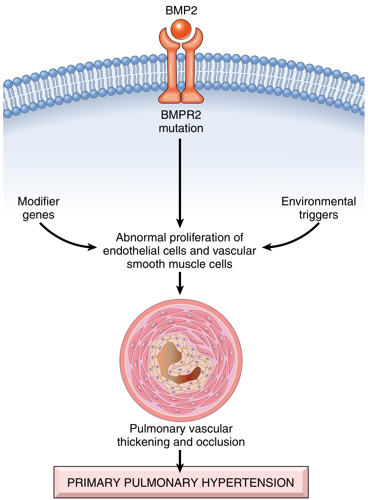
- Robbins, Cotran & Kumar Pathologic Basis of Disease, p.657
Morphology / Pathology
- Medial hypertrophy of pulmonary muscular and elastic arteries
- Right ventricular hypertrophy (cor pulmonale)
- Large vessel - atherosclerosis-like intimal plaques (in severe/chronic cases)
- Arterioles (40-300 μm) - marked medial hypertrophy + intimal fibrosis → near-complete luminal obliteration (pinpoint channels)
- Plexiform lesions (hallmark of advanced Group 1 PAH) - tuft of capillary formations forming a network spanning dilated thin-walled small arteries
Plexiform lesions are most prominent in: idiopathic/familial PAH (Group 1), congenital heart disease with left-to-right shunts (Group 2), and HIV/drug-related PAH (Group 1).
Clinical Features
| Symptom | Notes |
|---|---|
| Dyspnea on exertion | Most common presenting complaint |
| Fatigue | Early and prominent |
| Chest pain (anginal type) | From RV ischemia |
| Syncope | From fixed low cardiac output, especially on exertion |
| Peripheral edema | Late sign of RV failure |
| Cyanosis | Advanced disease |
Signs become evident only in advanced disease - hence late diagnosis is common.
Diagnostic Workup
Step-by-Step Approach
- Clinical suspicion - unexplained exertional dyspnea, signs of RV strain
- ECG - RV strain pattern: right axis deviation, RBBB, P pulmonale
- Echocardiography - screening tool; estimates PASP via TR jet; RV hypertrophy/dilation, septal flattening ("D-sign")
- HRCT chest - lung parenchymal disease (Group 3), mosaic attenuation
- V/Q scan - essential to rule out CTEPH (Group 4) - mismatched segmental defects
- Right Heart Catheterization (RHC) - GOLD STANDARD for diagnosis and hemodynamic characterization
- Vasoreactivity testing (during RHC with IV adenosine or inhaled NO) - positive response: fall in mPAP ≥10 mmHg to ≤40 mmHg → qualifies for CCB therapy (Group 1.5)
- Additional workup: ANA, anti-Scl-70, HIV serology, LFTs (portal HTN), sleep study, 6-minute walk test
Management
General Principles
- Treat the underlying cause in Groups 2, 3, 4, 5
- Group 1 (PAH) has specific vasodilator therapies
Non-Pharmacological
- Supplemental O2 (keep SpO2 > 91%)
- Supervised exercise/rehabilitation
- Diuretics for RV volume overload
- Anticoagulation (especially CTEPH and idiopathic PAH)
- Avoid pregnancy (mortality up to 30%)
Pharmacological - 3 Key Pathways for Group 1 PAH
| Pathway | Drug Class | Agents |
|---|---|---|
| Prostacyclin pathway | Prostanoids | Epoprostenol (IV - most potent), Treprostinil, Iloprost |
| Endothelin pathway | ERA (endothelin receptor antagonists) | Bosentan, Ambrisentan, Macitentan |
| NO/cGMP pathway | PDE-5 inhibitors | Sildenafil, Tadalafil |
| sGC stimulator | Riociguat |
ERAs (bosentan, ambrisentan) are teratogenic - avoid in pregnancy. PDE-5 inhibitors and prostanoids are preferred in pregnant patients despite fetotoxicity risk if benefit outweighs risk.
- Rosen's Emergency Medicine, p.802
CTEPH (Group 4) - Special Treatment
- Pulmonary thromboendarterectomy (PTE) - potentially curative surgical option
- Balloon pulmonary angioplasty (BPA) - for inoperable patients
- Riociguat - approved specifically for inoperable/persistent CTEPH
Surgical / Advanced
- Lung transplantation - definitive treatment for refractory disease
- Atrial septostomy - palliative in selected refractory cases
Recent Evidence (2024-2026)
- DPP-1 inhibitors (e.g., brensocatib) are being studied in related airway conditions with neutrophilic inflammation
- A 2026 practice guideline on portopulmonary hypertension (PMID: 40094355) from the International Liver Transplantation Society provides updated guidance for this Group 1 PAH subtype
- EULAR 2023 scleroderma guidelines (PMID: 39874231) recommend proactive screening for SSc-PAH with annual echocardiography + DETECT algorithm
Key Exam Points Summary
| Point | Answer |
|---|---|
| Diagnostic gold standard | Right heart catheterization (RHC) |
| mPAP threshold for PH (6th WSPH) | >20 mmHg (previously 25 mmHg) |
| Most common PAH subtype | Idiopathic (~50% of Group 1) |
| Gene mutated in familial PAH | BMPR2 (75% familial, 25% sporadic) |
| Hallmark histology lesion | Plexiform lesion |
| Demographic of idiopathic PAH | Young women, 20-40 years |
| Only potentially curable form | CTEPH (Group 4) - via PTE surgery |
| Contraindicated in PH | Pregnancy (mortality 30%); ERAs in pregnancy (teratogenic) |
| Drug not to use in non-PAH PH | PAH-specific therapies worsen Group 2 disease |
| RV failure + PH = | Cor pulmonale |