
Cutaneous tb latest development
"cutaneous tuberculosis"[MeSH Terms]
cutaneous tuberculosis latest treatment diagnosis 2025 2026
PMID: 40613433
https://nmji.in/cutaneous-tuberculosis-prevention-management…
lupus vulgaris cutaneous tuberculosis skin lesion
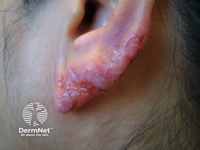
This is a high‑resolution clinical photograph of a left auricular skin lesion captured during external examination. Modality: Clinical photography; Technique: digital color image, standard exposure, lateral and frontal views of the pinna encompassing the helix and adjacent antihelix. Anatomical localization: auricle of the external ear, cutaneous layer with involvement of the skin and superficial subcutaneous tissue; no evident deep tissue penetration. Visual features: erythematous to violaceous plaque with irregular, undermined margins, surface scaling, crusting, and mild adjacent erythema; there is subtle cortical lichenification and focal atrophic change along the lesion’s edge; overall appearance suggests a chronic process with slow evolution. Clinical impression: findings are compatible with lupus vulgaris (cutaneous tuberculosis) presenting on the ear, a typically indolent plaque-type lesion that may ulcerate and scar over time. Significance: in TB‑endemic settings, lupus vulgaris should be considered in chronic auricular plaques; correlate with tuberculosis screening (Mantoux or IGRA), histopathology, culture, and PCR for Mycobacterium tuberculosis. Differential diagnoses include chronic eczema, discoid lupus erythematosus, granulomatous dermatitis, leprosy, keratoacanthoma. Potential clinical use: educational reference for dermatology, infectious disease, and medical training to recognize auricular lupus vulgaris and guide diagnostic workup and management. Clinical relevance: supports prompt antimicrobial therapy and lesion surveillance strategies.
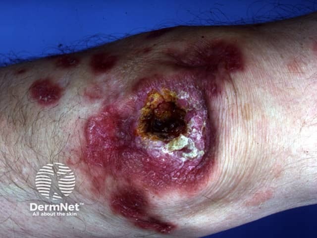
Clinical photography of a solitary cutaneous lesion on the distal leg consistent with lupus vulgaris (cutaneous tuberculosis). The image depicts a well-demarcated, annular to oval plaque with an erythematous to violaceous border and a central ulceration/crusted necrotic area. Surrounding skin shows coalescing erythema, mild edema, and postinflammatory hyperpigmentation. The surface center demonstrates crusting and a recessed, necrotic core with surrounding pale tissue, suggesting chronicity. The lesion size appears moderate and border is indurated. The anatomy is the integumentary system focusing on skin of the lower leg; the view is a single external surface close-up, taken in standard lighting without dermoscopic magnification. There is no visible active purulence. The morphological features, such as crusted ulcer with expanded border and plate-like scale, are compatible with lupus vulgaris, a form of cutaneous tuberculosis caused by Mycobacterium tuberculosis complex. The image emphasizes the chronic, slowly progressive nature and potential for secondary infection. Pathophysiology involves granulomatous inflammation with caseating necrosis; clinically, lupus vulgaris may present with apple-jelly nodules on diascopy, scarring, and deformity if untreated. The diagnostic significance lies in correlating dermatologic findings with microbiologic and histopathology confirmation, guiding antituberculous therapy and public health measures. Potential clinical use: dermatology education, TB cutaneous clinic, differential diagnosis workshops, teledermatology triage.
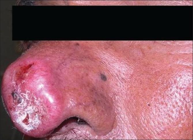
Clinical photograph of a patient's face in profile, showing a significant skin lesion on the nasal tip and ala, diagnosed as Lupus vulgaris, a form of cutaneous tuberculosis. The lesion presents as a large, erythematous, and violaceous plaque with prominent inflammation. Key morphological features include central ulceration with visible crusting and white scaling, indicating a chronic, destructive process of the nasal tissue. The surrounding skin of the cheek and bridge of the nose shows secondary changes, including diffuse erythema, thickened texture, and scattered hyperpigmented macules. This image serves as an educational example of the progressive and disfiguring nature of chronic cutaneous tuberculosis when it affects the central facial features, highlighting the characteristic 'apple-jelly' nodules (though not viewed under diascopy) and the resulting tissue destruction common in untreated lupus vulgaris.
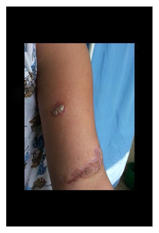
Clinical photograph of the right forearm of a 17-year-old female showing two distinct dermatological findings. The superior lesion is a 2x1 cm erythematous plaque characteristic of lupus vulgaris (cutaneous tuberculosis). This plaque features irregular, slightly raised borders and a central surface with grayish-white scaling or verrucous texture. Distally, on the lower forearm near the antecubital fossa, there is a secondary 4x2 cm linear hypertrophic scar resulting from previous trauma. The scar exhibits hyperpigmentation, irregular texture, and slight elevation. This image serves as a clinical example of cutaneous tuberculosis arising in a patient with a history of local skin injury, illustrating the chronic inflammatory presentation of lupus vulgaris and its morphological distinction from adjacent cicatricial tissue.
Cutaneous Tuberculosis: What's New
1. Diagnosis remains the main bottleneck
- A 2025 German review, Recognising cutaneous tuberculosis (Gramminger & Biedermann, JDDG 2025), stresses that CTB is under-suspected outside TB-endemic regions (notably in migrant populations) because it mimics common dermatoses. In paucibacillary forms (lupus vulgaris, tuberculosis verrucosa cutis), standard "gold-standard" tests (AFB smear, culture) frequently come back negative, so diagnosis depends on strong clinical suspicion plus repeated biopsy/PCR attempts.
- Molecular testing (PCR for mycobacterial DNA) is increasingly used. In multibacillary disease PCR sensitivity/specificity approaches 100%, but in paucibacillary disease it drops to roughly 55%, and even PCR-positive patients don't always respond to anti-TB therapy - underscoring that molecular tests supplement, not replace, clinical-histopathologic correlation (Fitzpatrick's Dermatology, 9780071837781, p. 2889-2890).
- WHO's Global TB Report 2025 research chapter notes an expanding TB diagnostic pipeline overall (~100 products in development as of August 2025), including point-of-care biomarker and nucleic-acid tests - relevant to extrapulmonary/cutaneous disease as these platforms mature, though none are CTB-specific yet.
2. Treatment: still standard multidrug therapy, with adjuncts under study
- First-line treatment continues to follow national TB program regimens (e.g., India's NTEP fixed-dose combination, weight-banded, in intensive + continuation phases), split by drug-susceptible vs. drug-resistant disease - per the National Medical Journal of India review on CTB management.
- Localized, non-disseminated forms (tuberculosis verrucosa cutis, limited lupus vulgaris) can sometimes be managed with isoniazid monotherapy for up to 12 months, continued at least 2 months past clinical resolution because viable organisms can persist in healed-appearing lesions (Fitzpatrick's Dermatology, p. 2890).
- Emerging/adjunctive options being discussed in recent reviews:
- Host-directed therapy - reducing TNF-alpha-driven granuloma formation and tissue damage to enhance bacillary clearance.
- Immunomodulators such as thalidomide for post-treatment inflammatory reactions in multibacillary disease.
- Topical isoniazid (compounded) reported successful for erythema induratum (a tuberculid).
- Surgical adjuncts - incision/drainage or excision for scrofuloderma, lupus vulgaris, and TB verrucosa cutis to reduce morbidity and shorten chemotherapy course; reconstructive surgery for disfiguring long-standing lupus vulgaris.
- Multidrug-resistant (MDR) and pre-XDR CTB, while rare, are increasingly reported and require second-line regimens of longer duration with higher toxicity burden - a 2026 case review (J Clin Med 2026;15:702, PMC12842349) highlights this alongside a concerning finding: chronic, long-standing lupus vulgaris carries a real (0.5-10.5%) risk of malignant transformation to squamous cell carcinoma, reinforcing the case for early diagnosis and complete treatment.
3. Epidemiological/awareness angle
- CTB is being reframed by some Indian authors as a "neglected tropical disease" within dermatology - underdiagnosed, underfunded relative to its morbidity, and disproportionately affecting TB-endemic, resource-limited settings.
- Recent case series (Cureus 2025, PMC case reports 2025) continue to emphasize the same message: pleomorphic presentations (in both HIV-positive and HIV-negative, immunocompetent patients) mean CTB should stay on the differential for any chronic, treatment-resistant granulomatous or ulcerative skin lesion, especially in or from endemic areas.