
Duration of degluttion apnea
duration of deglutition apnea physiology seconds
Deglutition Apnea - Duration
"Respiratory apnea associated with swallowing shows considerable variation across individuals, and in one study it ranged from 0.61 to 3.83 seconds."
Key Points
| Feature | Detail |
|---|---|
| Duration range | 0.61 - 3.83 seconds |
| Variation | Considerable inter-individual variation |
| Effect of bolus volume | Increases in some individuals, decreases in others |
| Spontaneous swallows | Tend to have a shorter apneic period |
| Timing in respiratory cycle | Most swallows occur during expiration or late inspiration |
| Post-swallow effect | Swallowing resets the respiratory rhythm (no compensatory modification afterward) |
Mechanism
-
Swallowing takes precedence over both respiration and mastication, briefly disrupting their rhythm.
-
The respiratory center in the medulla is directly inhibited by the swallowing (deglutition) center during the pharyngeal phase.
-
This apnea is essential for airway protection - the true vocal folds adduct simultaneously, preventing aspiration.
-
The central pattern generator for swallowing is located in the caudal nucleus of the solitary tract (NST) and the medullary reticular formation adjacent to the nucleus ambiguus.
-
Cummings Otolaryngology: Head and Neck Surgery, Oral Phase of Deglutition section
Physiology of high atm pressure
physiology high atmospheric pressure diving hyperbaric effects body
Physiology of High Atmospheric Pressure
1. Physical Principles (The Gas Laws)
| Law | Statement | Relevance |
|---|---|---|
| Boyle's Law | P₁V₁ = P₂V₂ (at constant temp) | Gas-containing spaces compress on descent, expand on ascent |
| Dalton's Law | Total P = sum of partial pressures | At depth, partial pressure of each gas rises proportionally |
| Henry's Law | Gas dissolved ∝ partial pressure | More gas dissolves into blood/tissues at high pressure |
- Medical Physiology (Boron & Boulpaep), p. 1791
2. Effect on Lung Volumes (Breath-Hold Diving)
- At 10 m (2 atm): lung volume → ½ of surface value
- At 20 m (3 atm): lung volume → ⅓
- At 30 m (4 atm): lung volume → ¼ (approximately residual volume)
3. Nitrogen Narcosis ("Rapture of the Deep")
- As depth increases, alveolar PN₂ rises and more N₂ dissolves into tissues (Henry's Law).
- N₂ is highly lipid-soluble and dissolves readily in neuronal membranes.
- It reduces ion conductance and neuronal excitability, mimicking general anesthesia.
- Onset: ~4-5 atm (about 30-40 m depth) breathing compressed air, after >1 hour.
- "Martini's Law": each 15 m of depth has the effect of drinking one additional martini.
- Loss of psychosocial inhibitions (mild euphoria/intoxication)
- Lethargy and drowsiness
- Rapid onset of fatigue
- Loss of consciousness (at extreme depth)
- Medical Physiology, p. 1795
4. Oxygen Toxicity
| Type | Threshold | Mechanism | Features |
|---|---|---|---|
| Pulmonary (Lorrain Smith) | >0.5 atm PO₂, prolonged exposure | Free radical damage to airway epithelium | Bronchiolar inflammation, pulmonary edema, atelectasis, fibrin formation, lung consolidation |
| CNS (Paul Bert) | ~1500 mm Hg PO₂ (~10 atm room air for >30-45 min) | Free radical oxidation of polyunsaturated fatty acids in cell membranes + energy metabolism enzymes | Muscle twitching, nausea, disorientation, irritability → seizures and coma |
- Medical Physiology, p. 1796; Tintinalli's Emergency Medicine, Chapter 214
5. Nitrogen Saturation and Decompression Sickness ("The Bends")
Saturation
- At 1 atm normally: ~1 L of N₂ dissolved in body tissues (equally in water and fat compartments).
- At 4 atm: ultimately 4 L of N₂ dissolves (fat equilibrates slowly due to poor perfusion - takes hours).
- The longer and deeper the dive, the more N₂ saturates tissues.
Decompression Sickness (DCS)
| Type | Features | Mechanism |
|---|---|---|
| Type I ("mild") | Deep, unrelieved joint pain (knees, shoulders); skin mottling | Bubbles in ligaments, fascia, tendon stretch receptors, long bone intramedullary spaces |
| Type II ("serious") | Neurologic - truncal constriction, ascending paralysis, transverse myelitis; vestibular (vertigo, tinnitus, hearing loss); pulmonary (cough, dyspnea, hemoptysis) | Venous bubble occlusion in spinal cord venous plexus; autochthonous in-situ spinal cord bubbles |
-
Symptoms begin minutes to several hours after surfacing (rarely, days later).
-
Commercial/military/caisson workers: predominantly joint pain. Sport divers: more spinal cord involvement.
-
Flying after diving can precipitate/worsen DCS (lower ambient pressure). Minimum wait: 12-24 hours.
-
Patent foramen ovale increases risk of paradoxical gas embolism and neurological DCS.
-
Treatment: Hyperbaric oxygen (HBO) recompression therapy - the only proven treatment.
-
Tintinalli's Emergency Medicine, p. 1410-1411; Medical Physiology, p. 1797
6. Prevention: Helium-Oxygen (Heliox) Mixtures
- Minimal narcotizing effect (far less lipid-soluble than N₂)
- Less tissue dissolution (lower water/lipid solubility)
- Lower density than N₂ - reduces airway resistance and work of breathing at depth
- Faster washout on decompression (lower partition coefficient)
- Medical Physiology, p. 1797-1798
7. Barotrauma
- Middle ear barotrauma - most prevalent diving injury. Failure to equalize middle ear pressure via Eustachian tube causes pain, hemorrhage, membrane rupture.
- Sinus barotrauma - mucosal engorgement/hemorrhage in paranasal sinuses.
- Pulmonary squeeze - rare in breath-hold divers at extreme depth.
- Pulmonary overinflation syndrome - if diver ascends without exhaling, air expands to >2x lung volume, causing alveolar rupture.
- Complications: pneumothorax, pneumomediastinum, arterial gas embolism (AGE) - the most dangerous form.
8. Threshold Summary
| Condition | Threshold / Onset |
|---|---|
| Nitrogen narcosis | ~4-5 atm (30-40 m depth), breathing air |
| CNS oxygen toxicity | ~10 atm room air (or 1.5-2 atm pure O₂) after 30-45 min |
| Pulmonary oxygen toxicity | >0.5 atm PO₂ over many hours |
| Decompression sickness | Rapid ascent from any significant depth/duration dive |
Functionnof type 1 pneumocutes
type 1 pneumocyte function alveolar cell physiology
Functions of Type I Pneumocytes (Type I Alveolar Cells)
Structure
Blood-Air Barrier - Electron Micrograph
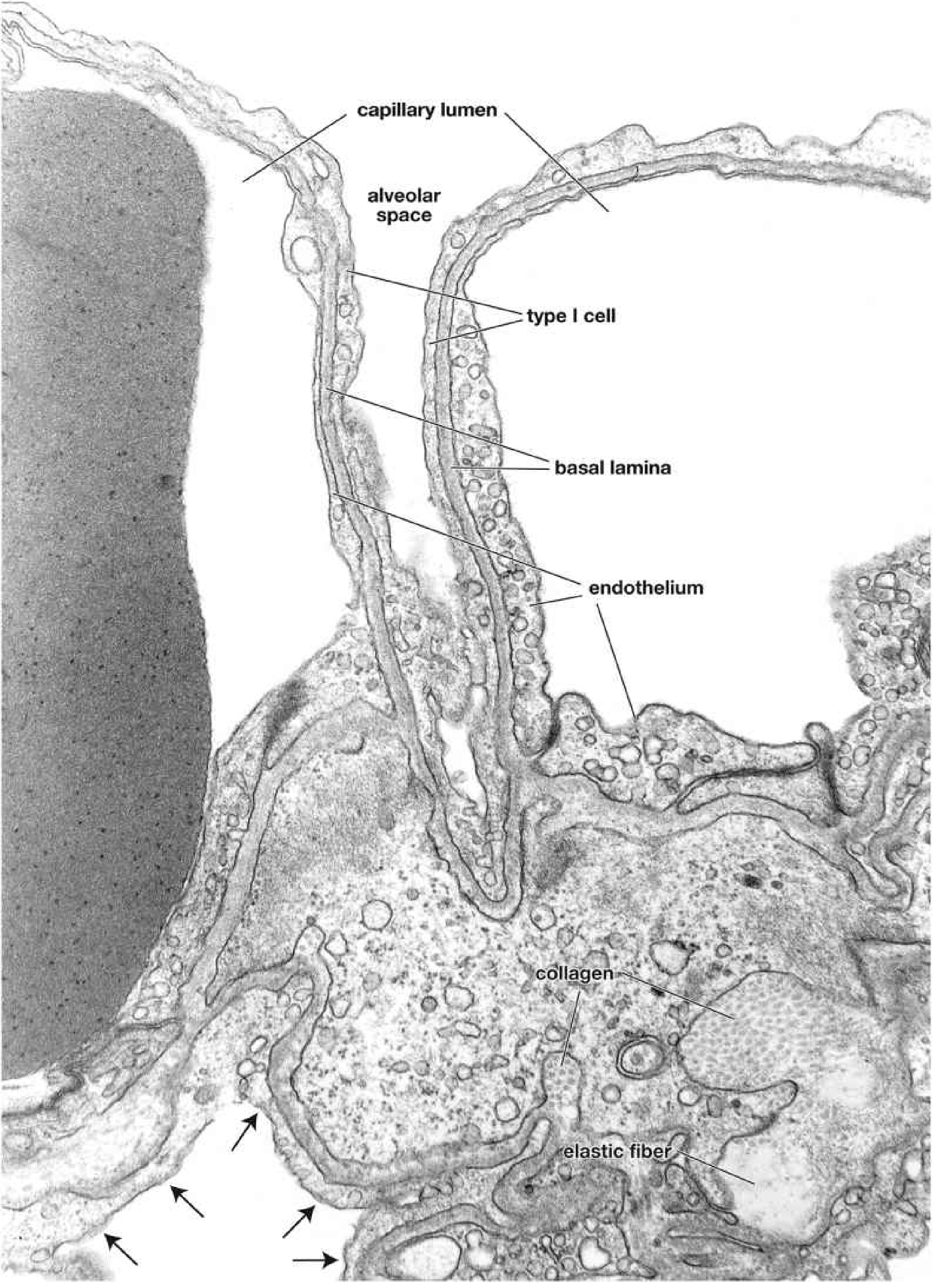
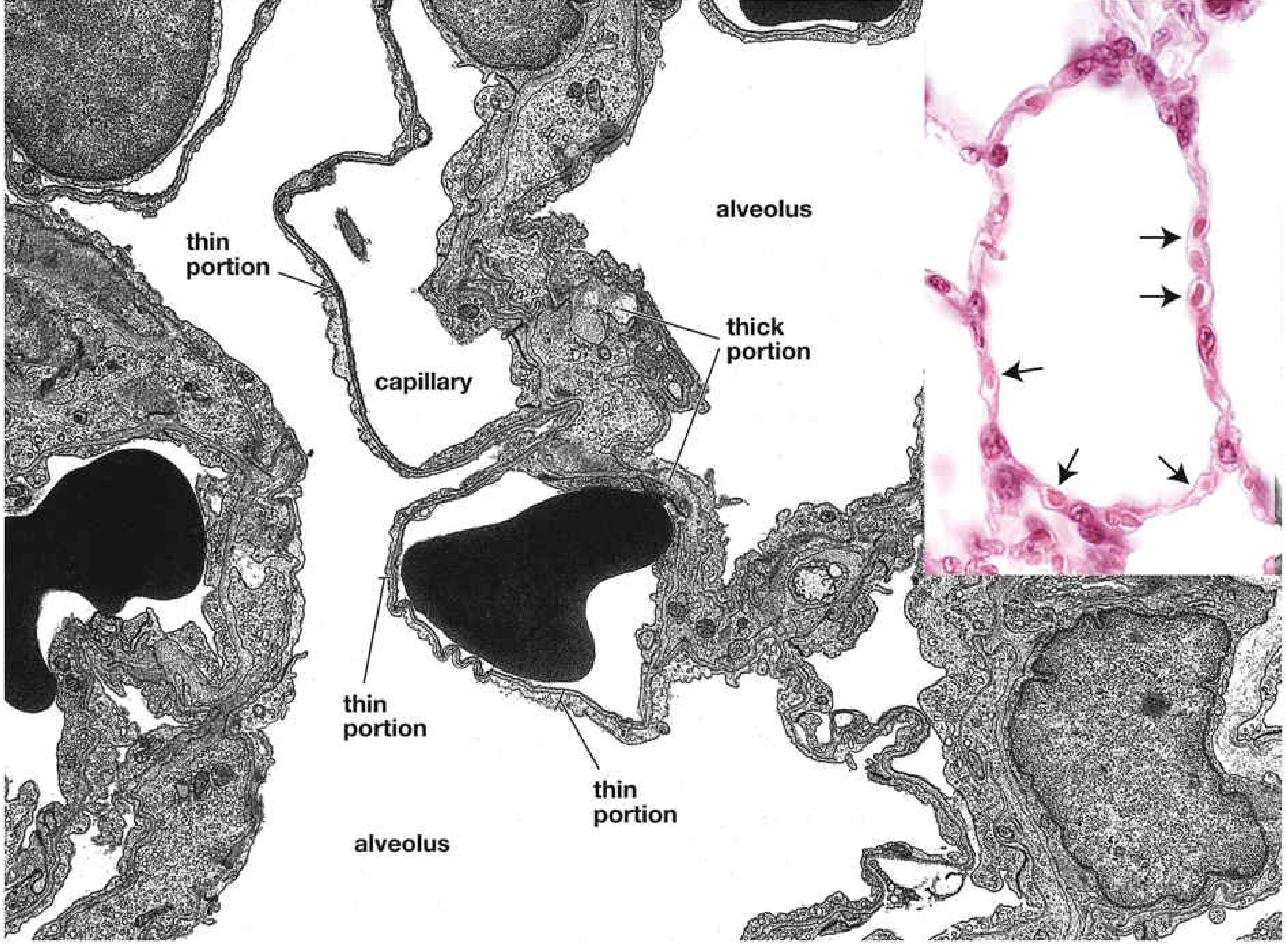
Functions
1. Gas Exchange (Primary Function)
- Type I alveolar cell (extremely thin cytoplasm, ~0.1-0.2 µm)
- Fused basal laminae of Type I cell and capillary endothelium
- Capillary endothelial cell (also thin)
2. Structural Integrity of the Alveolar Wall
- Type I cells provide the physical scaffold that maintains alveolar shape.
- Their wide, flat cytoplasmic extensions cover the alveolar surface and prevent collapse.
- The occluding (tight) junctions between Type I cells form an effective barrier between the air space and the septal wall components.
3. Fluid Barrier - Prevention of Pulmonary Edema
- The tight junctions between Type I cells prevent the passive movement of fluid and macromolecules from the interstitium into the alveolar air space.
- Type I cells also actively transport Na⁺ ions (via apical Na⁺ channels and basolateral Na⁺/K⁺-ATPase), which drives water reabsorption from the alveolar space - keeping alveoli dry and patent.
4. Passive Role in Surfactant Function
- Type I cells themselves do not produce surfactant.
- However, their flat surface provides the substrate on which the surfactant film (produced by Type II cells and club cells) spreads and functions to reduce surface tension.
5. Signal Transduction / Mechanosensing
- Type I cells contain numerous caveolae (small membrane invaginations) and are thought to play a role in mechanosensing - detecting stretch during lung inflation and potentially modulating downstream signaling.
Key Distinguishing Features vs. Type II Pneumocytes
| Feature | Type I Pneumocyte | Type II Pneumocyte |
|---|---|---|
| Shape | Flat/squamous (extremely thin) | Cuboidal |
| % of alveolar cells (by number) | ~40% | ~60% |
| % of surface covered | ~95% | ~5% |
| Function | Gas exchange, barrier | Surfactant production, stem cell |
| Lamellar bodies | Absent | Present (surfactant storage) |
| Cell division | Cannot divide | Can divide; regenerates Type I after injury |
| Tight junctions | Yes (with other alveolar cells) | Yes |
Important: Type I cells cannot replicate
What happens to vp ratio on moving from apex to lungs
V/Q ratio apex to base of lung changes physiology values
V/Q Ratio: Apex to Base of the Lung
Core Answer
The Classic West Data (Figure 10.19, Murray & Nadel)
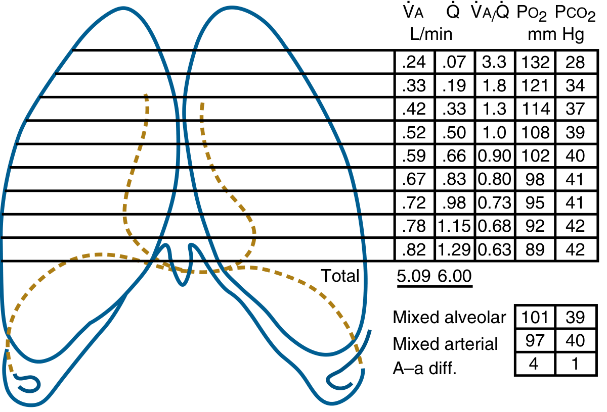
| Region | V̇A (L/min) | Q̇ (L/min) | V/Q | PO₂ (mmHg) | PCO₂ (mmHg) |
|---|---|---|---|---|---|
| Apex | 0.24 | 0.07 | 3.3 | 132 | 28 |
| ↓ | 0.33 | 0.19 | 1.8 | 121 | 34 |
| ↓ | 0.42 | 0.33 | 1.3 | 114 | 37 |
| ↓ | 0.52 | 0.50 | 1.0 | 108 | 39 |
| ↓ | 0.59 | 0.66 | 0.90 | 102 | 40 |
| ↓ | 0.67 | 0.83 | 0.80 | 98 | 41 |
| ↓ | 0.72 | 0.98 | 0.73 | 95 | 41 |
| ↓ | 0.78 | 1.15 | 0.68 | 92 | 42 |
| Base | 0.82 | 1.29 | 0.63 | 89 | 42 |
| Total | 5.09 | 6.00 | ~0.85 | - | - |
- Apex: V/Q = 3.3 (high - relatively over-ventilated, under-perfused)
- Base: V/Q = 0.63 (low - relatively under-ventilated, over-perfused)
- Overall mean: ~0.8
Why Does This Happen? (Mechanism)
Gravity Drives Both Differences
- In the upright lung, intrapleural pressure is less negative at the base than the apex (gravity pulls the lung down, compressing the base).
- Basal alveoli start each breath at a smaller resting volume and are therefore on a steeper (more compliant) part of the pressure-volume curve.
- They expand more per unit pressure change - so they receive more ventilation.
- Apical alveoli start inspiration already more expanded (more negative intrapleural pressure) and are on the flatter portion of the compliance curve - so less additional volume enters per breath.
- Pulmonary circulation is a low-pressure system (mean ~15 mmHg), so gravity has a proportionally larger effect than in systemic circulation.
- Blood is pulled down by gravity. Pressure increases ~1 cmH₂O per cm of lung height.
- At the apex, arterial pressure may barely exceed alveolar pressure - capillaries are narrowed or barely open.
- At the base, both arterial and venous pressures exceed alveolar pressure - capillaries are fully open and distended.
- Ganong's Review of Medical Physiology, p. 632; Murray & Nadel's, p. 240
West Zones of Pulmonary Perfusion
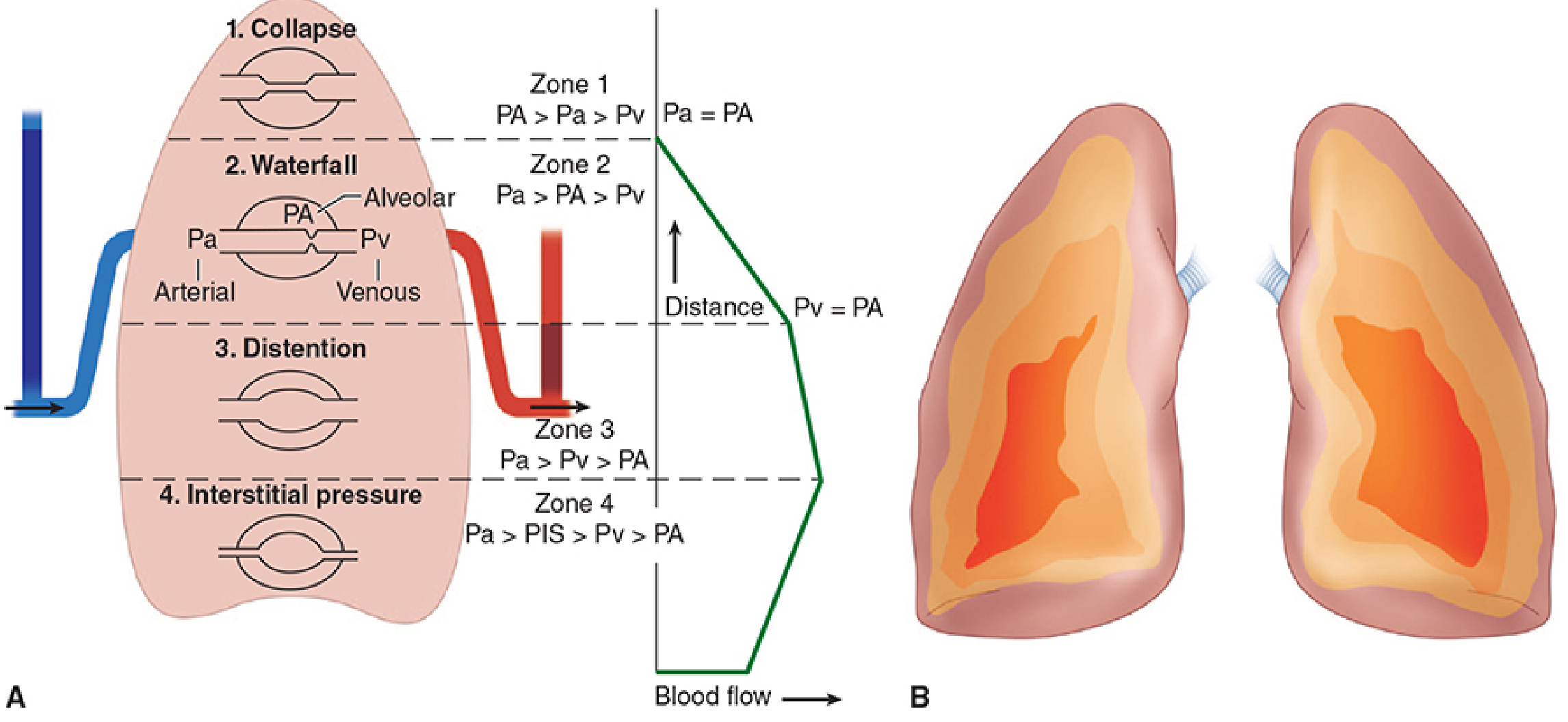
| Zone | Location | Pressure Relationship | Blood Flow | V/Q Effect |
|---|---|---|---|---|
| Zone 1 | Apex | PA > Pa > Pv | None (alveolar dead space) | V/Q = ∞ |
| Zone 2 | Middle upper | Pa > PA > Pv | Depends on Pa - PA (waterfall effect) | V/Q intermediate-high |
| Zone 3 | Middle-lower | Pa > Pv > PA | Full, independent of PA | V/Q intermediate-low |
| Zone 4 | Dependent base | Pa > PIS > Pv > PA | Reduced (interstitial compression) | V/Q slightly rises again |
- Morgan & Mikhail's Clinical Anesthesiology, p. 943-944
Consequences on Gas Exchange
| Region | V/Q | PO₂ | PCO₂ | Functional implication |
|---|---|---|---|---|
| Apex | High (3.3) | High (132 mmHg) | Low (28 mmHg) | Dead-space like; wastes ventilation; good TB site (high O₂) |
| Base | Low (0.63) | Low (89 mmHg) | High (42 mmHg) | Shunt-like; most blood flow but lower O₂ content |
- The net arterial PO₂ is determined largely by the base (which has most of the blood flow but lowest PO₂).
- The overall V/Q inequality causes arterial PO₂ to fall ~4 mmHg below what a perfectly homogeneous lung would achieve.
- A-a gradient exists partly because of this normal apex-to-base V/Q inequality.
"The base of the lung has most of the blood flow, but the PO₂ of the end-capillary blood is lowest there. As a result, the effluent pulmonary venous blood is loaded with moderately oxygenated blood from the base."
- Murray & Nadel's Textbook of Respiratory Medicine, p. 241
Note on Gravity Dependence
- Ganong's Review of Medical Physiology, p. 632
What causes V/Q inequality in ARDS or pneumonia 2,3 dpg
Are these two separate topics you want covered?
Topic 1: V/Q Inequality in ARDS and Pneumonia
The Core Mechanism
ARDS
- Capillary leak - protein-rich fluid floods alveoli (alveolar edema)
- Surfactant washout and inactivation - alveoli collapse (atelectasis)
- Alveoli are flooded or collapsed - no ventilation occurs
- Pulmonary blood flow continues to these non-ventilated units → V/Q = 0 (true shunt)
"Continued perfusion of alveoli that lack ventilation because of alveolar edema results in ventilation-perfusion ratios of zero, thereby defining physiologic shunt. Furthermore, the effects of this type of shunt are exacerbated by shunt-like contributions from alveoli with very low ventilation-perfusion ratios."
- Fishman's Pulmonary Diseases and Disorders, p. 2495
MIGET Studies (Multiple Inert Gas Elimination Technique) in ARDS:
- Only 52% of cardiac output flows to areas with normal V/Q
- Large shunt fraction + very low V/Q areas account for profound hypoxemia
- No diffusion limitation was found - all hypoxemia is V/Q-based
- Hypoxemia is resistant to supplemental O₂ (hallmark of shunt - you cannot ventilate collapsed/flooded alveoli)
Late ARDS (Proliferative/Fibroproliferative Phase):
- Dead space increases (fibrosis obliterates capillaries in some regions)
- Pulmonary hypertension develops
- High V/Q areas appear alongside existing low V/Q/shunt areas
Treatment Targets V/Q Directly:
- PEEP - opens atelectatic alveoli, converting shunt units to ventilated units → reduces shunt fraction
- Prone positioning - redistributes perfusion from collapsed dorsal segments to ventral ones, reducing shunt
- Inhaled vasodilators (e.g., NO) - selectively vasodilate ventilated regions → redirect blood away from shunt units
Pneumonia
- Consolidated lung (pus, fluid, inflammatory exudate filling alveoli) → no ventilation in affected region
- Surrounding, partially involved alveoli have reduced but not zero ventilation → low V/Q regions
- Blood flow to these areas continues → increased shunt + low V/Q units
- Hypoxic pulmonary vasoconstriction (HPV) is partially protective - it diverts blood away from hypoxic (consolidated) units. In pneumonia, inflammatory mediators partly blunt HPV, worsening shunt.
MIGET Data in Pneumonia (Murray & Nadel):
| Severity | Shunt fraction | Low V/Q flow |
|---|---|---|
| Mild-moderate (PaO₂ ~74 mmHg, spontaneous breathing) | 7.5 ± 1.8% | 4.2 ± 1.0% |
| Severe (mechanically ventilated, FiO₂ 0.46) | 21.9 ± 4.5% | 10.9 ± 4.6% |
- As consolidation worsens, shunt fraction rises progressively
- Response to supplemental O₂ depends on degree of shunt (mild shunt responds; large shunt does not)
Comparison: ARDS vs Pneumonia vs PE
| Disease | Primary V/Q Defect | O₂ Response | Mechanism |
|---|---|---|---|
| ARDS | Large true shunt | Poor | Diffuse flooding + atelectasis |
| Pneumonia | Shunt + low V/Q | Partial | Lobar/segmental consolidation |
| Pulmonary embolism | Dead space (high V/Q) | Good (for V/Q mismatch) | No perfusion to ventilated regions |
| Emphysema/asthma | Low V/Q (not shunt) | Good | Airway obstruction/airflow limitation |
Topic 2: 2,3-DPG (2,3-Diphosphoglycerate / 2,3-BPG)
What Is 2,3-DPG?
Mechanism of Action
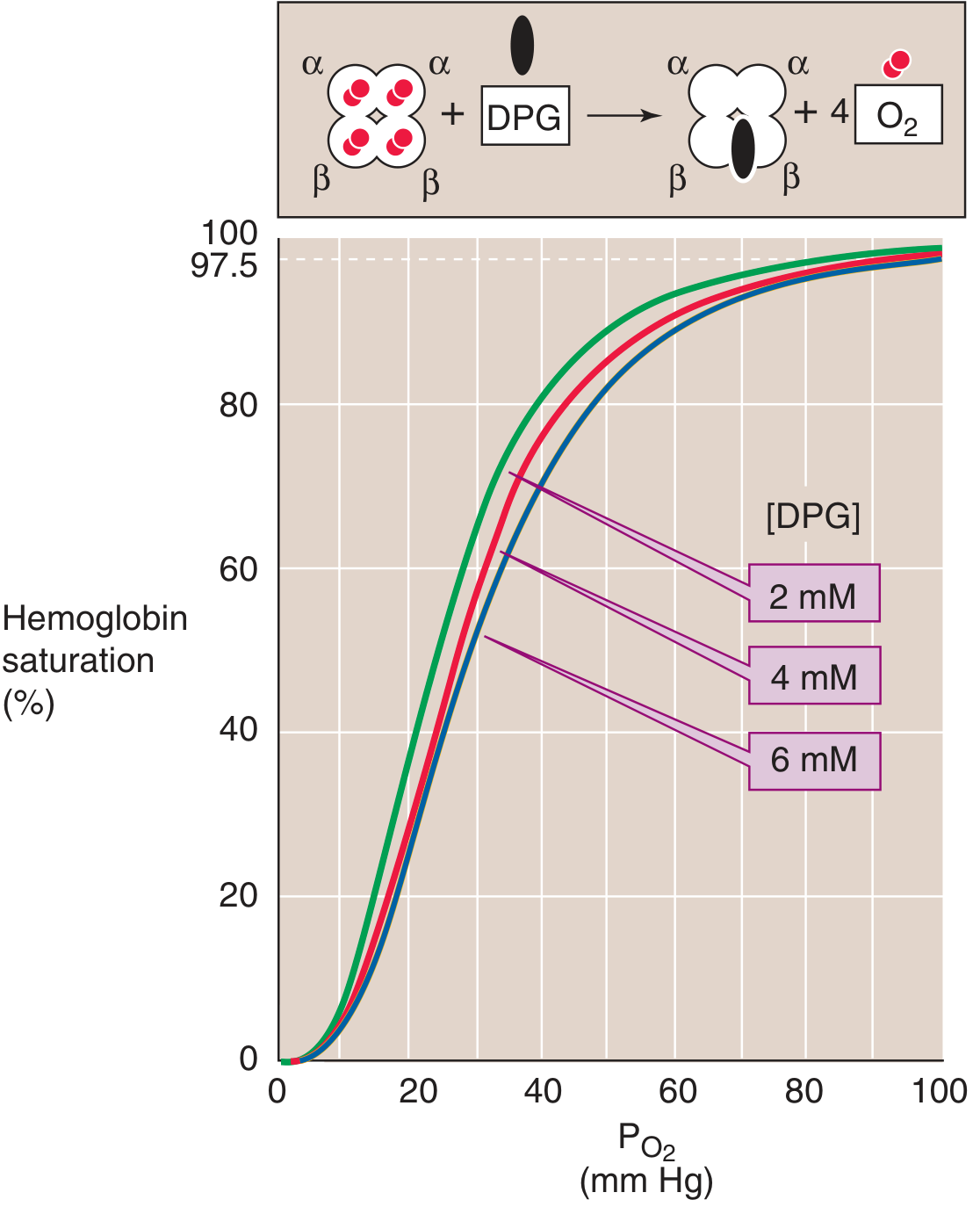
- 2,3-DPG has ~3.5 negative charges at physiological pH
- It binds to a central pocket formed by the two β-globin chains of deoxyhemoglobin, held by 8 positively charged amino acid residues
- This stabilizes the T (deoxy, low-affinity) state of hemoglobin
- O₂ binding changes the shape of this pocket, excluding 2,3-DPG → deoxy-Hb has 100× higher affinity for 2,3-DPG than oxy-Hb
- 2,3-DPG causes a right shift of the O₂-Hb dissociation curve
- P₅₀ (the PO₂ at which Hb is 50% saturated) increases
- At the tissue level (low PO₂), this markedly increases O₂ release
- At the alveolar level (high PO₂, flat part of curve), the reduction in O₂ loading is minimal
When Does 2,3-DPG Increase?
| Condition | Stimulus | Effect |
|---|---|---|
| Hypoxia (any cause) | Low RBC PO₂ stimulates glycolysis | ↑ 2,3-DPG → right shift → more O₂ delivery to tissues |
| Chronic anemia | Tissue hypoxia | ↑ 2,3-DPG → compensatory O₂ unloading |
| High altitude | Hypoxemia + respiratory alkalosis | ↑ 2,3-DPG (counteracts alkalosis-induced left shift) |
| Alkalosis | Directly stimulates glycolysis | ↑ 2,3-DPG |
"Chronic hypoxia, anemia, and acclimation to high altitude are all associated with an increase in 2,3-DPG levels and thus lower the O₂ affinity of Hb."
- Medical Physiology, p. 958
When Does 2,3-DPG Decrease?
| Condition | Effect |
|---|---|
| Stored blood (bank blood) | 2,3-DPG falls within hours of storage → left shift → poor O₂ delivery on transfusion (restored within 24h post-transfusion) |
| Acidosis | Inhibits glycolysis → ↓ 2,3-DPG (partially counteracts direct right shift of acidosis on Hb) |
| Fetal hemoglobin (HbF) | γ-chains bind 2,3-DPG less avidly than β-chains → effectively lower 2,3-DPG effect → left shift → HbF extracts O₂ from maternal HbA across the placenta |
Clinical Significance: The Two-Edged Sword
"Reducing the [O₂] affinity is a two-edged sword. At the relatively high PO₂ in alveoli, where the Hb-O₂ dissociation curve is fairly flat, this decrease in O₂ affinity reduces O₂ uptake - but only slightly. At the low PO₂ in systemic tissues, where the Hb-O₂ dissociation curve is steep, this decrease in O₂ affinity markedly increases the O₂ release. The net effect is enhanced O₂ unloading to metabolizing tissues."
- Medical Physiology, p. 958
Right and left shift in respiration.
Right and Left Shifts of the O₂-Haemoglobin Dissociation Curve
The Curve and P₅₀
- P₅₀ increases → right shift (lower affinity, more O₂ released)
- P₅₀ decreases → left shift (higher affinity, less O₂ released)
The Shifts - Classic Diagram
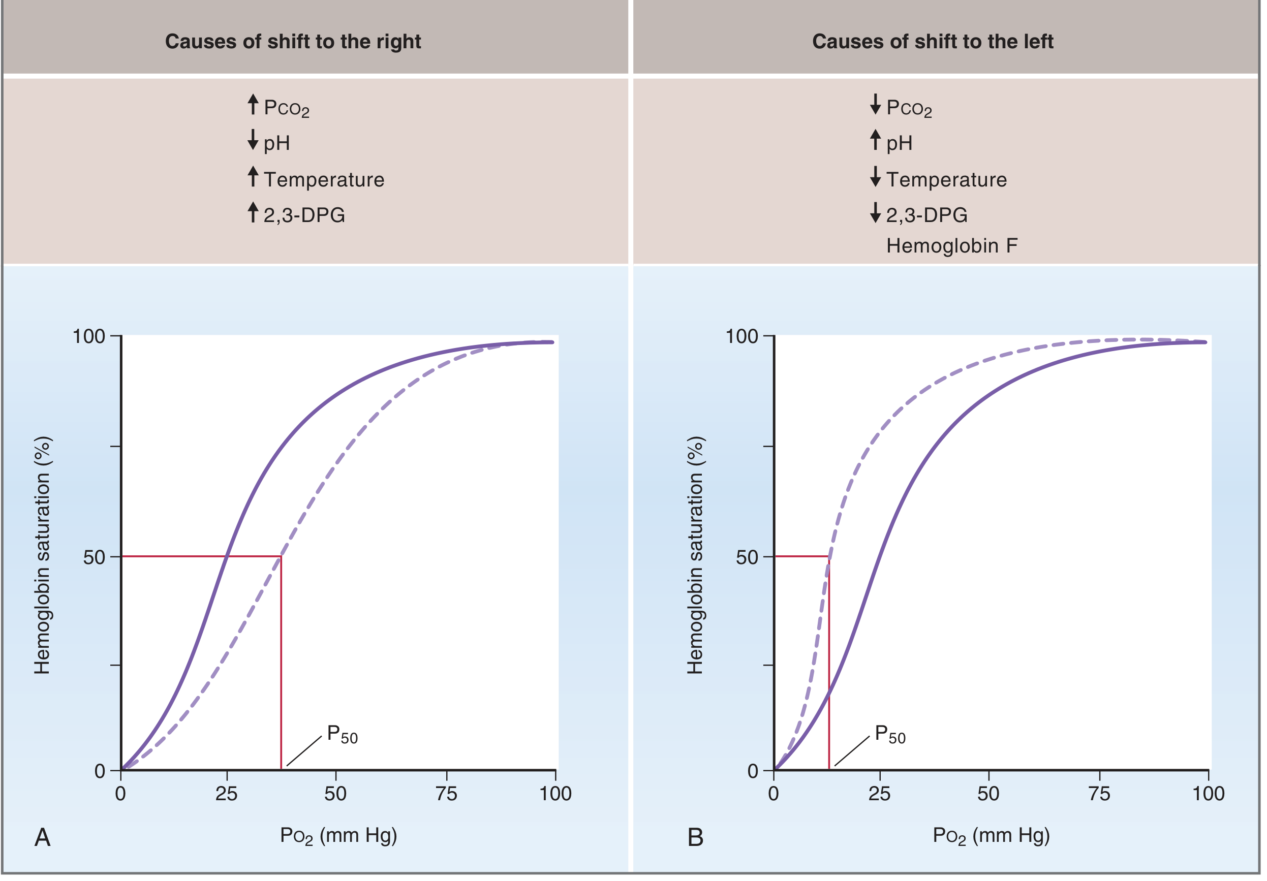
RIGHT SHIFT
| Cause | Mechanism | Where It Occurs |
|---|---|---|
| ↑ PCO₂ | CO₂ binds Hb at non-O₂ sites, allosterically reduces O₂ affinity (Bohr effect) | Tissues (CO₂ produced by metabolism) |
| ↓ pH (acidosis) | H⁺ binds histidine residues on Hb, stabilizes deoxy (T) state (Bohr effect) | Tissues (H⁺ generated from CO₂ + lactic acid) |
| ↑ Temperature | Heat destabilizes Hb-O₂ bond | Exercising muscle, fever |
| ↑ 2,3-DPG | Binds β-chains of deoxyHb, stabilizes T-state, reduces O₂ affinity | Hypoxia, anemia, high altitude |
LEFT SHIFT
| Cause | Mechanism | Clinical Context |
|---|---|---|
| ↓ PCO₂ | Opposite of Bohr effect | Hyperventilation, alkalosis, pulmonary capillaries |
| ↑ pH (alkalosis) | Fewer H⁺ to stabilize T-state | Respiratory alkalosis |
| ↓ Temperature | Stabilizes Hb-O₂ bond | Hypothermia, stored blood |
| ↓ 2,3-DPG | Less stabilization of T-state | Stored bank blood (depleted within hours), hypothyroidism |
| Fetal Haemoglobin (HbF) | γ-chains bind 2,3-DPG less avidly than β-chains → effectively acts as low-2,3-DPG state → left shift | Fetus (necessary to extract O₂ from maternal HbA across placenta) |
| Methaemoglobin | Fe³⁺ cannot bind O₂; remaining Fe²⁺ sites have increased affinity | Nitrite/dapsone toxicity |
The Bohr Effect (Most Important Right-Shift Mechanism)
"Both hydrogen ions and carbon dioxide bind at sites on the hemoglobin molecule that differ from the oxygen-binding sites. There are many more sites that bind hydrogen ions than bind CO₂ molecules. As the tertiary and quaternary configurations of the hemoglobin molecule change with release of oxygen, 16 of these hydrogen-binding sites change their physiologic buffering capacities."
- Fishman's Pulmonary Diseases and Disorders, p. 2374
- Cells produce CO₂ → CO₂ enters RBC → carbonic anhydrase converts to H⁺ + HCO₃⁻
- ↑ H⁺ + ↑ PCO₂ → right shift → Hb releases O₂ to the very tissue that needs it
- CO₂ expelled → ↓ PCO₂ → ↑ pH → left shift → Hb loads O₂ from alveolar air
Carbon Monoxide - A Special Case
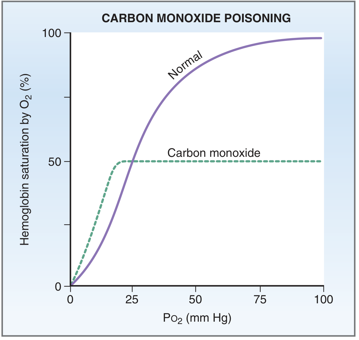
- CO binds heme with 250× greater affinity than O₂ (Costanzo) / 200× greater (Boron & Boulpaep)
- Blocks O₂ binding sites → functional anaemia (reduced O₂ capacity)
- The remaining unbound heme sites have increased O₂ affinity → left shift → O₂ that IS carried is not released to tissues
- Together these effects are catastrophic for O₂ delivery
Summary Table
| Factor | Direction of Shift | P₅₀ | Hb-O₂ Affinity | O₂ Released to Tissues |
|---|---|---|---|---|
| ↑ PCO₂ | Right | ↑ | ↓ | ↑ |
| ↓ pH / acidosis | Right | ↑ | ↓ | ↑ |
| ↑ Temperature | Right | ↑ | ↓ | ↑ |
| ↑ 2,3-DPG | Right | ↑ | ↓ | ↑ |
| ↓ PCO₂ | Left | ↓ | ↑ | ↓ |
| ↑ pH / alkalosis | Left | ↓ | ↑ | ↓ |
| ↓ Temperature | Left | ↓ | ↑ | ↓ |
| ↓ 2,3-DPG | Left | ↓ | ↑ | ↓ |
| HbF | Left | ↓ | ↑ | ↓ (fetal adaptation) |
| CO poisoning | Left + ↓ capacity | ↓ | ↑ (catastrophic) | ↓↓ |
Mnemonic: CADET (Right Shift)
Right and left shift. In lungs.
Right and Left Shifts In the Lungs
The Key Point
What Happens in the Pulmonary Capillaries
| Parameter | Change | Effect on Curve |
|---|---|---|
| PCO₂ | Falls (40 → ~40 mmHg, exhaled) | Left shift (↑ Hb-O₂ affinity) |
| pH | Rises (as H⁺ is released from HbH → Hb + H⁺ → combines with HCO₃⁻ → CO₂ exhaled) | Left shift (alkalosis → ↑ affinity) |
| Temperature | Slightly lower than exercising tissues | Mild left shift |
"All of the reactions previously described occur in reverse in the lungs. H⁺ is released from its buffering sites on deoxyhemoglobin, HCO₃⁻ enters the red blood cells in exchange for Cl⁻, H⁺ and HCO₃⁻ combine to form H₂CO₃, and H₂CO₃ dissociates into CO₂ and H₂O. The regenerated CO₂ and H₂O are expired by the lungs."
- Costanzo Physiology, 7th Ed., p. 231
The Bohr Effect: Tissues vs Lungs
| Location | PCO₂ | pH | Curve Shift | Purpose |
|---|---|---|---|---|
| Tissues | ↑ (CO₂ produced) | ↓ (acidosis) | RIGHT | O₂ unloading |
| Lungs | ↓ (CO₂ exhaled) | ↑ (alkalosis) | LEFT | O₂ loading |
The Haldane Effect - The Complementary Mechanism
"Binding of O₂ with hemoglobin in the lungs causes hemoglobin to become a stronger acid. This displaces CO₂ from the blood. The Haldane effect is quantitatively far more important in promoting CO₂ transport than the Bohr effect is in promoting O₂ transport."
- Guyton & Hall Textbook of Medical Physiology
- OxyHb is a stronger acid than deoxyHb
- As O₂ binds Hb in the lungs → Hb becomes more acidic → releases H⁺
- H⁺ + HCO₃⁻ → H₂CO₃ → CO₂ + H₂O → CO₂ exhaled
- OxyHb also binds CO₂ (carbamino) less avidly → more CO₂ released
- O₂ loading causes CO₂ unloading (Haldane)
- CO₂ unloading causes O₂ loading (reverse Bohr)
When a Right Shift Is Problematic in the Lungs
"The right shift is advantageous in the tissues because it is associated with increased P₅₀, decreased affinity, and increased unloading of O₂. However, the right shift is disadvantageous in the lungs because it becomes more difficult to load the pulmonary capillary blood with O₂."
- Costanzo Physiology, 7th Ed., p. 232
Summary
| Context | Shift | P₅₀ | Net Effect |
|---|---|---|---|
| Normal lungs (reverse Bohr) | LEFT | ↓ | Haemoglobin loads O₂ efficiently from alveolar air |
| Exercising tissues (Bohr effect) | RIGHT | ↑ | Haemoglobin releases O₂ to tissues |
| High altitude / ↑ 2,3-DPG | Right (even in lungs) | ↑ | Impairs O₂ loading slightly - disadvantageous at the lung |
| Hyperventilation / alkalosis | LEFT (in lungs) | ↓ | Enhanced O₂ loading - but may cause cerebral vasoconstriction |
Herriring breurer reflex and j receptors
Hering-Breuer Reflex and J Receptors
Part 1: The Hering-Breuer Reflex
Discovery and Basis
Receptor: Slowly Adapting Pulmonary Stretch Receptors (SAPSRs)
- Located in the smooth muscle of bronchi and bronchioles throughout the lung
- Myelinated afferent fibres
- Respond to stretch/lung inflation with sustained firing (slow adaptation - firing persists as long as lung remains inflated)
- Afferents travel to the dorsal respiratory group of neurons in the medulla via the vagus nerve (CN X)
The Hering-Breuer Inflation Reflex
- Lung inflates during inspiration → airways/bronchi stretch
- SAPSRs fire → signals travel via vagus to DRG (dorsal respiratory group) of medulla
- Inspiratory ramp is "switched off" → inspiration terminates
- Expiration begins
"When the lungs become overinflated, the stretch receptors activate a feedback response that 'switches off' the inspiratory ramp and stops further inspiration."
- Guyton & Hall Textbook of Medical Physiology
- ↑ Duration of expiration (longer expiratory pause)
- ↑ Respiratory frequency (by shortening inspiration time)
- Maintains constant alveolar ventilation
The Hering-Breuer Deflation Reflex
- Marked deflation of the lung → decreased SAPSR firing → decrease in duration of expiration → promotes next inspiration
- Protective against over-deflation
Threshold - Adults vs Infants
| Population | Threshold | Role |
|---|---|---|
| Human infants | Normal tidal volumes | Important in controlling tidal volume during eupnea |
| Human adults | Only when tidal volume > ~1.5 L (3× normal) | Mainly a protective mechanism against dangerous over-inflation, NOT a major control of normal quiet breathing |
"In humans, the Hering-Breuer reflex probably is not activated until the tidal volume increases to more than three times normal (~1.5 L/breath). Therefore, this reflex appears to be mainly a protective mechanism for preventing excess lung inflation rather than an important factor in normal control of ventilation."
- Guyton & Hall, p. 533
Proof - Vagotomy
Summary of Hering-Breuer
| Feature | Detail |
|---|---|
| Receptor | Slowly adapting pulmonary stretch receptors (SAPSRs) |
| Location | Smooth muscle of bronchi/bronchioles |
| Fibre | Myelinated, vagus nerve |
| Stimulus | Lung over-inflation/stretch |
| Reflex arc | Vagus → DRG medulla → stops inspiratory ramp |
| Effect | Terminates inspiration; ↑ expiratory duration; ↑ RR |
| Clinical importance (adults) | Protective, not routine breathing regulation |
| Abolished by | Vagotomy |
Part 2: J Receptors (Juxtacapillary Receptors)
What Are They?
"Alveolar C-fiber receptors become active very soon (1-2 seconds) after injection of certain chemicals into the blood of the right atrium of the heart, which suggests that the receptors may be very near pulmonary capillaries. Hence the name juxtacapillary or J receptors."
- Medical Physiology (Boron & Boulpaep), p. 1071
Location
- In the alveolar walls and interstitium
- Very close to pulmonary capillaries
- Afferent fibres are unmyelinated C fibres → conduct slowly (even at 4°C, unlike myelinated fibres which are blocked by cooling)
- Travel to medulla via vagus nerve
Stimuli (What Activates J Receptors?)
| Stimulus Type | Examples |
|---|---|
| Chemical - inflammatory mediators | Histamine, serotonin, bradykinin, prostaglandins |
| Chemical - exogenous | Capsaicin (IV), phenyl diguanide |
| Mechanical | Lung congestion (pulmonary oedema), vascular engorgement |
| Hyperinflation | Extreme lung distension |
| Pulmonary embolism | Via chemical mediator release |
Reflex Response - "Pulmonary Chemoreflex"
- Apnea (brief cessation of breathing) - first response
- Followed by rapid, shallow breathing (tachypnea)
- Bradycardia
- Hypotension
- Bronchoconstriction
- Increased mucus secretion
"The reflex response that is produced is apnea followed by rapid breathing, bradycardia, and hypotension (pulmonary chemoreflex)."
- Ganong's Review of Medical Physiology
Physiological and Clinical Significance
| Condition | Role of J Receptors |
|---|---|
| Pulmonary oedema | Activated by interstitial fluid congestion → rapid shallow breathing + dyspnoea sensation |
| Pulmonary embolism | Activated by released mediators (serotonin, bradykinin) → reflex apnea/tachypnea |
| Pneumonia/inflammation | Activated by histamine, prostaglandins |
| Exercise | May contribute to dyspnoea sensation |
| Heart failure | Pulmonary capillary congestion activates J receptors → orthopnoea, dyspnoea |
| Normal physiology | Role uncertain; likely a pathological defense mechanism |
Comparison: Three Types of Pulmonary Receptors
| Feature | Slowly Adapting PSRs (Hering-Breuer) | Rapidly Adapting (Irritant) Receptors | J Receptors / C-fibres |
|---|---|---|---|
| Fibre type | Myelinated | Myelinated | Unmyelinated (C fibres) |
| Location | Airway smooth muscle (bronchi/bronchioles) | Airway epithelium (trachea to bronchioles) | Alveolar wall, near capillaries |
| Adapts? | Slowly (sustained) | Rapidly (fast) | Slow |
| Stimulus | Inflation/stretch | Chemicals, irritants, sudden inflation | Congestion, inflammation, chemicals |
| Reflex | Hering-Breuer (stop inspiration) | Cough, bronchoconstriction, mucus secretion | Apnea → rapid breathing, bradycardia, hypotension |
| Blocked by | Cooling (~8-10°C) | Cooling | NOT blocked by cooling |
Bezold jarisch reflex
Bezold-Jarisch reflex mechanism clinical significance cardiology
Bezold-Jarisch Reflex
History and Definition
Bradycardia + Hypotension + Apnea (followed by rapid shallow breathing)
Reflex Arc
Receptor
- Chemoreceptors and mechanoreceptors in the left ventricular wall (inferoposterior region preferentially)
- Also in: atria, great veins, pulmonary artery, juxtacapillary (J receptor) region of alveoli
- These are collectively called cardiopulmonary chemosensitive receptors
Afferent Limb
- Unmyelinated vagal C fibres → travel to the nucleus tractus solitarius (NTS) in the medulla
Efferent Limb
- Increased parasympathetic (vagal) tone to the heart → bradycardia
- Withdrawal of sympathetic tone to resistance vessels → vasodilation and hypotension
- Coronary artery dilation
Stimuli That Activate the Reflex
| Type | Examples |
|---|---|
| Veratrum alkaloids (original stimulus) | Veratridine, veratrine |
| Serotonin (5-HT₃) | Released during ischaemia/reperfusion |
| Capsaicin | Substance P pathway |
| Phenylbiguanide / phenylguanide | Experimental |
| Histamine | Allergic/inflammatory states |
| Nicotine | |
| Snake and insect venoms | |
| Contrast media | During coronary angiography |
| Thrombolytic agents | During reperfusion |
| Oxygen free radicals | Myocardial ischaemia/reperfusion |
| Ventricular under-filling | Upright posture, hypovolaemia |
Classic Triad of Responses
| Response | Mechanism |
|---|---|
| Bradycardia | Increased parasympathetic tone → slowing of SA node |
| Hypotension | Vasodilation (loss of sympathetic tone) + decreased cardiac output |
| Apnea → then rapid shallow breathing | C-fibre activation in cardiopulmonary region |
| (Also) Coronary artery dilation | Possibly cardioprotective |
"The Bezold-Jarisch reflex responds to noxious ventricular stimuli sensed by chemoreceptors and mechanoreceptors within the left ventricular wall by inducing the triad of hypotension, bradycardia, and coronary artery dilatation."
- Miller's Anesthesia, 10th Ed.
Clinical Significance
1. Vasovagal Syncope (Most Common Clinical Context)
- Prolonged upright posture / dehydration → blood pools in lower limbs → reduced ventricular filling
- Under-filled but vigorously contracting left ventricle → activates ventricular mechanoreceptors
- Paradoxical reflex: instead of tachycardia, ventricular receptors trigger bradycardia + vasodilation
- Massive parasympathetic discharge + sympathetic withdrawal → profound fall in BP and HR
- Cerebral perfusion drops → loss of consciousness
"Pressure receptors in the wall of the left ventricle respond by sending signals that trigger paradoxical bradycardia and decreased contractility, resulting in sudden marked hypotension."
- Ganong's Review of Medical Physiology
2. Inferior/Posterior Myocardial Infarction
- Occlusion of the right coronary artery (which supplies the inferoposterior LV wall) → activation of ventricular receptors
- Produces bradycardia + hypotension seen in inferior MI (the classic "vagal MI")
- Thought to be cardioprotective - coronary dilation may improve collateral perfusion
3. Coronary Angiography
- Injection of radiocontrast into coronary arteries → activates ventricular receptors → bradycardia + hypotension during the procedure
4. Thrombolysis / Reperfusion
- Release of oxygen radicals and serotonin during reperfusion injury activates the reflex
- Contributes to reperfusion hypotension and bradycardia
5. Spinal Anaesthesia
- Sympathetic blockade → venous pooling → reduced ventricular filling → Bezold-Jarisch activation
- Important cause of severe bradycardia/asystole during spinal anaesthesia (estimated 2-3 per 10,000 anaesthetic procedures)
Relationship to Vasovagal Syncope Mechanism
Emotional stress / standing / blood loss
↓
Reduced ventricular filling
↓
Vigorous contraction of under-filled LV
↓
Activation of LV inferoposterior mechanoreceptors
↓
C-fibre vagal afferents → NTS → medulla
↓
↑ Parasympathetic + ↓ Sympathetic output
↓
Bradycardia + Vasodilation + ↓ Cardiac output
↓
↓ Mean arterial pressure → Cerebral hypoperfusion
↓
Syncope (loss of consciousness in ~10 seconds)
Modulation
- ANP/BNP (natriuretic peptides): Stimulate receptors that can modulate the reflex
- Cardiac hypertrophy / atrial fibrillation: Reflex may be attenuated
- Atropine: Blocks the bradycardia component (but does not reliably prevent vasovagal syncope since the vasodilatory component persists)
- β-blockers / disopyramide: Used to reduce syncope episodes
- IV fluids / salt loading / fludrocortisone: Prevent by increasing ventricular filling
Summary Table
| Feature | Detail |
|---|---|
| Receptor | LV chemoreceptors/mechanoreceptors (inferoposterior wall) |
| Afferent | Unmyelinated C fibres → vagus nerve → NTS |
| Efferent | ↑ Parasympathetic + ↓ Sympathetic |
| Triad | Bradycardia + Hypotension + Apnea → tachypnea |
| Also | Coronary vasodilation |
| Key clinical scenario | Vasovagal syncope, inferior MI, spinal anaesthesia, coronary angiography |
| Protective role | Coronary dilation in inferoposterior ischaemia |
Homometeric and heterometeric regulation
Heterometric and Homometric Regulation of the Heart
1. Heterometric Regulation (Length-Dependent)
Definition
"When cardiac output is regulated by changes in cardiac muscle fiber length, this is referred to as heterometric regulation."
- Ganong's Review of Medical Physiology
Basis: Frank-Starling Law
- The heart muscle behaves like skeletal muscle: the more it is stretched before contraction (preload), the greater the force of contraction
- Starling stated: "The energy of contraction is proportional to the initial length of the cardiac muscle fibre"
- The key variable is end-diastolic volume (EDV) → determines fibre length before systole
Molecular Mechanism
- ↑ Preload → ↑ EDV → ↑ sarcomere length at end-diastole
- Greater sarcomere stretch → ↑ myofilament Ca²⁺ sensitivity (troponin C binds Ca²⁺ more avidly)
- Narrowing of interfilament spacing at longer lengths may enhance cross-bridge formation
- More cross-bridges formed → ↑ force generated per beat → ↑ stroke volume
"There is also a substantial increase in myofilament Ca²⁺ sensitivity with an increase in sarcomere length... the myocyte is at constant volume, so as the cell shortens it must thicken, and conversely, when stretched, the cell becomes thinner and filament spacing becomes narrower."
- Braunwald's Heart Disease
Examples in Physiology
| Situation | ↑ Preload → ↑ EDV → |
|---|---|
| Increased venous return (exercise, fluid loading) | ↑ Stroke volume (self-compensating) |
| Sudden volume load | ↑ Force to eject the extra volume |
| Aortic regurgitation | Dilated LV → ↑ EDV → ↑ contractile force |
| Right heart receiving more blood from leg veins (standing → lying) | ↑ RV output automatically |
Frank-Starling Curve
- X-axis: End-diastolic volume (or filling pressure/preload)
- Y-axis: Stroke volume (or cardiac output)
- Rising limb: Optimal filament overlap and Ca²⁺ sensitivity
- Descending limb: Excessive stretch, reduced overlap (seen in severe heart failure)
2. Homometric Regulation (Length-Independent)
Definition
"Regulation due to changes in contractility independent of length is sometimes called homometric regulation."
- Ganong's Review of Medical Physiology
"Conditions in which contraction is strengthened independent of sarcomere length (e.g., typically by increased Ca²⁺ transient amplitude) are referred to as positive inotropic states or enhanced contractility."
- Braunwald's Heart Disease
Mechanism
- Changes in the amplitude of the intracellular Ca²⁺ transient (how much Ca²⁺ is released per beat from the SR)
- More Ca²⁺ per beat → more troponin C binding → more cross-bridges → more force - at the same fibre length
- Measured as a shift of the Frank-Starling curve upward (better performance at the same preload)
Examples of Homometric Regulation
A. Sympathetic / Catecholamine Stimulation (most important)
- Noradrenaline/adrenaline → β₁ receptors → ↑ cAMP → PKA activation
- PKA phosphorylates L-type Ca²⁺ channels → ↑ Ca²⁺ influx per action potential
- PKA phosphorylates phospholamban → ↑ SR Ca²⁺ reuptake and storage → larger Ca²⁺ store
- Net: ↑ Ca²⁺ transient amplitude → ↑ force at same length = positive inotropy
- Chronotropic action (↑ HR) is a separate effect
B. Bowditch Effect / Treppe Phenomenon (Force-Frequency Relationship)
"An increased HR progressively enhances the force of ventricular muscle contraction... the Bowditch staircase phenomenon (treppe = German for 'steps')"
- Braunwald's Heart Disease
- ↑ Heart rate → more action potentials per minute → more Ca²⁺ entry per unit time, less time to extrude Ca²⁺ → ↑ intracellular [Ca²⁺] → ↑ force per beat
- Peaks at ~150-180 bpm; declines at very high HR (insufficient diastolic filling time)
- Clinically important during exercise
C. Anrep Effect (Homeometric Adaptation to Afterload)
"When the aortic pressure is elevated abruptly, ejection is limited, tending to increase EDV, which acutely increases force by the Frank-Starling effect. However, in a slower adaptation (seconds to minutes), the inotropic state of the heart increases (larger Ca²⁺ transients). This slow force response is referred to as the Anrep effect."
- Braunwald's Heart Disease
- Acute ↑ afterload → initially heterometric response (Starling) - brief
- Then slower homometric response: stretch-induced autocrine/paracrine signalling → ↑ Ca²⁺ transients via NOS1/CaMKII pathway
- Allows heart to maintain output against higher pressure without persistently increasing fibre length
- Important for RV adaptation to pulmonary hypertension: RV hypertrophies and increases contractility without enlarging (homeometric/homometric)
D. Pharmacological Inotropes
| Drug | Mechanism | Effect |
|---|---|---|
| Digoxin | Inhibits Na⁺/K⁺-ATPase → ↑ intracellular Na⁺ → ↑ Ca²⁺ via NCX | ↑ Contractility |
| Dobutamine | β₁ agonist | ↑ cAMP → ↑ Ca²⁺ |
| Milrinone | PDE inhibitor → ↑ cAMP | ↑ Ca²⁺ |
| β-blockers | ↓ sympathetic tone | ↓ Contractility (negative inotropy) |
Comparison Summary
| Feature | Heterometric Regulation | Homometric Regulation |
|---|---|---|
| Also called | Frank-Starling mechanism, preload regulation | Inotropic regulation, contractility regulation |
| Depends on | Change in fibre/sarcomere length (preload/EDV) | Change in Ca²⁺ transient - independent of length |
| Frank-Starling curve | Movement along the curve | Shift of the curve (up = positive, down = negative inotropy) |
| Key variable | End-diastolic volume | Intracellular [Ca²⁺] per beat |
| Molecular basis | ↑ Sarcomere length → ↑ myofilament Ca²⁺ sensitivity | ↑ Ca²⁺ release from SR |
| Classic examples | Starling's law, increased venous return | Sympathetic stimulation, Bowditch/treppe, Anrep effect |
| Clinical context | Volume loading, heart failure compensation (initial) | Catecholamines, inotropic drugs, heart rate changes |
| Limits | Cannot compensate indefinitely (descending limb) | Limited by SR Ca²⁺ stores and receptor downregulation |
Clinical Relevance
| Condition | Dominant Regulatory Mechanism |
|---|---|
| Normal resting heart | Both; heterometric maintains baseline output |
| Exercise | Both: ↑ venous return (heterometric) + sympathetic activation (homometric) |
| Heart failure | Initially compensated by heterometric (dilated heart) → eventually fails; drugs target homometric |
| Pulmonary hypertension (RV) | Homeometric (Anrep): RV hypertrophies and increases contractility without dilation |
| Inotropic drug therapy | Pure homometric - improve contractility without increasing preload |
Heart blocks
Heart Blocks (AV Blocks)
Anatomy of AV Conduction
- AV node → Bundle of His → Left and Right bundle branches → Purkinje fibres
Causes of AV Block
| Cause | Examples |
|---|---|
| Ischaemia | Coronary artery disease; inferior MI (RCA) → AV nodal block; anterior MI → infranodal block |
| Inflammation | Rheumatic fever, myocarditis, endocarditis, Lyme disease, Chagas disease |
| Degeneration | Lenègre disease (fibrosis of conduction system), Lev disease (calcification), ageing |
| Drugs | Digoxin, β-blockers, calcium channel blockers, amiodarone |
| Compression | Scar tissue, calcified areas |
| Vagal excess | Carotid sinus syndrome, vasovagal reactions |
| Congenital | Associated with maternal SLE (anti-Ro antibodies) |
| Infiltrative | Sarcoidosis, amyloidosis, haemochromatosis |
Classification
1st Degree AV Block
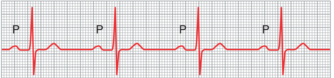
- Every P wave is followed by a QRS (1:1 conduction maintained)
- PR interval typically 0.20-0.35 sec (can reach 0.45 sec)
- QRS usually narrow (normal)
- Rhythm: regular
- Normal variant in up to 2% of healthy young adults
- May be caused by increased vagal tone (athletes)
- Alone: benign, no specific treatment
- Indicates increased susceptibility if caused by drugs/disease
2nd Degree AV Block
Mobitz Type I (Wenckebach)
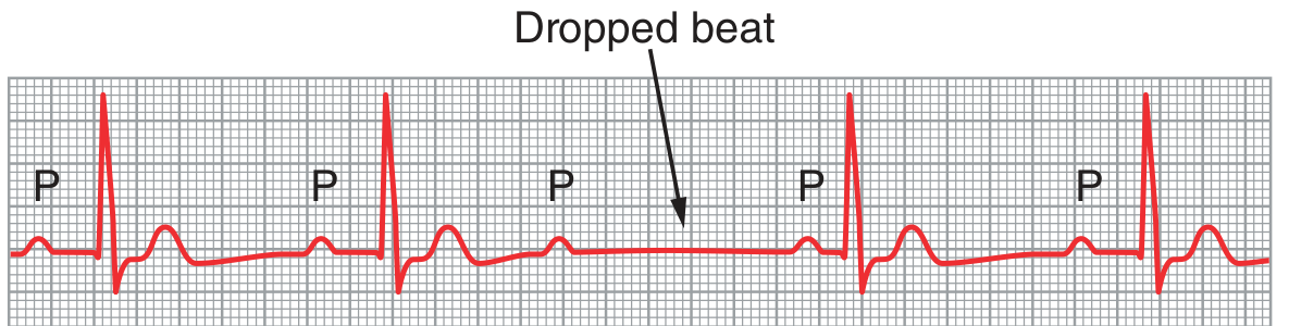
- Progressive PR interval lengthening beat-to-beat
- Until one P wave is not followed by a QRS (dropped beat)
- After the dropped beat, PR interval resets to shortest value → cycle repeats
- QRS: narrow (usually)
- Grouped beating pattern (e.g., 3:2, 4:3, 5:4)
- RP/PR reciprocity: shorter RP → longer PR
Mobitz Type II
- Fixed PR interval (does NOT progressively prolong)
- Sudden, unexpected dropped beats - P waves not followed by QRS
- Conduction ratio fixed: 2:1, 3:1, 3:2, etc.
- QRS often wide (bundle branch block present)
2:1 AV Block - Diagnostic Challenge
- Every alternate P wave is blocked - cannot tell if Mobitz I or II from rhythm alone
- Wide QRS → likely Mobitz II (infranodal)
- Prolonged PR → likely Mobitz I (nodal)
- May require electrophysiology study to determine level
3rd Degree (Complete) AV Block
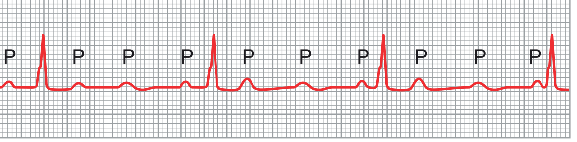
- P waves and QRS complexes are completely independent - no fixed relationship
- Atrial rate: normal (~60-100 bpm)
- Ventricular rate: slow escape rhythm (determined by site of escape pacemaker)
- R-R interval > P-P interval (ventricular rate slower than atrial rate)
| Site of escape pacemaker | Rate | QRS |
|---|---|---|
| AV node / His bundle (above bifurcation) | 45-60 bpm | Narrow (QRS ≤0.12 sec) |
| Bundle branches / Purkinje (infranodal) | 30-45 bpm | Wide (QRS >0.12 sec) |
| Ventricular myocardium | <30 bpm | Very wide, bizarre |
Stokes-Adams Syndrome
- Sudden loss of consciousness (syncope)
- Seizure-like activity (cerebral anoxia)
- Pallor then flushing on recovery
- No warning, immediate loss then rapid recovery
- Managed with permanent pacemaker
Comparison Table: All Degrees
| Feature | 1st Degree | 2nd Degree Mobitz I | 2nd Degree Mobitz II | 3rd Degree |
|---|---|---|---|---|
| PR interval | Prolonged (fixed) | Progressively ↑ | Fixed (normal) | No relationship |
| Dropped beats | None | Yes (periodic) | Yes (sudden) | All P waves blocked |
| QRS | Narrow | Narrow | Often wide | Narrow or wide (escape) |
| Site | AV node | AV node | Infranodal | Any level |
| Ventricular rate | Normal | Slightly reduced | Reduced | Very slow (escape) |
| Risk of progression | Low | Low | High | - |
| Atropine response | - | Helps | Worsens | Minimal effect |
| Treatment | None | Usually none | Pacemaker | Pacemaker |
Bundle Branch Blocks (Related Conduction Defects)
| RBBB | LBBB | |
|---|---|---|
| QRS duration | >0.12 sec | >0.12 sec |
| V1 | rSR' ("M" pattern) | QS or rS |
| V6 | Wide S wave | Broad R, no q |
| Significance | Often benign; RV strain | Usually significant LV disease |
Management Principles
| Block Type | Management |
|---|---|
| 1st degree | Treat cause; avoid offending drugs; no pacing needed |
| Mobitz I | Treat cause; atropine if symptomatic; usually no pacemaker |
| Mobitz II | Pacemaker (high risk of sudden complete block) |
| Complete (3rd degree) | Emergency temporary pacing → permanent pacemaker |
| Acute inferior MI + AV block | Often transient; atropine; temporary pacing if needed |
| Acute anterior MI + AV block | Serious (large infarct); emergency pacing |
Acregaly and gigantism characteristic features
Acromegaly and Gigantism
The Fundamental Distinction
| Gigantism | Acromegaly | |
|---|---|---|
| Age of onset | Before epiphyseal closure (childhood/adolescence) | After epiphyseal closure (adults) |
| Epiphyses | Open → bones grow in length | Fused → bones grow in width/thickness |
| Result | Proportional tall stature (up to 8 feet) | Soft tissue + acral + membranous bone changes |
| GH mediator | IGF-1 (somatomedin C) | Same |
"All body tissues grow rapidly, including the bones. If the condition occurs before adolescence, before the epiphyses of the long bones have fused, height increases so that the person may develop gigantism and become up to 8 feet tall." - Guyton & Hall
Pathophysiology
- Pituitary somatotroph adenoma → uncontrolled GH hypersecretion
- GH → liver → IGF-1 (Insulin-like Growth Factor-1) - the main effector
- IGF-1 promotes growth of bone, soft tissue, and viscera
- Normal feedback (IGF-1 → inhibits GH via somatostatin) is bypassed
- Symptoms are subtle and insidious - average diagnostic delay of 10+ years
- 75% are macroadenomas by time of presentation
- First clue may be "large, enveloping, doughy handshake" or voice changes
- Ectopic GHRH production: bronchial/abdominal carcinoid tumors, small cell lung cancer, medullary thyroid cancer, phaeochromocytoma
- Ectopic GH: islet cell tumors, lymphomas
- MEN1 syndrome (menin mutation) - 10% of tumors secrete GH
Gigantism - Clinical Features
| Feature | Details |
|---|---|
| Stature | Proportional tall stature (>8 feet possible) |
| Growth rate | Abnormally rapid from childhood |
| Body proportions | Proportional (unlike Marfan's which has disproportionate limbs) |
| Hyperglycaemia | Present; beta cells overworked |
| Diabetes mellitus | Develops in ~10% of cases |
| Panhypopituitarism | Eventually - as expanding tumor destroys normal pituitary |
| Prognosis | Death in early adulthood if untreated; panhypopituitarism is the usual cause |
Acromegaly - Characteristic Features (System by System)
Facial and Head Features (Hallmarks)
| Feature | Mechanism |
|---|---|
| Frontal bossing | Enlarged supraorbital ridges (membranous bone growth) |
| Prognathism | Mandibular overgrowth - lower jaw protrudes forward (sometimes up to ½ inch) |
| Malocclusion / splayed teeth | Mandibular enlargement; widened lower incisor spacing |
| Macroglossia | Enlarged tongue |
| Large fleshy nose | May double in size |
| Coarse facial features | Thickened, reddened, wrinkled forehead; exaggerated nasolabial grooves |
| Cutis verticis gyrata | Skin folds on scalp (~30% of patients) |
| Deep/hollow-sounding voice | Enlarged vocal cords and pharyngeal soft tissue |
Hands and Feet (Classic Acral Changes)
- Increased ring, shoe, glove, and hat size (patients often notice this first)
- Fingers broad and spade-shaped - fingertips resemble drumsticks
- Feet require size 14+ shoes
- Hands almost twice normal size
- Increased heel pad thickness (visible on lateral X-ray)
Skin Changes
| Feature | Notes |
|---|---|
| Hyperhidrosis | Very common; sweating at rest; persists after treatment in most |
| Oily skin | GH/IGF-1-driven sebaceous gland hypertrophy |
| Skin tags (acrochordon) | Multiple; strongly associated with colonic polyps |
| Acanthosis nigricans | Insulin resistance marker |
| Hypertrichosis | Excess hair growth |
| Hyperpigmentation | Present in many patients |
| Diffuse skin thickening | Collagen deposition in dermis; measurable on heel pad X-ray |
Musculoskeletal
| Feature | Notes |
|---|---|
| Arthropathy | Large and small joints; cartilage overgrowth, then destruction → OA |
| Kyphosis | Vertebral changes → hunched back |
| Carpal tunnel syndrome | Nerve entrapment by soft tissue/bony overgrowth (25% of patients) |
| Proximal muscle weakness | Myopathy |
| Paresthesias | Peripheral neuropathy |
Cardiovascular (Most Important for Prognosis)
- Left ventricular hypertrophy (biventricular cardiomegaly)
- Cardiomyopathy - systolic and diastolic dysfunction
- Hypertension (common) - volume expansion + direct vascular GH effects
- Arrhythmias
- Heart failure
- Premature coronary artery disease - via hypertension, dyslipidaemia, insulin resistance
- Overall mortality increased threefold, primarily from CV and cerebrovascular disease
- Survival reduced by ~10 years if GH levels uncontrolled
Respiratory
- Obstructive sleep apnoea (OSA) - occurs in >60% of patients
- Soft tissue pharyngeal/laryngeal overgrowth
- Central sleep dysfunction component
- Macroglossia contributes to upper airway obstruction
- Subglottic narrowing possible → important for anaesthetists (difficult intubation)
Metabolic and Endocrine
| Complication | Frequency/Notes |
|---|---|
| Glucose intolerance | Most patients; GH is counter-regulatory (anti-insulin) |
| Diabetes mellitus | ~25% |
| Hyperlipidaemia | Raised triglycerides |
| Hypercalciuria / kidney stones | GH stimulates renal 1α-hydroxylase → ↑ calcitriol → ↑ Ca²⁺ absorption |
| Goitre/thyroid enlargement | ~50% |
| Hyperprolactinaemia | ~25% of tumours co-secrete prolactin; galactorrhea in women |
| Hypogonadism | Tumour mass effect on gonadotrophs; menstrual irregularity, reduced libido |
GI and Oncological
- Colonic polyps - up to 1/3 of patients
- Colon cancer - increased mortality; skin tags are a marker
- Visceromegaly - liver, kidneys, thyroid, salivary glands, spleen all enlarged
Neurological
- Headache - in up to 60% (direct tumour effect, not just mass)
- Bitemporal hemianopia - chiasmal compression by macroadenoma (10% at presentation)
- Cranial nerve palsies - with cavernous sinus invasion
- Peripheral neuropathy
- Hypersomnia
Summary Comparison
| Feature | Gigantism | Acromegaly |
|---|---|---|
| Timing | Pre-pubertal | Post-pubertal |
| Height | ↑↑↑ proportional | Normal or slightly ↑ |
| Hands/feet | Proportionally large | Spade-shaped, acral enlargement |
| Facial changes | Less obvious | Prominent (prognathism, frontal bossing, coarse features) |
| Bone growth | Longitudinal | Periosteal/membranous thickening |
| Joints | Variable | Arthropathy, carpal tunnel |
| Metabolic | Hyperglycaemia, DM in 10% | DM in 25%, hypertension, dyslipidaemia |
| Cause of death | Panhypopituitarism | Cardiovascular disease |
| Prognosis | Death in early adulthood if untreated | Survival ↓ ~10 years if uncontrolled |
Diagnosis
- Normal: GH suppresses to <0.4 µg/L (ultrasensitive assay) or <1 µg/L (immunoassay)
- Acromegaly: GH fails to suppress (remains elevated or paradoxically rises in ~20%)
- Random GH is unreliable due to pulsatile secretion
Treatment
| Modality | Details |
|---|---|
| Transsphenoidal surgery | First-line for most; microadenomas: cure in 80-90%; macroadenomas: <30% cure but GH significantly reduced |
| Somatostatin analogues | Octreotide LAR / Lanreotide - monthly IM depot injections; suppress GH in ~70%; shrink tumour; used pre-op, post-op, or if surgery not feasible |
| GH receptor antagonist | Pegvisomant - blocks GH receptor peripherally; normalises IGF-1 in >90%; used for resistant cases |
| Dopamine agonists | Bromocriptine / Cabergoline - adjunctive; suppress GH in some; especially useful when co-secreting prolactin |
| Radiotherapy | Last resort; GH falls slowly (may take 5-10 years); risk of hypopituitarism and secondary tumours; stereotactic radiosurgery (SRS/Gamma Knife) preferred |