
Middle year simplfied anatomy mbbs 1st year
Reading File
The query is broad - "middle year simplified anatomy MBBS 1st year" could mean many things. Let me clarify before diving in.
Asking for Preferences
What specific anatomy topic or system would you like covered? · What kind of content do you need?
Middle ear hnf
The middle ear (tympanic cavity) is an air-filled space in the temporal bone containing the ossicular chain (malleus, incus, stapes) that transmits sound vibrations from the tympanic membrane to the oval window. It communicates anteriorly with the nasopharynx via the Eustachian tube and posteriorly with the mastoid air cells.
Searching sources...
Reading File
Finding Sources
Finding Sources
Reading File
Finding Sources
Reading File
Loading Image
Reading File
Loading Image
Reading File
Reading File
Loading Image
Here is a complete, simplified Head & Neck anatomy (HNF = Head Neck Final) note on the Middle Ear for MBBS 1st year, sourced from Gray's Anatomy for Students.
Middle Ear - Simplified Anatomy Notes (MBBS 1st Year)
(Source: Gray's Anatomy for Students)
What is the Middle Ear?
The middle ear is an air-filled, mucous membrane-lined space inside the temporal bone. It sits between:
- The tympanic membrane (laterally)
- The lateral wall of the internal ear (medially)
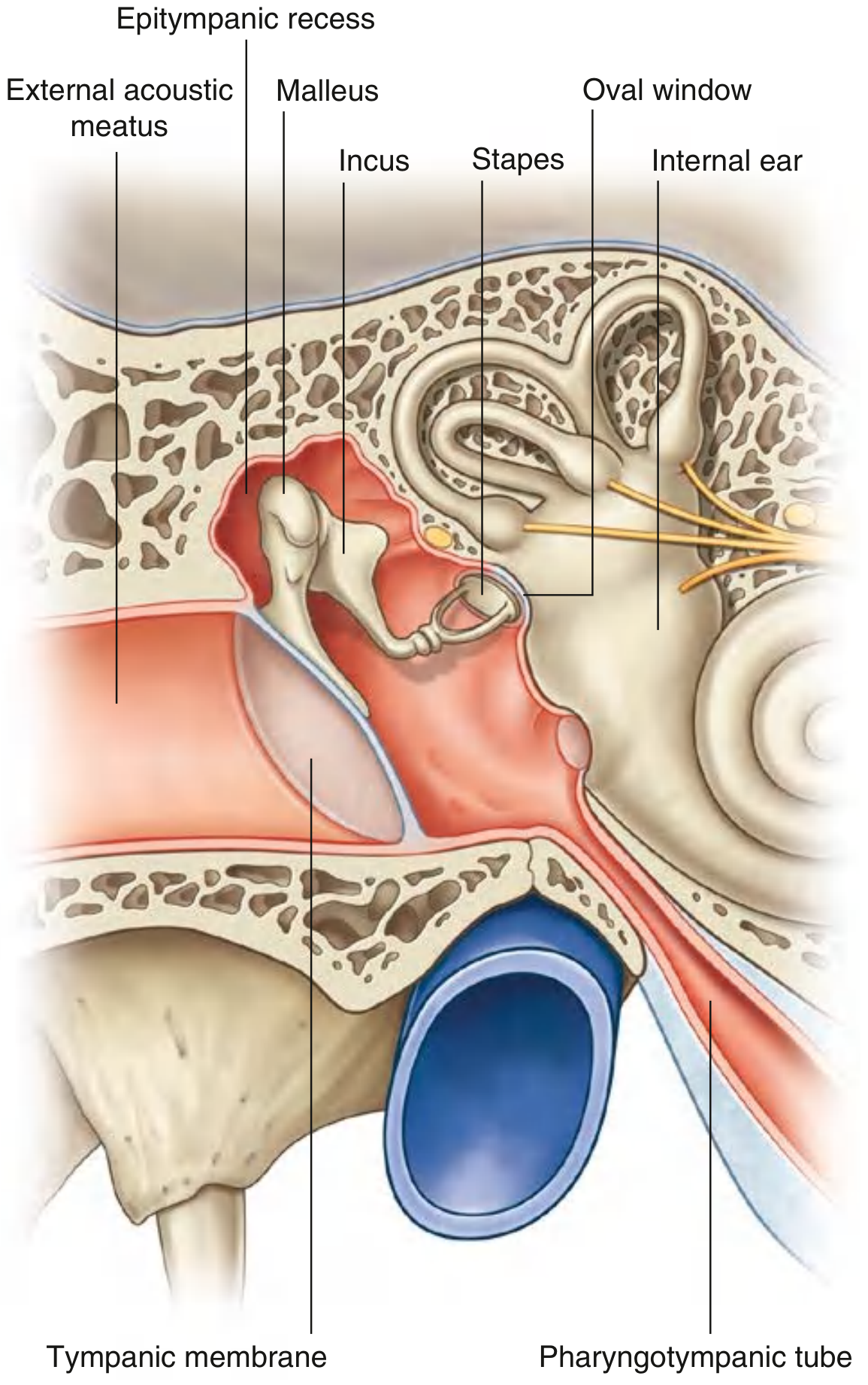
It has two parts:
- Tympanic cavity - immediately adjacent to the tympanic membrane
- Epitympanic recess - the part above/superiorly
Function: Transmit sound vibrations from the tympanic membrane to the internal ear via the ossicular chain.
Communications:
- Anteriorly - nasopharynx (via the pharyngotympanic / Eustachian tube)
- Posteriorly - mastoid air cells (via the aditus to mastoid antrum)
6 Walls of the Middle Ear (Easy Memory Aid: "TJ-MAL")
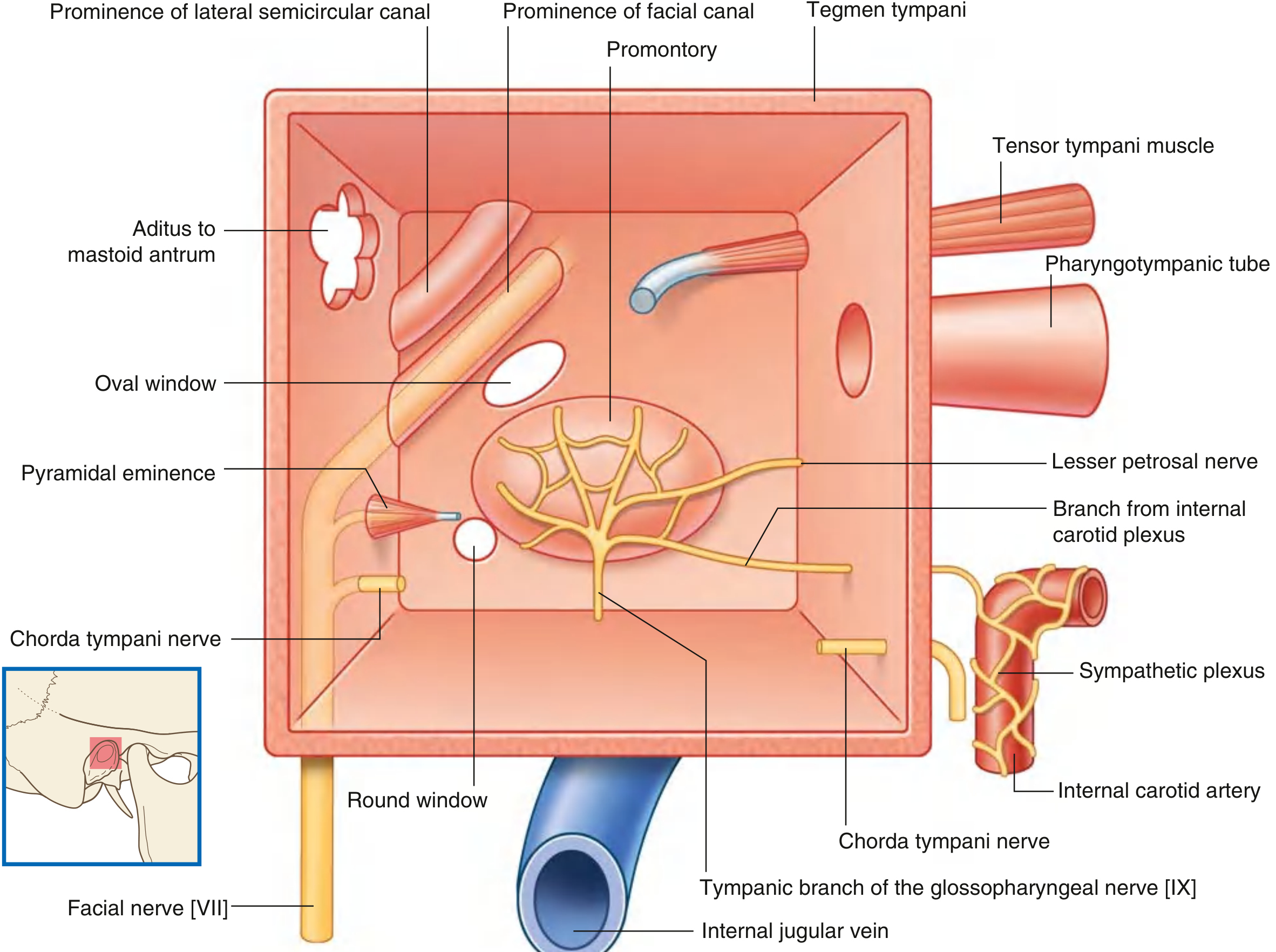
| Wall | Name | Key Contents / Relations |
|---|---|---|
| Roof | Tegmental wall | Tegmen tympani (thin bone) - separates middle ear from middle cranial fossa |
| Floor | Jugular wall | Separates from internal jugular vein; tympanic branch of CN IX enters here |
| Lateral | Membranous wall | Mostly the tympanic membrane; upper part = bony lateral wall of epitympanic recess |
| Medial | Labyrinthine wall | Promontory (basal coil of cochlea); oval window (posterosuperior); round window (posteroinferior); prominence of facial canal (CN VII); prominence of lateral semicircular canal |
| Posterior | Mastoid wall | Aditus to mastoid antrum (superiorly); pyramidal eminence (stapedius tendon exits here); chorda tympani enters here |
| Anterior | Carotid wall | Related to internal carotid artery; openings for pharyngotympanic tube and tensor tympani canal |
Auditory Ossicles (3 Bones)
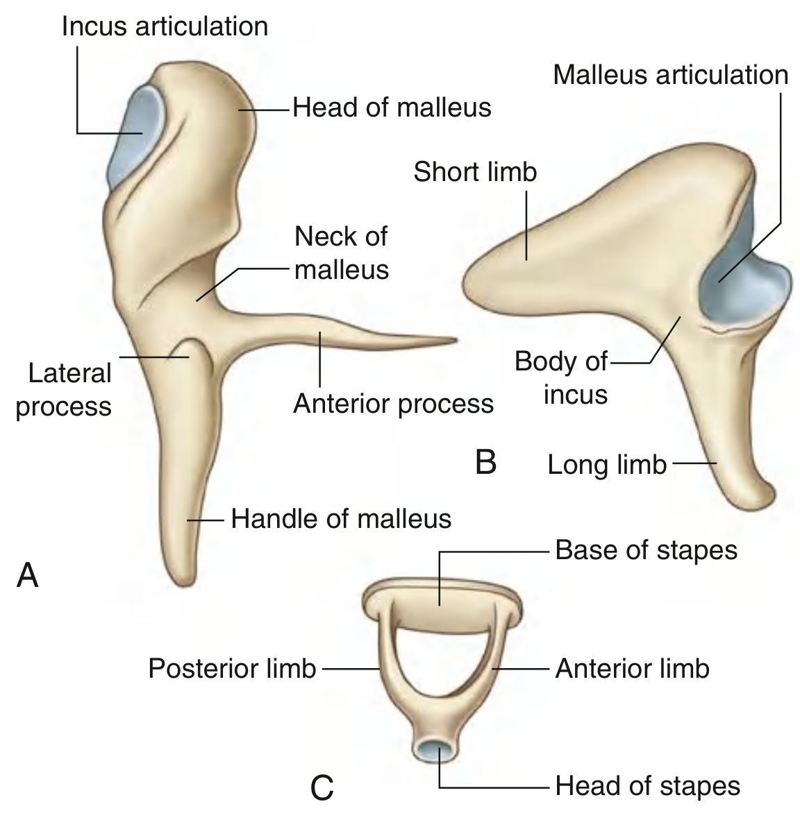
They form a chain from the tympanic membrane to the oval window:
1. Malleus (largest)
- Attached to the tympanic membrane (by its handle)
- Parts: Head, neck, anterior process, lateral process, handle
- Head articulates with the incus
2. Incus
- Parts: Body (articulates with malleus head), long limb (goes down, bends medially to meet stapes), short limb (projects posteriorly, anchored by ligament)
3. Stapes (smallest bone in the body)
- Most medial; its base fits into the oval window
- Parts: Head, anterior + posterior limbs, base
Two Muscles of the Middle Ear
| Muscle | Origin | Insertion | Nerve | Action |
|---|---|---|---|---|
| Tensor tympani | Cartilaginous pharyngotympanic tube, greater wing of sphenoid | Handle of malleus | CN V3 (mandibular) | Pulls malleus medially - tenses tympanic membrane - dampens loud sounds |
| Stapedius | Inside pyramidal eminence | Neck of stapes | CN VII (facial) | Pulls stapes posteriorly - prevents excessive ossicle movement |
Clinic tip: Paralysis of the facial nerve (CN VII) abolishes the stapedius reflex, causing hyperacusis (abnormal sensitivity to loud sounds).
Pharyngotympanic (Eustachian) Tube
- Connects the middle ear to the nasopharynx
- Equalises pressure on both sides of the tympanic membrane
- 1/3 bony (near middle ear) + 2/3 cartilaginous (near pharynx)
- Nerve supply: Tympanic plexus (mainly from CN IX)
- Arterial supply: Ascending pharyngeal artery + branches of maxillary artery
Clinic tip: Children have a shorter, more horizontal Eustachian tube - this is why they get more frequent middle ear infections (otitis media).
Key Nerves in the Middle Ear
| Nerve | Source | Function |
|---|---|---|
| Tympanic branch (Jacobson's nerve) | CN IX (glossopharyngeal) | Enters floor; forms tympanic plexus; supplies mucosa of middle ear, mastoid, Eustachian tube |
| Lesser petrosal nerve | Tympanic plexus | Leaves via foramen ovale to otic ganglion - parasympathetic to parotid gland |
| Chorda tympani | CN VII (facial) | Enters via mastoid wall, exits via anterior wall; taste from anterior 2/3 tongue + secretomotor to sublingual & submandibular glands |
Mastoid Area (Clinical Importance)
- The mastoid antrum is connected to the epitympanic recess via the aditus
- Mastoid air cells are lined by mucous membrane continuous with the middle ear
- Infection in the middle ear can spread to mastoid air cells = mastoiditis
- Mastoiditis can spread to:
- Middle cranial fossa (meningitis)
- Facial nerve (facial palsy)
Quick Revision Summary
Tympanic membrane → Malleus → Incus → Stapes → Oval window → Internal ear
- 6 walls: Roof (tegmen), Floor (jugular), Lateral (tympanic membrane), Medial (promontory + oval/round windows), Posterior (mastoid/aditus), Anterior (carotid/Eustachian tube)
- 3 ossicles: Malleus (CN V3 via tensor tympani), Incus, Stapes (CN VII via stapedius)
- Key nerves through middle ear: CN VII (facial canal - medial wall), Chorda tympani (branch of VII), Jacobson's nerve (branch of IX)
- Eustachian tube: Equalises pressure; 1/3 bone + 2/3 cartilage; supplied by CN IX
This is a shared conversation. Sign in to Orris to start your own chat.