
Stylet - part pictures, indication and contraindication in MD Anaesthesia
endotracheal tube stylet parts diagram anaesthesia
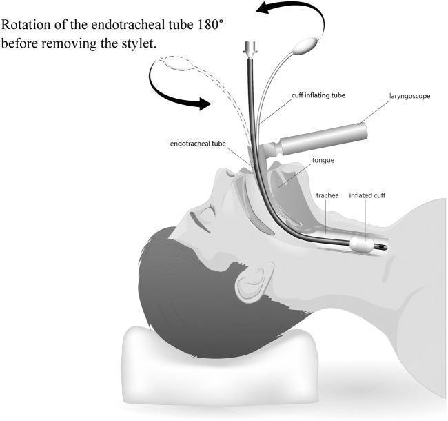
This medical illustration demonstrates the 'ETT 180-degree rotation' intubation technique in a sagittal view of a patient in the supine position. The diagram shows an endotracheal tube (ETT) with an internal stylet passing through the oral cavity, over the tongue, and into the trachea. A laryngoscope is depicted positioned to displace the tongue for glottic visualization. The primary educational focus is the clockwise rotation of the ETT by 180 degrees once the distal tip, featuring an inflated cuff, passes the vocal cords but before the stylet is removed. A curved arrow illustrates this rotational maneuver. This specific airway management technique is designed to align the ETT and stylet with the posterior angulation of the trachea, thereby minimizing trauma to the anterior tracheal wall during advancement. Labeled structures include the endotracheal tube, stylet-loaded cuff inflating tube, laryngoscope, tongue, trachea, and inflated cuff. This resource is relevant for anesthesiology and emergency medicine training regarding optimized endotracheal intubation maneuvers.
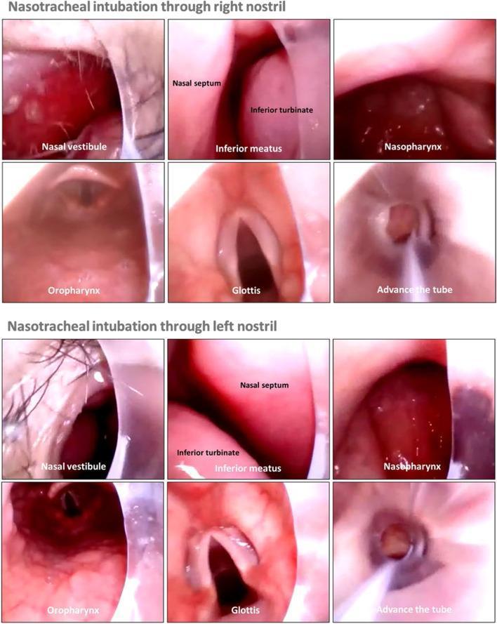
This clinical endoscopic comparison chart illustrates the anatomical progression of video rigid stylet-guided nasotracheal intubation through both the right and left nostrils. The image is organized into two rows of six frames each, documenting the view from initial entry to endotracheal tube advancement. Key anatomical landmarks labeled include the nasal vestibule, nasal septum, inferior turbinate, and inferior meatus within the nasal cavity. As the stylet-tube assembly progresses, the frames show the transition into the nasopharynx and oropharynx. The final stages depict the direct visualization of the glottis with clearly visible vocal cords, followed by the successful advancement of the beveled endotracheal tube into the trachea. This sequence demonstrates the clinical utility of video-assisted intubation in maintaining direct visualization of mucosal surfaces and laryngeal structures, regardless of the nostril selected, to minimize trauma and ensure accurate placement.
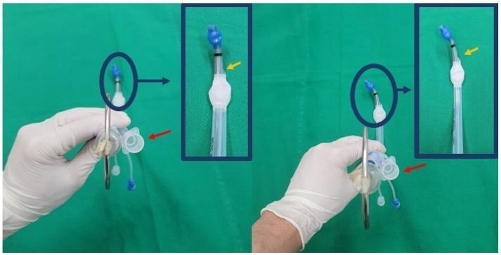
Clinical photograph demonstrating the orientation of a left-sided double-lumen endotracheal tube (DLT) with a customized J-shaped stylet, illustrating the non-rotatory maneuver technique for intubation. The image is split into two panels (initial setup on the left, insertion state on the right) with magnified insets of the distal tip. The bronchial lumen is oriented to the left, while the tracheal lumen's proximal orifice (indicated by a red arrow) and distal orifice (indicated by a yellow arrow in the insets) are oriented to the right side. The distal tip features a blue-colored cuff connected to the clear main body of the silicone tube. The right panel demonstrates how the tube maintains its orientation during the non-rotatory maneuver, with a slight adjustment in the curvature of the stylet's tip relative to the main axis. This visual provides clinical guidance on DLT positioning to facilitate faster intubation and minimize mucosal trauma compared to conventional rotatory techniques.
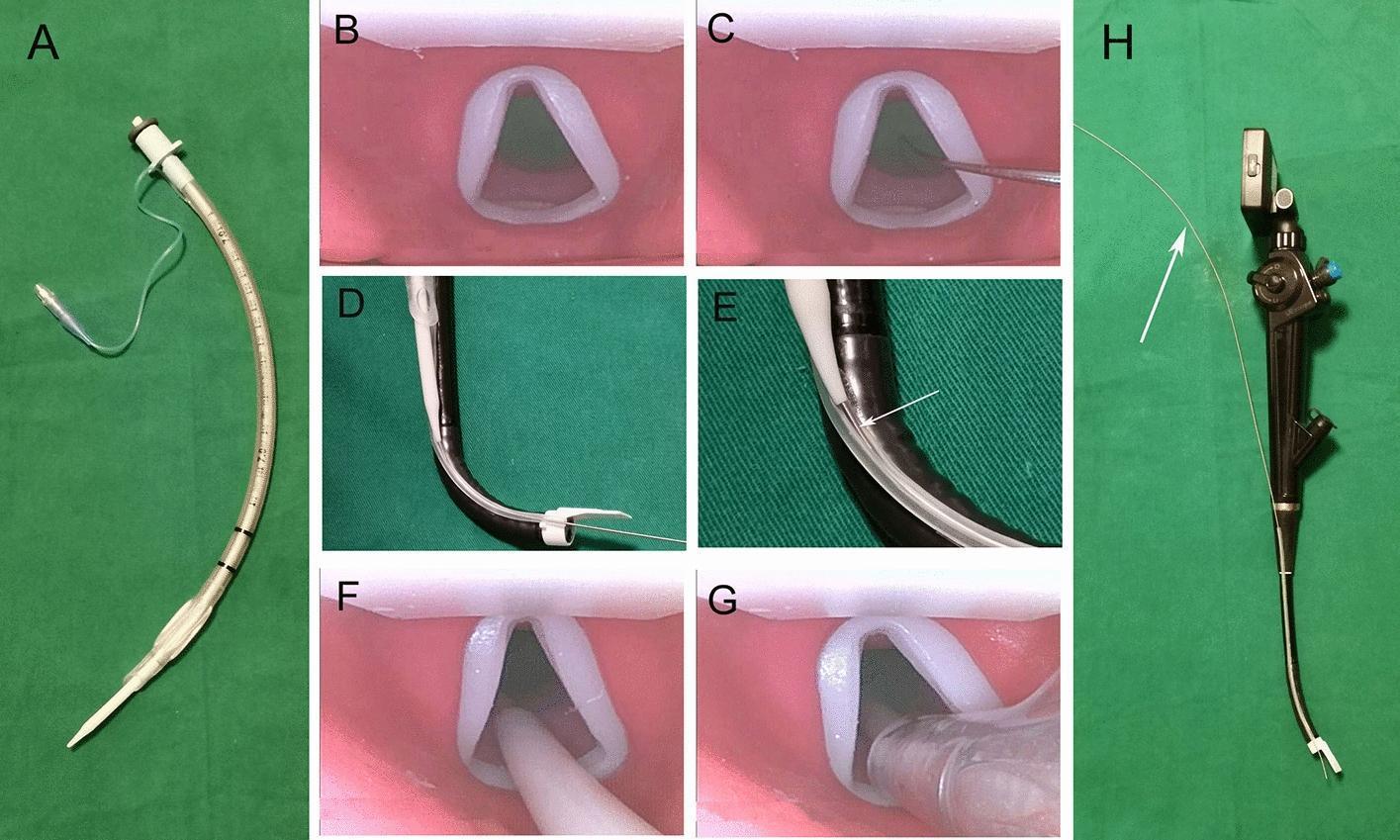
A multi-panel figure illustrating the components and clinical application of a Flexible Video Laryngoscope (FVL) for endotracheal intubation. Image A shows a 7.0 mm endotracheal tube (ETT) mounted over a white guiding tube. Image H displays the fully assembled FVL system, featuring a proximal digital display, a control handle with a thumb lever, and a flexible insertion tube with a side-mounted guiding channel and a pre-installed metal stylet (white arrow). Panels B, C, F, and G provide a first-person endoscopic view of a simulated glottis. Sequential steps are demonstrated: visualization of the glottis (B); insertion of the thin stylet through the glottic opening (C); advancement of the white guiding tube along the stylet (F); and final passage of the ETT into the trachea (G). Panels D and E show the mechanical relationship between the black flexible insertion tube and the guiding components, illustrating how the stylet and ETT slide along the external guiding channel. This clinical series demonstrates difficult airway management techniques using a specialized FVL system.
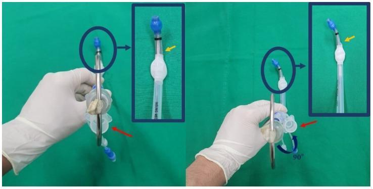
A clinical comparison photograph demonstrating the rotatory maneuver for inserting a left-sided double-lumen endotracheal tube (DLT). The image consists of two panels (left and right) showing a gloved hand holding a DLT with an internal rigid J-shaped stylet against a green surgical drape. In the left panel, the tube is shown in its initial orientation: the bronchial lumen and stylet tip are aligned anteriorly (12 o'clock position), while the proximal (red arrow) and distal (yellow arrow) tracheal lumen orifices are oriented posteriorly. The right panel illustrates the 90-degree counterclockwise rotation maneuver typically performed as the bronchial tip passes through the vocal cords. Post-rotation, the tracheal orifices are re-positioned to the lateral (right-side) orientation. Blue circular highlights and inset magnifications emphasize the tip's spatial relationship. This visual serves as an educational guide for anesthesiology residents and clinical staff on proper DLT manipulation techniques during thoracic surgery intubation procedures to ensure correct anatomical placement into the left mainstem bronchus.
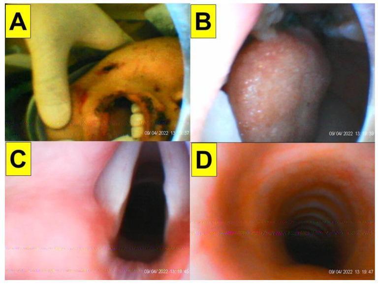
This sequence of clinical and intraoral images demonstrates the stages of endotracheal intubation using a video-assisted intubating stylet in a trauma patient. The images provide a first-person perspective of the airway progression. Panel A shows an external view of the mouth being manually opened, revealing teeth and minor perioral trauma. Panel B depicts the stylet's entry into the oral cavity, focusing on the posterior portion of the tongue. Panel C provides full glottic visualization, clearly showing the vocal cords, arytenoid cartilages, and the laryngeal inlet. Panel D shows successful tracheal entry, with the characteristic cartilaginous tracheal rings visible within the lumen. This series illustrates the clinical utility of optical stylets in navigating the airway and confirming tube placement in patients with restricted neck mobility, such as those in a cervical collar. The educational focus is on the sequential visualization of anatomical landmarks—from the oral space to the glottis and finally the trachea—to ensure safe airway management.
intubating stylet malleable wire parts labelled
This sequence of clinical and intraoral images demonstrates the stages of endotracheal intubation using a video-assisted intubating stylet in a trauma patient. The images provide a first-person perspective of the airway progression. Panel A shows an external view of the mouth being manually opened, revealing teeth and minor perioral trauma. Panel B depicts the stylet's entry into the oral cavity, focusing on the posterior portion of the tongue. Panel C provides full glottic visualization, clearly showing the vocal cords, arytenoid cartilages, and the laryngeal inlet. Panel D shows successful tracheal entry, with the characteristic cartilaginous tracheal rings visible within the lumen. This series illustrates the clinical utility of optical stylets in navigating the airway and confirming tube placement in patients with restricted neck mobility, such as those in a cervical collar. The educational focus is on the sequential visualization of anatomical landmarks—from the oral space to the glottis and finally the trachea—to ensure safe airway management.
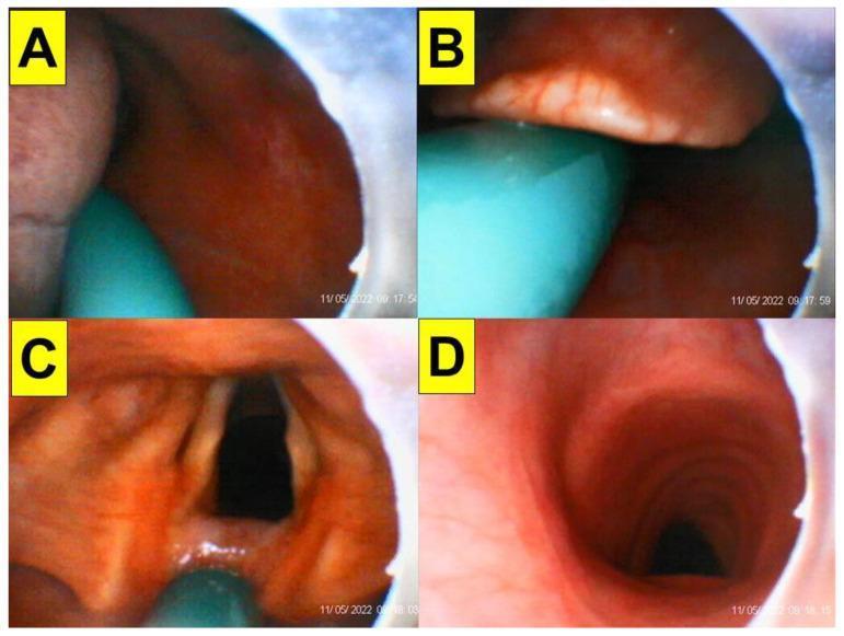
This endoscopic clinical image series consists of four panels (A-D) demonstrating the sequential progression of endotracheal intubation using a video-assisted intubating stylet. Panel A shows the initial entry into the oral space, with the tongue and posterior oropharyngeal mucosa visible alongside the teal-colored distal end of the intubating stylet. Panel B illustrates the visualization of the epiglottis, with the stylet positioned just inferior to it to facilitate lifting or bypassing the structure. Panel C provides a clear, 'full glottic' view, identifying key laryngeal landmarks including the vocal cords, arytenoid cartilages, and the glottic opening. Panel D captures the successful entry into the trachea, characterized by the distinct visual appearance of the tracheal rings (cartilaginous arches). The series serves as an educational tool for anesthesiology and emergency medicine, highlighting airway anatomy and the use of optical stylets in managing patients with potentially restricted neck mobility, such as those in stereotactic headframes.
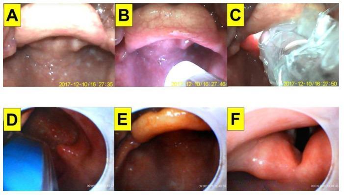
This clinical image sequence demonstrates the 'video-twin' intubation technique, combining videolaryngoscopy (VL) with a video-assisted intubating stylet (VS) in a patient with a cervical spine injury and Mallampati class III airway. Panels A–C show the external videolaryngoscopic perspective. (A) Initial view of the posterior pharynx and base of the tongue. (B) Insertion of the intubating stylet into the oropharyngeal space, facilitating the lifting of the tongue root. (C) Further advancement of the stylet toward the laryngeal inlet, though the view is partially obscured by secretions. Panels D–F provide the internal perspective from the tip of the intubating stylet. (D) Approaching the epiglottis with the blue stylet tip visible. (E) Clearer visualization of the epiglottis as the stylet is maneuvered. (F) Direct visualization of the glottic opening and vocal cords, allowing the endotracheal tube to be advanced into the trachea under direct vision. This dual-view approach is used to manage difficult airways by improving laryngeal exposure and confirming tube placement in real-time.
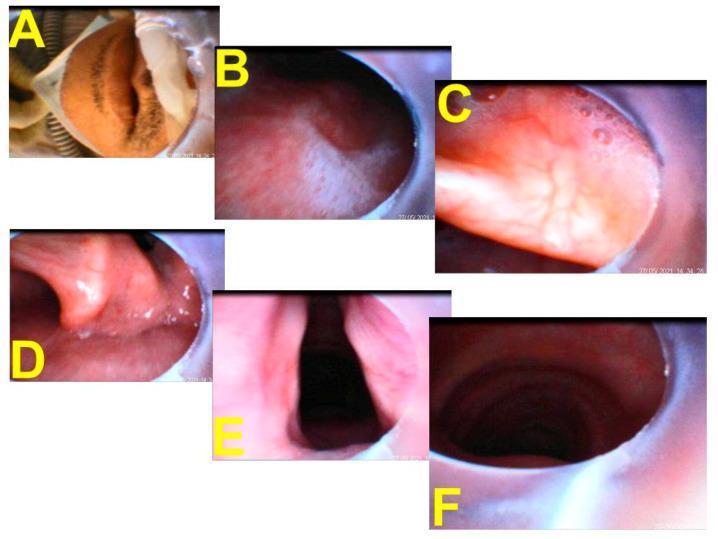
This composite of endoscopic images illustrates a serial pictorial progression of tracheal intubation using a video-assisted intubating stylet (Shikani technique). (A) Shows the clinical context of the patient receiving high-flow nasal cannula (HFNC) treatment just prior to insertion. (B) Displays the pharyngeal view with visible mucus and saliva at the start of the procedure. (C) Provides a close-up of the vallecula as the stylet advances. (D) Visualizes the corniculate and cuneiform cartilages of the posterior larynx. (E) Offers a clear, full view of the glottic opening and vocal cords, representing the target for endotracheal tube (ETT) placement. (F) Confirms successful placement by visualizing the internal tracheal rings and the distal tip of the ETT within the trachea. The images demonstrate the necessary anatomical landmarks for safe airway management, including the transition from the oropharynx through the glottis and into the subglottic space. This series is an educational example of real-time visualization used to guide airway management in high-risk patients, such as those with severe respiratory infections requiring rapid ventilator support.
lighted stylet lightwand trachlight parts anaesthesia
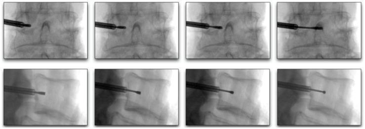
This diagnostic image sequence consists of eight fluoroscopic frames arranged in two rows, illustrating the progression of a curved stylet during a minimally invasive spinal procedure, such as basivertebral nerve ablation or vertebral augmentation. The top row displays an anterior-posterior (AP) view, showing the stylet advancing from a lateral transpedicular entry point toward the 25-40% midline of the vertebral body. Key landmarks visible include the pedicles, vertebral endplates, and spinous process. The bottom row displays a lateral view of the same lumbar vertebral level, demonstrating the stylet's trajectory as it moves from the posterior cortex into the anterior two-thirds of the vertebral body, remaining centered between the superior and inferior endplates. The sequence highlights the spatial relationship between the surgical instrument and the bony anatomy of the spine, emphasizing the precise placement required to reach the target zone while avoiding cortical perforation. This material is designed for intermediate to advanced medical educational indexing in the fields of interventional pain management, radiology, and orthopedic surgery.
This sequence of clinical and intraoral images demonstrates the stages of endotracheal intubation using a video-assisted intubating stylet in a trauma patient. The images provide a first-person perspective of the airway progression. Panel A shows an external view of the mouth being manually opened, revealing teeth and minor perioral trauma. Panel B depicts the stylet's entry into the oral cavity, focusing on the posterior portion of the tongue. Panel C provides full glottic visualization, clearly showing the vocal cords, arytenoid cartilages, and the laryngeal inlet. Panel D shows successful tracheal entry, with the characteristic cartilaginous tracheal rings visible within the lumen. This series illustrates the clinical utility of optical stylets in navigating the airway and confirming tube placement in patients with restricted neck mobility, such as those in a cervical collar. The educational focus is on the sequential visualization of anatomical landmarks—from the oral space to the glottis and finally the trachea—to ensure safe airway management.
![Recommendation Table 18. Recommendation for depth of anaesthesia monitoring
<table><thead><tr><th>Recommendation</th><th>Class<sup>a</sup></th><th>Level<sup>b</sup></th><th>Ref<sup>c</sup></th></tr></thead><tbody><tr><td>The use of processed EEG monitoring should be considered to reduce the incidence of intraoperative awareness and excess in depth of anaesthesia.</td><td>IIa</td><td>B</td><td>[231-234]</td></tr></tbody></table>](https://cdn.orris.care/cdss_images/GLGCA_4542999_1766781036019_b81b990d-b477-4cd4-be5d-4f49fb079f29_53c9eda3-6178-4b66-b461-17f141a2f565.png)
Recommendation Table 18. Recommendation for depth of anaesthesia monitoring <table><thead><tr><th>Recommendation</th><th>Class<sup>a</sup></th><th>Level<sup>b</sup></th><th>Ref<sup>c</sup></th></tr></thead><tbody><tr><td>The use of processed EEG monitoring should be considered to reduce the incidence of intraoperative awareness and excess in depth of anaesthesia.</td><td>IIa</td><td>B</td><td>[231-234]</td></tr></tbody></table>
endotracheal tube stylet parts indication contraindication anaesthesia MD exam
https://anesthguide.com/topic/airway-management
Bonfils Shikani optical stylet rigid fiberoptic intubation scope
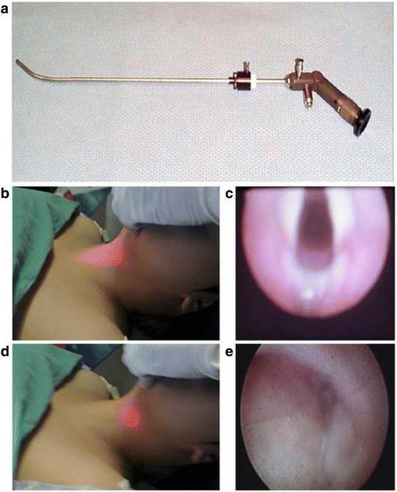
This composite educational image illustrates the transillumination-assisted intubation technique using a Bonfils optical stylet. Panel (a) shows the medical device, a rigid, slender optical stylet with a curved distal tip and a proximal eyepiece and light source connector. Panels (b) through (e) demonstrate clinical application and endoscopic correlation. Image (b) shows a centered, bright transillumination light on the anterior neck, indicating the stylet tip is positioned correctly at the midline. This corresponds to the endoscopic view in (c), which clearly depicts the laryngeal inlet, including the vocal cords and glottis. In contrast, image (d) displays a darker, laterally deviated light spot on the neck. The associated endoscopic view in (e) shows non-target anatomy, such as the pyriform sinus and pharyngeal walls, indicating the stylet is off-axis. This figure teaches clinicians how to use external visual cues (transillumination) to guide internal airway visualization, facilitating successful tracheal intubation by aligning the device with the glottic opening.
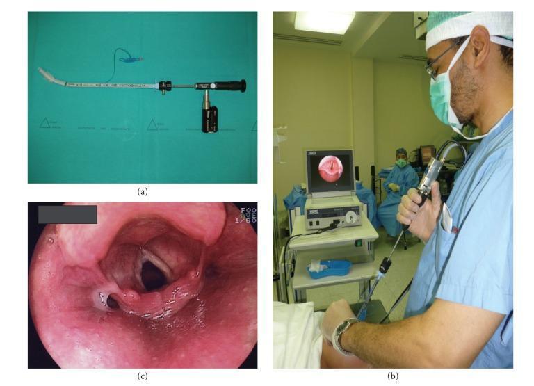
This composite educational image illustrates the components and clinical application of the Bonfils Intubation Fiberscope (BIF) in anesthesiology. Panel (a) shows the medical hardware: a rigid, straight silver/grey fiberscope with a distal curved tip, armed with a 7.5 mm inner diameter endotracheal tube. Key features include measurement graduations on the insertion shaft and a black handle with an attached battery power source or camera module. Panel (b) depicts an intraoperative clinical scenario where a practitioner in surgical attire performs a video-assisted intubation. The BIF is held at the eyepiece, with the live feed displayed on an external monitor. Panel (c) provides an endoscopic view of the laryngeal aditus obtained during the procedure. It displays anatomical landmarks including the vocal cords forming a glottic opening, the epiglottis, and surrounding aryepiglottic folds. The mucosal tissue appears pink and moist with visible vascularity. This image serves as a teaching tool for difficult airway management and the technical setup of rigid fiberoptic intubation devices.
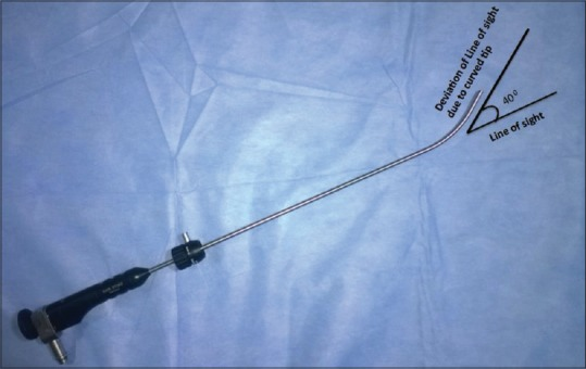
**Imaging Modality:** Clinical photograph of medical instrumentation with schematic annotations. **Device Classification:** Rigid fiberscope (Bonfils Intubation Fiberscope) designed for airway management and indirect laryngoscopy. **Technical Specifications and Key Features:** * **Shaft Design:** The image displays a long, slender, semi-rigid metallic shaft connected to a handle containing an eyepiece and light source connector. * **Distal Configuration:** The distal tip of the scope exhibits a fixed cephalad curvature. * **Optical Alignment:** Annotations indicate a 40-degree deviation of the line of sight relative to the long axis of the shaft. This angulation is specifically designed to facilitate visualization of anteriorly displaced laryngeal structures. * **Clinical Application:** Utilized in anesthesia and emergency medicine for "retromolar" intubation techniques or managing patients with difficult airways where direct laryngoscopy is restricted. **Diagnostic Relevance:** The photograph highlights the characteristic distal angulation that distinguishes this device from standard rigid endoscopes, serving as a key visual cue for its use in overcoming anatomical challenges such as a limited mouth opening or a high-anterior glottis.
Stylet in Anaesthesia - MD Level
1. Introduction
2. Types of Stylets
A. Malleable (Standard) Stylet
B. Lighted Stylet (Lightwand / Trachlight)
C. Optical / Video Stylet
3. Parts of a Stylet (Malleable Type)
| Part | Description |
|---|---|
| Proximal end (handle/stop piece) | Bent at 90 degrees or fitted with a flanged stop to prevent the stylet from advancing too far into the tube and protruding beyond the tip |
| Shaft | Malleable aluminium/steel core; allows shaping of the ETT into "hockey-stick" (35-degree distal bend), "straight-to-cuff," or "J-shape" as needed |
| Distal end (tip) | Smooth, blunt tip; must NEVER extend beyond the Murphy eye (distal eyelet) of the ETT to avoid mucosal trauma or perforation |
| Lubrication coat | Most disposable stylets have a smooth or waxed surface to allow easy withdrawal after intubation |
- Lubricate the stylet and insert into the ETT
- Bend the ETT + stylet into a "straight-to-cuff" shape with a 35-degree distal bend (or "hockey-stick" shape for anteriorly placed larynx)
- The bend is placed at the cuff-tube junction
4. Parts of an Optical Stylet (Bonfils / Shikani Type)
| Part | Description |
|---|---|
| Proximal eyepiece / LCD screen | Allows visualization of glottis; can connect to video monitor |
| Handle / control body | Houses battery/light source connection, suction port (working channel in some models) |
| Shaft | 40 cm rigid or malleable stainless steel with fiberoptic bundle; ETT is pre-loaded onto the shaft |
| Distal tip | Fixed 40-degree anterior curvature (Bonfils); malleable in Shikani; steerable 3 cm in SensaScope |
| Working channel | Present in some models - used for suction, oxygen insufflation (max 3 L/min to avoid barotrauma), or "spray-as-you-go" (SAYGO) local anaesthesia |
5. Flexible Video Laryngoscope Stylet System
- Proximal digital display for real-time visualization
- Thumb lever on handle for tip deflection
- Flexible insertion tube with side-mounted guiding channel
- Pre-installed metal stylet (white arrow) that passes first through cords, then the guiding tube is railroaded, then the ETT is advanced
6. Optical Stylet - Video-Assisted Intubation Sequence
7. Indications
Malleable Stylet
| Indication | Rationale |
|---|---|
| Routine intubation (Cormack-Lehane grade 1-2) | Provides directional control of ETT tip when view is adequate |
| Anterior larynx | Hockey-stick or "J" shape allows the tube to be directed anteriorly |
| Rapid Sequence Induction (RSI) | Allows quick, single-attempt intubation by pre-shaping the tube |
| Video laryngoscopy (VL) | Mandatory with hyperangulated blades (e.g., GlideScope); rigid GlideRite stylet pre-shaped to match blade curvature is used |
| Obese patients | Restricted manoeuvrability of hands makes tube control with a stylet easier |
| Double-lumen tube (DLT) insertion | J-shaped rigid stylet to facilitate bronchial tube placement (rotatory and non-rotatory techniques) |
| Maximum control of ETT tip required | Any situation needing precise directional placement |
Lighted Stylet (Lightwand / Trachlight)
| Indication | Rationale |
|---|---|
| Limited mouth opening | Does not require laryngoscopy |
| Cervical spine immobility | Causes minimal cervical movement |
| Anticipated difficult laryngoscopy | Transillumination guides placement blindly |
| Restricted access to oral cavity | Nasotracheal or oral approach possible |
Optical / Video Stylet (Bonfils, Shikani, SensaScope)
- Limited neck mobility (e.g., cervical spine disease, halos, collars)
- Small mouth opening (trismus, ankylosis)
- Abnormal airway anatomy (tumours, previous surgery, burns)
- Anticipated difficult laryngoscopy
- Awake intubation - can be used as an alternative or adjunct to flexible bronchoscopy
- In combination with direct laryngoscopy (DL) or video-assisted laryngoscopy (VAL) for augmented control
8. Contraindications
Malleable Stylet
| Contraindication | Reason |
|---|---|
| Extending stylet beyond Murphy eye | This is a technical contraindication - stylet must NEVER protrude past the ETT tip (risk of laryngeal/tracheal perforation, mucosal laceration) |
| Active airway infection/epiglottitis (relative) | Stylet increases tube rigidity; risk of trauma to friable inflamed mucosa |
| When flexible scope or bougie is preferred (grade 2b-3 view) | Stylet is less effective than a bougie when the glottic view is poor; a bougie is preferred for grade 2b-3 Cormack-Lehane |
| Base of skull fractures (nasal route) | Absolute contraindication to nasal intubation with any adjunct |
| Suspected foreign body in airway (relative) | Rigid instrument may dislodge or impact the foreign body further |
Lighted Stylet (Trachlight)
| Contraindication | Reason |
|---|---|
| Morbid obesity / short neck | Excessive subcutaneous tissue prevents transillumination from being visible |
| Neck tumours / infections / trauma | Distorts anatomy; transillumination unreliable |
| Foreign body in airway | Blind technique risks impaction |
| Bright ambient light environments | Transillumination glow is not visible (relative) |
| Oropharyngeal/laryngeal tumours | Trauma and potential bleeding |
Optical/Video Stylet (Bonfils, Shikani)
| Contraindication | Reason |
|---|---|
| Airway secretions / blood in airway | Obscures fiberoptic/video view (relative); suction via working channel can overcome this partially |
| Completely obstructed airway | Cannot navigate through total obstruction |
| Patient unable to cooperate (awake intubation, without topicalization) | Requires adequate topical anaesthesia for awake use |
| Oxygen insufflation > 3 L/min via working channel | Risk of barotrauma - a specific technical contraindication for the Bonfils working channel |
9. Practical Tips for MD Anaesthesia Viva
-
Stylet shape for video laryngoscopy: Hyperangulated blades (GlideScope, C-MAC D-blade) require the ETT+stylet to be bent to match the blade curve (approx. 60-70 degrees); standard blades use the 35-degree "straight-to-cuff" bend.
-
Withdraw the stylet 1-2 cm as the tip enters the glottis during video laryngoscopy - this reduces resistance as the tube angles from the hyperangulated approach to the more vertical trachea, as described in [Barash Clinical Anesthesia 9e].
-
180-degree rotation trick: When using a stylet and the ETT tip catches on the anterior tracheal wall, rotating the tube 180 degrees before advancing aligns the bevel posteriorly and reduces hang-up.
-
Bougie vs. Stylet: Stylet = good view but need control (CL grade 1-2). Bougie = poor/partial view (CL grade 2b-3). A practical viva rule: "Stylet for precision, bougie for rescue."
-
ETT tube changers (Airway Exchange Catheters) are long hollow stylet-like devices used to maintain access during extubation in high-risk airways - they allow reintubation and can deliver O2 (at low flow to avoid barotrauma).