I now have comprehensive data from multiple authoritative neurology textbooks. Let me compile the full exam-oriented answer:
GUILLAIN-BARRÉ SYNDROME (GBS)
Comprehensive Exam-Oriented Review — Harrison's / Adams & Victor's Based
1. DEFINITION & OVERVIEW
Guillain-Barré Syndrome (also called Landry-Guillain-Barré-Strohl Syndrome) refers to a group of acquired, immune-mediated, acute inflammatory polyradiculoneuropathies sharing:
- Acute onset (maximal deficit within 4 weeks)
- Cytoalbuminologic dissociation in CSF (elevated protein, low WBC)
- Monophasic course with spontaneous recovery
GBS is the most common cause of acute or subacute generalized paralysis in clinical practice (Adams & Victor's).
2. EPIDEMIOLOGY
| Parameter | Detail |
|---|
| Annual incidence | 1-2 per 100,000 (range 0.4-1.7/100,000) |
| Sex | Males > Females (1.4:1) |
| Age | All ages; attack rates highest in 50-74 years |
| Seasonality | Generally non-seasonal (except AMAN in rural China - summer outbreaks) |
| Antecedent event | 60% have preceding respiratory or GI infection (1-3 weeks prior) |
- Goldman-Cecil Medicine (9780323930345)
- Adams & Victor's Principles of Neurology, 12th Ed
3. ETIOLOGY & PRECEDING INFECTIONS
| Agent | Notes |
|---|
| Campylobacter jejuni | Most common identifiable antecedent; serotype HS:O19 linked to AMAN variant |
| Cytomegalovirus (CMV) | Common viral trigger |
| Epstein-Barr Virus (EBV) | "Large viruses of herpes family" |
| HIV | GBS at time of seroconversion |
| Mycoplasma pneumoniae | Antigalactocerebroside antibodies |
| Hepatitis E | 5-10% in Belgium/Netherlands |
| Zika virus | Significantly increased risk of all GBS forms |
| Influenza vaccine (A/NJ swine, 1976) | Classic exam association |
| Hodgkin's lymphoma | Less certain association |
SARS-CoV-2: GBS does not appear substantially associated with COVID-19 or most COVID vaccines (slight increase with ChADOx1nCoV-19: 0.6 cases/100,000 doses). GBS incidence actually declined during the pandemic.
4. PATHOGENESIS
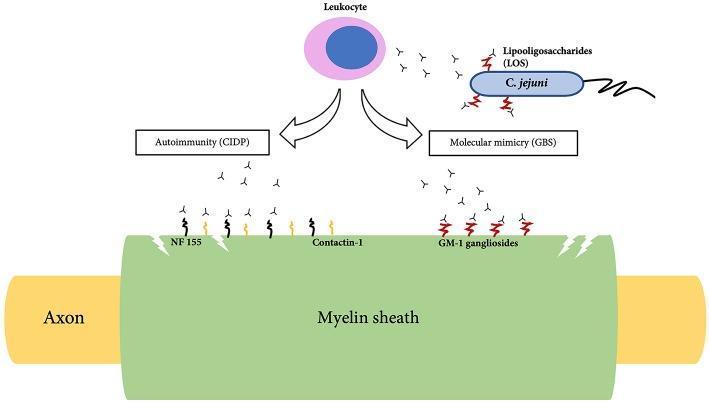
Molecular Mimicry is the central mechanism:
- Immune system attacks peripheral nerve antigens that resemble microbial antigens (especially C. jejuni)
- The HS/O:19 serotype of C. jejuni has lipooligosaccharides (LOS) that mimic GM1 gangliosides on nerve axons
- This triggers production of cross-reactive antibodies against nerve components
Earliest immunologic event: Complement deposition on the myelin surface (Asbury et al., 1969 established perivascular mononuclear inflammatory infiltration as the essential lesion).
Pathology:
- AIDP: Segmental demyelination of spinal roots and peripheral nerves + perivascular lymphocytic infiltration
- AMAN/AMSAN: Primary axonal injury without demyelination; antibodies target gangliosides on axolemma
5. SUBTYPES / VARIANTS
| Subtype | Full Name | Key Features | Antibodies |
|---|
| AIDP | Acute Inflammatory Demyelinating Polyneuropathy | Most common in North America/Europe (97%); demyelinating | No specific ganglioside Ab |
| AMAN | Acute Motor Axonal Neuropathy | Pure motor; prevalent in northern China (summer epidemics in children); preceded by C. jejuni | Anti-GM1, anti-GD1a (IgG) |
| AMSAN | Acute Motor-Sensory Axonal Neuropathy | Both motor and sensory axons; severe; poor recovery | Anti-GD1b, anti-GalNAc-GD1a |
| MFS | Miller-Fisher Syndrome | Triad: Ophthalmoplegia + Ataxia + Areflexia | Anti-GQ1b (95-98%) |
| Pharyngeal-cervical-brachial | PCB variant | Weakness of face, throat, neck and arms | Anti-GT1a |
| Bickerstaff brainstem encephalitis | BBE | MFS + altered consciousness/signs | Anti-GQ1b |
Key exam point: MFS = GQ1b antibody positive in 95-98% of cases; accounts for 6% of GBS in Western countries, 18% in Taiwan.
6. CLINICAL FEATURES
Classic Presentation
- Ascending symmetric weakness beginning in legs, spreading to arms, face
- Areflexia / hyporeflexia (universal finding)
- Paresthesias (tingling, numbness in hands and feet) - often present before weakness
- Pain (back pain, limb pain) is common and often underappreciated
Progression Pattern
- Maximum deficit reached in <4 weeks (50% by 2 weeks, 75% by 3 weeks, >90% by 4 weeks)
- Followed by plateau phase, then gradual recovery
Specific Features
Motor:
- Bilateral facial weakness in 50% of patients
- Bulbar weakness (dysphagia, dysarthria)
- Proximal weakness is common (not only distal/ascending)
- 5% begin with cranial nerve involvement then descend
Sensory:
- Slight sensory loss in most patients
- Sensory symptoms are typically mild compared to motor deficits
Autonomic dysfunction (in ~65% of cases):
- Cardiac arrhythmias (bradycardia, tachycardia, heart block)
- Labile blood pressure (hypertension and hypotension alternating)
- Urinary retention
- Ileus
- Abnormal sweating
Respiratory:
- ~25% require mechanical ventilation
- Rule of 20-30-40 for predicting ventilatory failure:
- VC < 20 mL/kg
- Maximum inspiratory pressure < 30 cm H2O
- Maximum expiratory pressure < 40 cm H2O
SIADH can occur, causing hyponatremia.
7. DIAGNOSTIC CRITERIA (Brighton/Asbury-Cornblath Criteria)
Features REQUIRED for Diagnosis
- Progressive weakness of both legs and arms
- Areflexia or hyporeflexia
Clinical Features SUPPORTIVE of Diagnosis
- Progression over days to 4 weeks
- Relative symmetry of symptoms/signs
- Mild sensory symptoms or signs
- Bifacial palsies
- Autonomic dysfunction
- Absence of fever at onset
- Recovery beginning 2-4 weeks after progression ceases
Laboratory Features SUPPORTIVE
- Elevated CSF protein with <10 cells/μL (cytoalbuminologic dissociation)
- Electrodiagnostic features of nerve conduction slowing or block
8. INVESTIGATIONS
CSF Analysis (MOST IMPORTANT for Exam)
| Finding | AIDP |
|---|
| Protein | Elevated (>45 mg/dL; may reach >1000 mg/dL) |
| WBC | <10 cells/μL (normal) |
| Opening pressure | Normal |
| Glucose | Normal |
- CSF protein may be normal in first 7-10 days (10% remain normal throughout)
- CSF WBC >50 cells/μL - suggests HIV seroconversion or Lyme disease instead
Nerve Conduction Studies (NCS) / EMG
| Finding | AIDP (Demyelinating) | AMAN/AMSAN (Axonal) |
|---|
| Conduction velocity | Reduced (<60% of normal) | Normal or mildly reduced |
| Distal latency | Prolonged | Normal or mildly prolonged |
| F-wave latency | Prolonged/absent | May be normal |
| CMAP amplitude | Reduced (distal block) | Markedly reduced |
| H-reflex | Absent | Absent |
| SNAPs | Abnormal | Abnormal (AMSAN) / Normal (AMAN) |
- Early finding: Prolonged or absent H-reflex and F-wave abnormalities (reflect proximal/root involvement)
- Absent F-waves: a key early electrophysiological clue
Antibody Testing
| Antibody | Subtype |
|---|
| Anti-GQ1b IgG | Miller-Fisher Syndrome (95-98%) |
| Anti-GM1 IgG | AMAN (post-C. jejuni) |
| Anti-GD1a IgG | AMAN |
| Anti-GD1b IgG | AMSAN |
| Anti-GT1a | Pharyngeal-cervical-brachial variant |
MRI
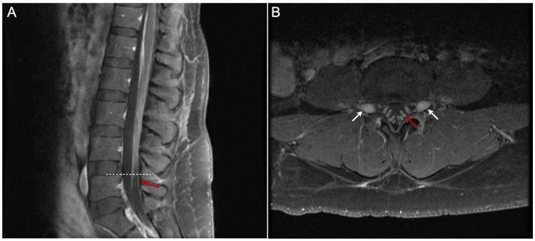
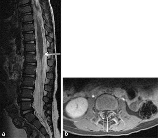
- Lumbar MRI with gadolinium: Enhancement of cauda equina nerve roots (especially in children)
- Brain/spine MRI: primarily to exclude CNS causes (brainstem stroke, transverse myelitis)
MRI lumbar spine in GBS: Post-contrast T1 showing prominent smooth enhancement of cauda equina nerve roots - classic radiological finding
9. DIFFERENTIAL DIAGNOSIS
Warning Signs Suggesting Alternative Diagnosis
| Warning Sign | Alternative Diagnosis |
|---|
| Sensory predominant | Sensory neuronopathy |
| Prominent bowel/bladder symptoms | Myelopathy |
| Spinal sensory level | Myelopathy (transverse myelitis) |
| Persistently asymmetrical weakness | Enteroviral encephalomyelitis, vasculitic mononeuritis multiplex |
| Distal predominant pattern | Toxic neuropathy (arsenic) |
| Slow progression (>4 weeks) | CIDP |
| CSF WBC >50/μL | HIV seroconversion, Lyme disease |
| Fever at onset | Infectious myelitis |
Full Differential Diagnosis Categories (Bradley & Daroff's)
Muscle Disorders: Polymyositis, dermatomyositis, necrotizing autoimmune myopathy, rhabdomyolysis, critical illness myopathy
Muscle Membrane Disorders: Familial periodic paralysis, hypokalemic paralysis (thyrotoxicosis, barium poisoning)
Neuromuscular Junction: Myasthenia gravis, botulism, drug-induced NMJ blockade, organophosphate toxicity, tick paralysis
Neuropathies: Porphyria, diphtheria, vasculitic neuropathy, Lyme radiculopathy
Spinal cord: Transverse myelitis, NMO, vascular myelopathy, cord compression
Key distinction - Botulism vs GBS:
- Botulism: ophthalmoplegia + unreactive pupils + dry mouth + constipation + orthostatic hypotension - NO sensory symptoms - descending paralysis
- GBS: reactive pupils, ascending paralysis, sensory symptoms present
Tick paralysis: mimics GBS in children; removal of tick causes dramatic improvement within hours.
10. MANAGEMENT
A. Monitoring & ICU Criteria
Admit ALL patients except mildest cases for observation. ICU admission indications:
- Vital capacity <1 L (or <15 mL/kg)
- Negative inspiratory force < -70 cm H2O (< 30 cm H2O in some criteria)
- Rapidly deteriorating respiratory function
- Severe autonomic instability
Rule of 20/30/40 for elective intubation:
- VC < 20 mL/kg
- Max. inspiratory pressure < 30 cmH2O
- Max. expiratory pressure < 40 cmH2O
If vital capacity <10 mL/kg → intubate.
B. Specific Immunotherapy (within 2 weeks of onset)
| Treatment | Dose/Regimen | Notes |
|---|
| IVIG (preferred) | 2 g/kg IV divided over 2-5 days | Generally preferred (easier to complete full course) |
| Plasma Exchange (PE) | 5 plasma volumes over 10 days | Equivalent efficacy to IVIG |
| Corticosteroids | NOT recommended | No benefit; methylprednisolone + IVIG shows no long-term advantage |
| Second dose IVIG | Not recommended | No improved outcome; higher complication rate |
Key exam point: IVIG and PE are equivalent in efficacy; combining them does NOT provide additional benefit. Steroids alone are NOT effective in GBS.
Timing: Treatment most effective when initiated within 2 weeks of onset.
C. Autonomic Management
- Hypotension: IV saline, vasopressors (short-term)
- Hypertension: IV labetalol (short-acting, titratable) - use cautiously as BP fluctuates rapidly
- Bradycardia: atropine, temporary pacing in severe cases
- Avoid provocative maneuvers (e.g., suctioning) that can trigger arrhythmias
D. Supportive Care
- DVT prophylaxis: subcutaneous heparin or pneumatic compression boots
- Nutrition: nasogastric feeding if bulbar involvement; watch for adynamic ileus
- SIADH: fluid restriction; distinguish from cerebral salt-wasting (salt replacement)
- Pain management: gabapentin, pregabalin, or carbamazepine for neuropathic pain
- Communication: establish early (letter board, eye-blinking codes) before intubation
- Bladder care, pressure ulcer prevention, physiotherapy
11. PROGNOSIS
| Parameter | Data |
|---|
| Mortality (modern ICU care) | 3-5% (was 33% before positive-pressure ventilation) |
| Full/near-full recovery | ~80% |
| Persistent disability | ~20% |
| Time to maximum deficit | 50% by 2 weeks, 75% by 3 weeks, >90% by 4 weeks |
| Recovery onset | 2-4 weeks after progression ceases |
| Recovery duration | Weeks to months |
Poor prognostic factors:
- Older age
- Preceding diarrheal illness (C. jejuni)
- Axonal variants (AMAN/AMSAN)
- Severe weakness at onset (requiring ventilation)
- Low CMAP amplitudes in upper limbs (axonal loss)
- Rapid onset to nadir
12. GBS vs CIDP - Key Distinctions
| Feature | GBS (Acute) | CIDP (Chronic) |
|---|
| Time to maximum deficit | <4 weeks | >8 weeks (or relapsing) |
| Antecedent infection | Common (60%) | Less common |
| CSF protein | Elevated | Elevated |
| Treatment | IVIG / PE (short course) | IVIG / PE / steroids (long-term) |
| Steroids | No benefit | Beneficial |
| Prognosis | Monophasic, usually good | Chronic, requires maintenance |
13. HIGH-YIELD EXAM MNEMONICS & SUMMARY POINTS
"GBS PACED" Mnemonic (Key Features)
-
Gastrointestinal / respiratory infection precedes by 1-3 weeks
-
Bilateral facial weakness (50%), Bifacial palsy
-
Symmetric ascending weakness + areflexia
-
Protein elevated in CSF (cytoalbuminologic dissociation)
-
Autonomic dysfunction (65%)
-
Campylobacter jejuni = most common identifiable antecedent
-
Electrodiagnostics confirm demyelination
-
Don't give steroids; give IVIG or plasmapheresis
Classic Exam "ONE-LINERS"
- Most common cause of acute flaccid paralysis in an unvaccinated adult = GBS
- CSF finding = albuminocytologic dissociation (protein ↑, cells normal)
- MFS triad = Ophthalmoplegia + Ataxia + Areflexia + anti-GQ1b antibody
- Antibody for MFS = anti-GQ1b (95-98% positive)
- Drug of choice = IVIG 2g/kg over 2-5 days (or PE; steroids are NOT used)
- Autonomic dysfunction in = 65%
- Ventilation needed in = 25%
- Maximal deficit at 4 weeks (diagnostic; if >8 weeks → CIDP)
- Post-C. jejuni variant = AMAN (axonal, motor-only, poor prognosis)
- Swine flu vaccine (1976 A/NJ) = classic GBS-vaccine association
Pathogenesis diagram: C. jejuni molecular mimicry triggers cross-reactive antibodies against GM1 gangliosides (GBS/AMAN) vs direct autoimmunity in CIDP
14. QUICK REFERENCE - SUBTYPES COMPARISON TABLE
| Feature | AIDP | AMAN | AMSAN | MFS |
|---|
| Prevalence | 97% (US/Europe) | Common in China | Rare | 6% (West), 18% (Taiwan) |
| Pathology | Demyelination | Axonal (motor) | Axonal (motor+sensory) | Demyelination (CN) |
| NCS | Slow CV, prolonged DL | Normal CV, low CMAP | Normal CV, low CMAP+SNAP | Normal |
| Antibody | None specific | Anti-GM1, Anti-GD1a | Anti-GD1b | Anti-GQ1b |
| Preceding infection | Mixed | C. jejuni | C. jejuni | Various |
| Prognosis | Good | Variable | Poor | Excellent |
| Clinical | Classic ascending | Pure motor | Motor + sensory | OAA triad |
Sources used: Adams & Victor's Principles of Neurology (12th Ed), Bradley & Daroff's Neurology in Clinical Practice, Goldman-Cecil Medicine (International Ed), Robbins & Kumar Basic Pathology