
membranoproliferative glomerulonephritis
Reading File
Finding Sources
Finding Sources
Searching PubMed
"membranoproliferative glomerulonephritis"
Searching the Web
KDIGO membranoproliferative glomerulonephritis management guideline complement mediated glomerulopathy
Reading File
Reading File
Reading File
Searching Images
membranoproliferative glomerulonephritis light microscopy tram track double contour immunofluorescence
Membranoproliferative glomerulonephritis (MPGN)
MPGN is a pattern of glomerular injury, not one single disease. It is characterized by mesangial proliferation, capillary-wall remodeling, and basement-membrane thickening. Modern classification is based mainly on immunofluorescence and the underlying mechanism, rather than the old types I, II, and III.
Classification
| Category | Main mechanism | Typical associations |
|---|---|---|
| Immune complex-mediated MPGN (IC-MPGN) | Immune-complex deposition with complement activation | Hepatitis C or B, chronic infection such as endocarditis, mixed cryoglobulinemia, SLE and other autoimmune disease, monoclonal gammopathy |
| Complement-mediated MPGN / C3 glomerulopathy | Alternative-complement pathway dysregulation | C3 nephritic factor, antibodies to complement-regulatory proteins, mutations involving factor H, factor I, C3, etc. |
| MPGN pattern without prominent immune deposits or complement | Chronic endothelial injury / thrombotic microangiopathy-like processes | Antiphospholipid syndrome, TTP, drugs, radiation, transplant-related injury, prothrombotic disorders |
Historically, “type I” largely corresponds to immune-complex MPGN, while “type II” is now called dense deposit disease, a form of C3 glomerulopathy. - Robbins & Kumar Basic Pathology, p. 507
Pathology
- Light microscopy: enlarged hypercellular glomeruli, mesangial and endocapillary proliferation, lobular accentuation.
- Classic feature: double-contoured glomerular basement membrane, the “tram-track” appearance.
- Mechanism of tram-tracking: mesangial-cell interposition, matrix formation, and immune-deposit-related remodeling of the capillary wall.
- Immunofluorescence:
- IC-MPGN: immunoglobulins plus C3, often granular.
- C3 glomerulopathy: dominant C3 staining with little or no immunoglobulin.
- Electron microscopy: often subendothelial deposits; dense intramembranous deposits suggest dense deposit disease.
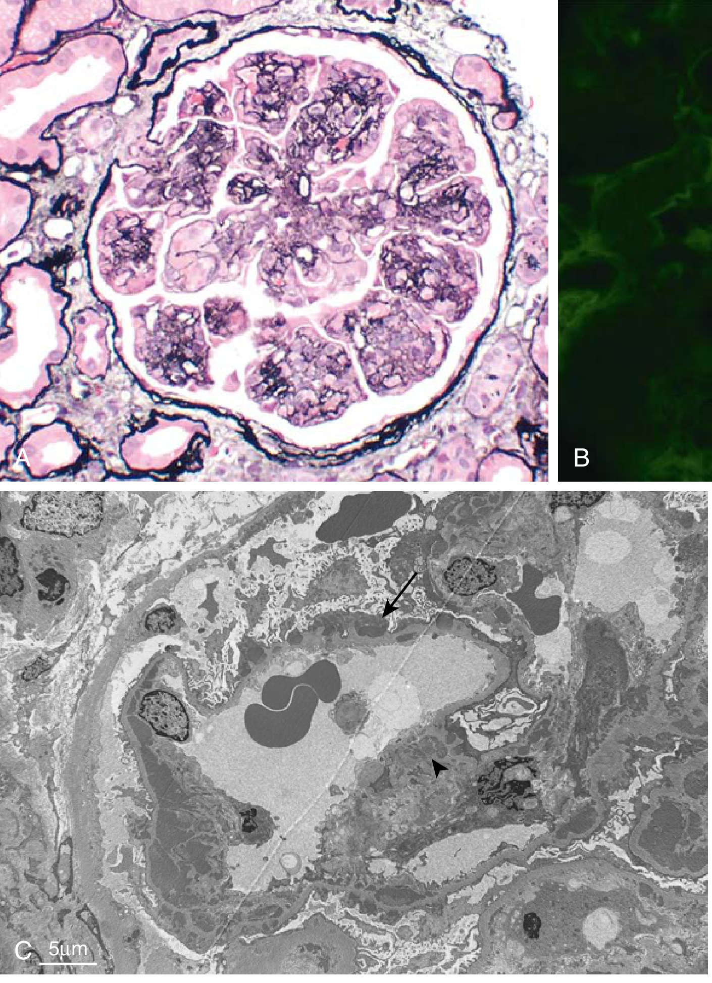
- Robbins & Kumar Basic Pathology, p. 507
Clinical presentation
Patients may present with a mixed nephritic-nephrotic syndrome:
- Hematuria, sometimes with RBC casts
- Proteinuria, potentially nephrotic-range
- Edema and hypertension
- Reduced GFR / elevated creatinine
- Low complement levels:
- Low C3 and C4 can occur with immune-complex disease
- Predominantly low C3 suggests alternative-pathway complement activation / C3 glomerulopathy
Diagnostic work-up
- Urinalysis, urine protein quantification, serum creatinine/eGFR, albumin, lipids.
- Complement testing: C3, C4, CH50/AH50.
- Evaluate secondary causes:
- Hepatitis B, hepatitis C, HIV
- Blood cultures if endocarditis or chronic infection is possible
- ANA, anti-dsDNA, cryoglobulins
- Serum and urine electrophoresis, immunofixation, and serum free light chains for monoclonal gammopathy
- Kidney biopsy with light microscopy, immunofluorescence, and electron microscopy is needed to define the category.
Treatment principles
Treatment is directed at the cause and severity, plus kidney-protective care.
- All patients: blood-pressure control, ACE inhibitor or ARB when appropriate to reduce proteinuria, salt restriction, diuretics for edema, and management of dyslipidemia and CKD complications.
- IC-MPGN: identify and treat the driver, such as hepatitis C with direct-acting antivirals, infection with appropriate antimicrobial therapy, autoimmune disease with disease-specific therapy, or a monoclonal gammopathy with clone-directed hematologic treatment.
- C3 glomerulopathy: specialist nephrology assessment, complement testing and selected genetic/autoantibody studies. Immunosuppression such as mycophenolate plus glucocorticoids may be used in active moderate-to-severe disease; complement-directed therapies may be considered in selected cases or clinical trials.
- Do not assume that a biopsy labeled “MPGN” automatically needs steroids. The underlying immune-complex, infection-related, or complement-mediated cause determines treatment.
KDIGO emphasizes that MPGN on biopsy is a histologic pattern reflecting immune-complex deposition and/or complement dysregulation, rather than a stand-alone diagnosis. The KDIGO 2021 guideline summary uses this pathophysiologic framework.
Prognosis
The course varies substantially. Persistent heavy proteinuria, impaired kidney function at presentation, hypertension, low eGFR, and chronic scarring on biopsy predict poorer renal outcomes. Treating a reversible underlying condition can improve or stabilize disease.
A recent systematic review highlights that proteinuria remains closely associated with adverse outcomes in C3 glomerulopathy and primary IC-MPGN (PMID 40971335).
This is a shared conversation. Sign in to Orris to start your own chat.