
applied anatomy of rectus sheath. For 1st mbbs students easy to learn
Reading File
The rectus sheath is formed by the aponeuroses of the three flat muscles of the anterior abdominal wall (external oblique, internal oblique, transversus abdominis). Its composition differs above and below the arcuate line - above it, the internal oblique aponeurosis splits to enclose the rectus abdominis, while below the arcuate line all three aponeuroses pass anterior to the muscle.
Searching sources...
Finding Sources
Searching the Web
rectus sheath applied anatomy 1st MBBS easy explanation arcuate line
Good - I have Gray's Anatomy for Students and the THIEME Atlas. Let me read those sections plus check for more detailed anatomy content.
Reading File
Reading File
Finding Sources
Excellent - great content from Gray's and THIEME. Let me now read the Schwartz's Surgery section (which has surgical/applied content) and fetch the diagram image from Gray's.
Reading File
Loading Image
Loading Image
I now have excellent, comprehensive content from authoritative sources. Let me also quickly get the contents inside the rectus sheath and applied importance sections.
Finding Sources
Finding Sources
I have all the information needed. Let me now read the Sabiston section on rectus sheath hematoma for the applied part.
Reading File
Reading File
I now have all the content needed from authoritative textbooks. Let me write a comprehensive, well-organized, student-friendly answer.
Applied Anatomy of Rectus Sheath - Easy Notes for 1st MBBS
What is the Rectus Sheath?
The rectus sheath is an aponeurotic (fibrous) envelope that wraps around the rectus abdominis and pyramidalis muscles. It is formed by the fusion of the aponeuroses (flat tendons) of the three flat muscles of the anterolateral abdominal wall:
- External oblique
- Internal oblique
- Transversus abdominis
Think of it as a "jacket" made by splitting and interweaving these three aponeuroses around the rectus abdominis.
- Gray's Anatomy for Students, p. 345
Formation - The KEY Rule (Above vs Below Arcuate Line)
This is the single most important concept - and the most common exam question.
Here is the cross-section diagram from Gray's Anatomy showing both levels:
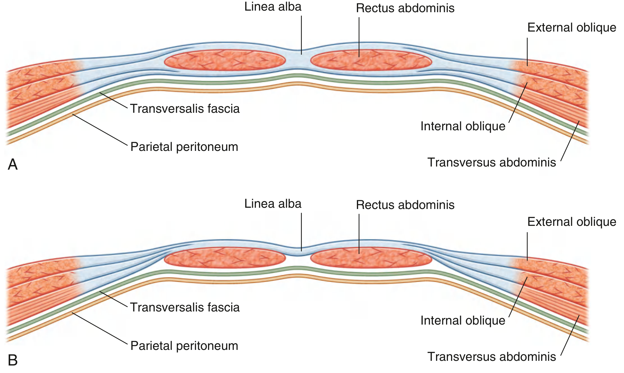
Fig A = above arcuate line; Fig B = below arcuate line. Note how in B, the posterior wall is absent and only transversalis fascia lies behind the muscle.
And the detailed labeled diagram from the THIEME Atlas:
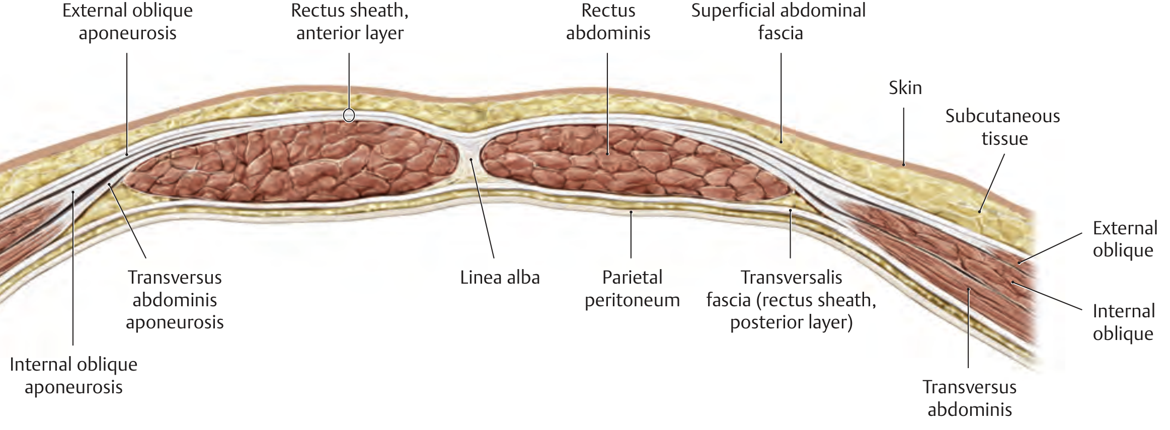
The Arcuate Line (of Douglas)
- Located midway between the umbilicus and pubic symphysis (roughly 3-5 cm below the umbilicus)
- It is the lower curved free edge of the posterior wall of the rectus sheath
- Below this line, the posterior wall simply disappears
ABOVE the Arcuate Line (Upper 3/4 of rectus)
| Wall | Composition |
|---|---|
| Anterior wall | Aponeurosis of External oblique + Anterior half of Internal oblique aponeurosis |
| Posterior wall | Posterior half of Internal oblique aponeurosis + Aponeurosis of Transversus abdominis |
Easy memory trick: "Internal oblique splits - half goes front, half goes back"
BELOW the Arcuate Line (Lower 1/4 of rectus)
| Wall | Composition |
|---|---|
| Anterior wall | All three aponeuroses together (EO + IO + TA) |
| Posterior wall | ABSENT - only transversalis fascia covers the muscle |
Easy memory trick: "Below the line, all three go to the front"
Why does this happen? The THIEME Atlas explains it well: "the two layers blend together approximately 3-5 cm below the umbilicus to form a single, more stable sheet that passes in front of the rectus abdominis muscle." - THIEME Atlas of Anatomy, p. 200
Quick Summary Table
| Feature | Above Arcuate Line | Below Arcuate Line |
|---|---|---|
| Anterior wall | EO + anterior IO | EO + IO + TA (all 3) |
| Posterior wall | Posterior IO + TA | Absent |
| Posterior cover | Posterior rectus sheath | Transversalis fascia only |
| Muscle contact | Posterior sheath | Transversalis fascia directly |
Contents of the Rectus Sheath
Inside the sheath you will find:
- Rectus abdominis muscle - the main occupant (paired, vertical muscles)
- Pyramidalis muscle - small triangular muscle in front of lower rectus, may be absent; attached to pubis below and linea alba above
- Superior epigastric artery - terminal branch of internal thoracic artery, enters from above
- Inferior epigastric artery - branch of external iliac artery, enters from below
- Venae comitantes (companion veins) of above arteries
- Lymphatics
- Lower 5 intercostal nerves (T7-T11) and subcostal nerve (T12) - pierce the sheath laterally to supply the rectus and skin
The superior and inferior epigastric arteries anastomose within the sheath - this is clinically important (see below).
Boundaries and Relations
| Border | Structure |
|---|---|
| Medial | Linea alba (meets the opposite sheath) |
| Lateral | Linea semilunaris (semilunar line) - a curved line visible on surface |
| Superior | Costal margin (5th, 6th, 7th costal cartilages, xiphoid) |
| Inferior | Pubic crest and pubic symphysis |
Applied Anatomy (Clinical Importance)
This is what makes the topic interesting for surgeons and clinicians.
1. Rectus Sheath Hematoma
-
Most common applied complication - frequently asked in exams
-
Blood collects within the sheath, usually from damage to the inferior epigastric artery or its branches
-
Why around the arcuate line? Because below the arcuate line, the posterior wall is absent and the artery is relatively fixed - it is vulnerable to shearing forces from muscle contraction. Above the arcuate line, the posterior sheath provides a tamponade effect; below it, blood can spread more freely.
-
Causes: Trauma, vigorous coughing/sneezing, anticoagulation therapy, trocar placement during laparoscopy, pregnancy
-
Presentation: Acute abdominal pain + palpable abdominal wall mass
-
Fothergill's Sign: A mass in the abdominal wall that remains palpable and does NOT disappear (or does not change position) when the rectus is contracted - this distinguishes it from an intraperitoneal mass which would become impalpable on contraction
-
Diagnosis: Ultrasound or CT scan with contrast
-
Treatment: Conservative (rest, reverse anticoagulation) for stable cases; angiographic embolization or surgical ligation for severe cases
-
Schwartz's Principles of Surgery, p. 1584
2. Surgical Incisions Through the Rectus Sheath
- Paramedian incision: Made just lateral to the linea alba, through the anterior wall of the sheath; the rectus is retracted laterally, and the posterior wall is incised - good access, strong closure
- Midline (linea alba) incision: Between the two sheaths; avascular, no muscle cutting - fastest and most commonly used
- Pfannenstiel incision: Horizontal cut through both anterior walls of the rectus sheath just above the pubis; rectus muscles are retracted sideways - used in gynecological and obstetric surgery; cosmetically placed in skin crease
3. Rectus Sheath Block (Analgesia)
- Local anesthetic is injected into the potential space between the rectus muscle and the posterior sheath
- Used for post-operative pain relief after midline abdominal surgery
- Effective because the terminal branches of T7-T11 nerves run in this plane
- Morgan and Mikhail's Clinical Anesthesiology, p. 3932
4. Epigastric Anastomosis - Collateral Circulation
- The superior and inferior epigastric arteries anastomose within the sheath
- This forms a collateral pathway between the internal thoracic artery (subclavian) and external iliac artery
- Important in aortic occlusion (Leriche syndrome) - collateral flow can reconstitute below the obstruction
5. Linea Semilunaris - Surface Anatomy
- The lateral border of the rectus sheath, curved like a half-moon
- Clinically used to mark the lateral edge for surgical planning and nerve blocks
6. Spigelian Hernia
- An uncommon hernia that occurs through a defect in the linea semilunaris (at the junction of the semilunar line and the arcuate line)
- Often interparietal (between the abdominal wall layers) so may be difficult to palpate
- More common below the arcuate line because the posterior sheath is absent there, providing a weak point
7. Diastasis Recti
- Abnormal widening of the linea alba, causing the two rectus muscles to separate
- Common after pregnancy or obesity (raised intra-abdominal pressure)
- NOT a true hernia (midline fascia is intact; no hernial sac) - no risk of incarceration
- Presents as a fusiform bulge along the midline that worsens with raising the head or doing sit-ups
- Schwartz's Principles of Surgery, p. 1584
Memory Aid - MBBS Summary
RECTUS SHEATH - Quick Recall
Formation:
Above arcuate line → EO + (½ IO in front) + (½ IO + TA behind)
Below arcuate line → All 3 aponeuroses in FRONT only
Contents: Rectus + Pyramidalis + Superior & Inferior Epigastric A&V + T7-T12 nerves
Arcuate line = midway between umbilicus and pubic symphysis
Applied:
Hematoma → damaged inferior epigastric A, Fothergill's sign
Spigelian H → at linea semilunaris + arcuate line junction
Diastasis → NOT a hernia, no incarceration
Sheath block → anaesthesia for T7-T11 dermatomes
Sources:
- Gray's Anatomy for Students (Gray's), p. 344-345
- Schwartz's Principles of Surgery, 11e, p. 1580-1584
- THIEME Atlas of Anatomy - General Anatomy and Musculoskeletal System, p. 200
- Sabiston Textbook of Surgery, p. 2235
- Morgan and Mikhail's Clinical Anesthesiology, 7e, p. 3932
Usse vishram singh anatomy as reference
Finding Sources
Finding Sources
Unfortunately, Vishram Singh's Anatomy is not available in our textbook library. The library has these anatomy books:
- Gray's Anatomy for Students
- THIEME Atlas of Anatomy (General Anatomy & Musculoskeletal System)
- Netter's Atlas of Human Anatomy
- Color Atlas of Human Anatomy (Vols 1, 2, 3)
- Color Atlas of Ultrasound Anatomy
- Imaging Anatomy (Bones, Joints, Vessels & Nerves)
Vishram Singh is a very popular reference for Indian MBBS students - I completely understand why you prefer it. Since it is not in the library, I have two options for you:
-
Use Netter's + Gray's - both follow similar content structure to Vishram Singh for rectus sheath, and I can present it in Vishram Singh's style (point-wise, with applied anatomy highlighted separately the way he does)
-
You upload the relevant pages from Vishram Singh's book as an image/PDF, and I will use that exact content to build the notes
Which would you prefer?
Create notes using Netter's & Gray's in Vishram Singh's style
Finding Sources
Finding Sources
Reading File
Finding Sources
Reading File
Finding Sources
Reading File
Finding Sources
Loading Image
Loading Image
Loading Image
Here are the notes written in Vishram Singh's style - numbered points, bold headings, separate applied anatomy box, clinical correlations highlighted, and a clean table format exactly as he presents it.
RECTUS SHEATH
(Ref: Gray's Anatomy for Students, p. 344-345; Netter's Atlas of Human Anatomy)
INTRODUCTION
The rectus sheath is an aponeurotic compartment formed by the interlacing aponeuroses of the three flat muscles of the anterolateral abdominal wall. It encloses the rectus abdominis and pyramidalis muscles along with their vessels and nerves.
FORMATION
The rectus sheath is formed by the aponeuroses of:
- External oblique
- Internal oblique
- Transversus abdominis
Key Point: The internal oblique aponeurosis splits at the lateral border of the rectus abdominis into two lamellae - this is the reason the sheath has different compositions above and below the arcuate line.
ARCUATE LINE (Line of Douglas)
- It is a curved free lower border of the posterior wall of the rectus sheath
- Located midway between the umbilicus and pubic symphysis
- Below this line, the posterior wall of the sheath is completely absent
- The rectus abdominis below the arcuate line is in direct contact with the transversalis fascia
COMPOSITION OF THE RECTUS SHEATH
Fig. 4.33 - Gray's Anatomy for Students: A = Above arcuate line; B = Below arcuate line
Above the Arcuate Line (Upper 3/4)
| Wall | Formed by |
|---|---|
| Anterior wall | Aponeurosis of external oblique + Anterior lamella of internal oblique |
| Posterior wall | Posterior lamella of internal oblique + Aponeurosis of transversus abdominis |
Below the Arcuate Line (Lower 1/4)
| Wall | Formed by |
|---|---|
| Anterior wall | Aponeuroses of all three muscles (EO + IO + TA) |
| Posterior wall | Absent - only transversalis fascia present |
Memory Aid:
- Above arcuate line: IO splits - half front, half back ("IO shares")
- Below arcuate line: All three go to front ("All go front")
CONTENTS OF THE RECTUS SHEATH
The rectus sheath contains:
-
Rectus abdominis muscle
- Long flat paired muscle
- Origin: Pubic crest, pubic symphysis
- Insertion: Costal cartilages of ribs V-VII and xiphoid process
- Has 3-4 tendinous intersections along its length (visible in well-built individuals)
-
Pyramidalis muscle (may be absent)
- Small triangular muscle lying in front of lower rectus
- Origin: Front of pubis
- Insertion: Linea alba
- Nerve supply: T12 (subcostal nerve)
- Function: Tenses the linea alba
-
Superior epigastric artery - terminal branch of internal thoracic artery; enters sheath from above
-
Inferior epigastric artery - branch of external iliac artery; enters sheath from below
-
Venae comitantes of both epigastric arteries
-
Lower 5 intercostal nerves (T7-T11) and subcostal nerve (T12) - run in the plane between rectus and posterior sheath
BLOOD SUPPLY WITHIN THE SHEATH
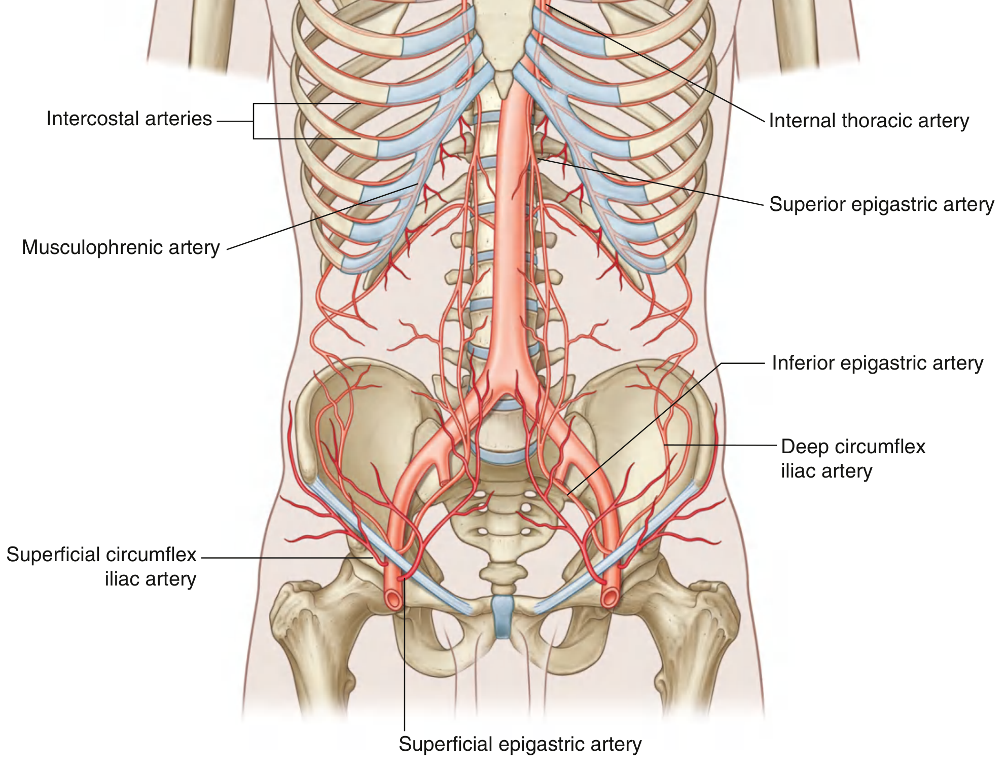
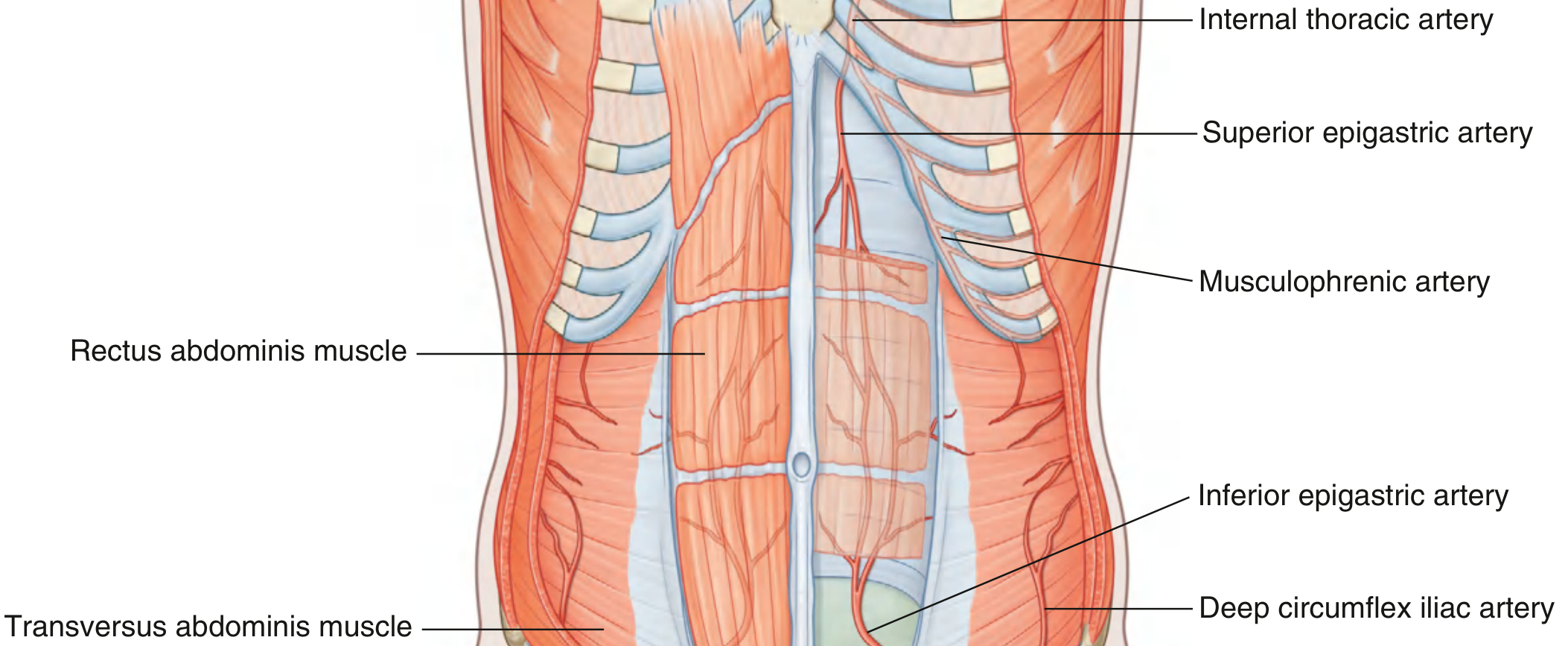
- The superior and inferior epigastric arteries both run posterior to the rectus abdominis muscle
- They anastomose with each other near the umbilicus
- This anastomosis forms a collateral pathway between:
- Internal thoracic artery (subclavian system) above
- External iliac artery below
NERVE SUPPLY OF RECTUS ABDOMINIS
- Anterior rami of T7-T12 (lower 6 intercostal and subcostal nerves)
- These nerves pierce the posterior wall of the rectus sheath laterally and enter the sheath to supply the muscle
- Cutaneous distribution: T10 = umbilicus level (important landmark)
SURFACE MARKING
- Medial border: Linea alba (meets opposite sheath in midline)
- Lateral border: Linea semilunaris (semilunar line) - a curved line seen on the surface of a muscular abdomen, marking where the aponeuroses begin
APPLIED ANATOMY
(This section corresponds to Vishram Singh's "Clinical Correlations" boxes)
1. Rectus Sheath Hematoma ⭐ (Most Important)
- Definition: Collection of blood within the rectus sheath due to rupture of the inferior epigastric artery or its branches
- Why near arcuate line? Below the arcuate line, posterior sheath is absent - the artery is exposed and fixed, making it vulnerable to shearing during sudden muscle contraction. Above the arcuate line, the posterior sheath acts as a tamponade.
- Causes:
- Vigorous coughing, sneezing, straining (especially in patients on anticoagulants)
- Trauma / direct blow
- Laparoscopic trocar injury
- Pregnancy
- Clinical features: Acute abdominal pain + tender, palpable abdominal wall mass
- Fothergill's Sign: The mass remains palpable (and does NOT disappear) when the rectus is contracted - because it is within the abdominal wall, not inside the peritoneal cavity. An intraperitoneal mass becomes impalpable on rectus contraction.
- Diagnosis: Ultrasound or CECT abdomen
- Treatment: Conservative (rest, reverse anticoagulation) for stable cases; angiographic embolization for large/unstable hematomas
2. Surgical Incisions
| Incision | Description | Common Use |
|---|---|---|
| Midline (linea alba) | Between the two sheaths, through avascular linea alba | Laparotomy - fastest, no muscle cutting |
| Paramedian | Through anterior sheath, rectus retracted laterally, posterior sheath incised | Gives strong closure |
| Pfannenstiel | Horizontal cut through both anterior sheaths above pubis; muscles retracted apart | Gynaecology, obstetrics, Caesarean section |
3. Rectus Sheath Block (Anaesthesia)
- Local anaesthetic injected into the plane between rectus muscle and posterior wall of the sheath
- Blocks T7-T11 anterior cutaneous branches
- Used for post-operative analgesia after midline abdominal surgery
4. Epigastric Anastomosis - Collateral Circulation
- The anastomosis between superior and inferior epigastric arteries is an important collateral channel connecting the subclavian system (above) and the femoral system (below)
- This becomes clinically significant in aortic occlusion - blood can bypass the obstruction via: internal thoracic → superior epigastric → inferior epigastric → external iliac
5. Spigelian Hernia
- Hernia through the linea semilunaris (lateral border of rectus sheath)
- Most commonly occurs at the junction of the semilunar line and arcuate line - because here the posterior sheath is absent, creating a weak point
- Often interparietal (between layers), so not easily visible on the surface - can be missed on clinical examination
6. Diastasis Recti
- Abnormal separation/widening of the linea alba, causing the two rectus muscles to be unusually far apart
- NOT a true hernia - no hernial sac, no risk of strangulation or incarceration
- Causes: Pregnancy, obesity, chronic raised intra-abdominal pressure
- Signs: A fusiform bulge along the midline, worst when the patient raises their head (contracts the rectus)
- Treatment: Weight loss, physiotherapy; surgery only if severe functional disability
QUICK REVISION BOX
RECTUS SHEATH - AT A GLANCE
Formed by aponeuroses of EO, IO, TA
Above arcuate line:
- Ant wall = EO + anterior IO
- Post wall = posterior IO + TA
Below arcuate line:
- Ant wall = EO + IO + TA (ALL THREE)
- Post wall = ABSENT (transversalis fascia only)
Arcuate line = midway umbilicus to pubic symphysis
Contents = Rectus + Pyramidalis + Sup/Inf epigastric A&V + T7-T12 nerves
Applied:
- Hematoma → inferior epigastric injury, Fothergill's sign
- Spigelian hernia → at linea semilunaris + arcuate line
- Diastasis recti → NOT a hernia
- Epigastric anastomosis → collateral in aortic occlusion
Sources: Gray's Anatomy for Students (Gray's), pp. 344-345, 349; Netter's Atlas of Human Anatomy; Schwartz's Principles of Surgery, 11e
This is a shared conversation. Sign in to Orris to start your own chat.