
Nutcracker and sma
nutcracker syndrome left renal vein compression aorta SMA
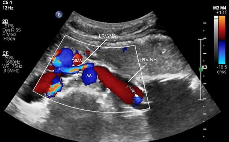
This color Doppler ultrasound image in the transverse plane demonstrates the anatomical relationship between the abdominal aorta (AA), superior mesenteric artery (SMA), and left renal vein (LRV), illustrating Nutcracker syndrome (renal vein compression syndrome). The SMA is positioned anterior to the AA, creating an acute angle where the meso-aortic segment of the left renal vein (LRV-MA) is seen compressed between these two arterial structures. The color Doppler indicates flow direction relative to the transducer, with the color bar ranging from +18.5 cm/s (red, toward) to -18.5 cm/s (blue, away). The AA and SMA show blue signals, indicating flow away from the transducer. The hilar segment of the left renal vein (LRV-NH) is significantly dilated compared to the compressed LRV-MA segment, a hallmark diagnostic feature of anterior Nutcracker syndrome. The LRV-NH exhibits a predominantly red signal, indicating venous return toward the transducer before it enters the narrow meso-aortic space. This imaging is critical for assessing venous hypertension and potential collateral circulation in patients presenting with hematuria or pelvic pain.
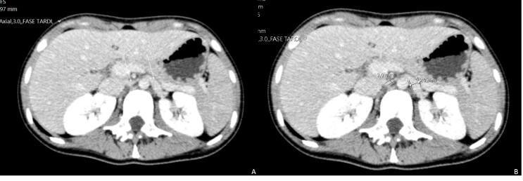
This diagnostic image consists of two side-by-side axial contrast-enhanced abdominal CT scans (labeled A and B) demonstrating the classic radiographic features of Nutcracker Syndrome. The images show the anatomical relationship between the abdominal aorta, the superior mesenteric artery (SMA), and the left renal vein. In image A, the left renal vein is seen passing through the narrow aortomesenteric angle. Image B provides quantitative measurements of this compression: the caliber of the left renal vein is significantly reduced to 3.0 mm within the aortomesenteric space, while the pre-stenotic segment of the vein near the renal hilum is dilated to 9.0 mm. Key anatomical landmarks visible include the liver (right), stomach (left), and both kidneys flanking the spine. This comparison highlights the vascular 'nutcracker' effect where the SMA compresses the left renal vein against the aorta, potentially leading to renal venous hypertension and associated clinical symptoms.
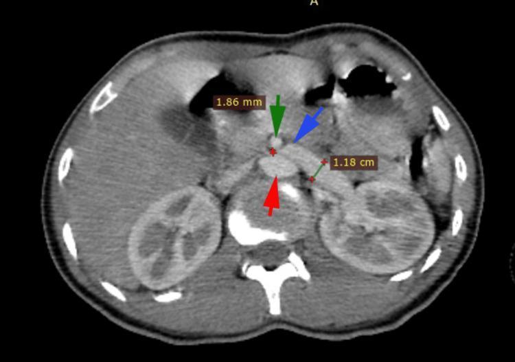
This diagnostic image is an axial contrast-enhanced abdominal CT scan demonstrating the anatomical hallmarks of Nutcracker Syndrome (renal vein entrapment). The image highlights the critical relationship between major abdominal vessels. An abdominal aorta (indicated by a red arrow) is positioned posteriorly. Anterior to it, the superior mesenteric artery (SMA, indicated by a green arrow) is shown with a measured diameter of 1.86 mm. The left renal vein (LRV, indicated by a blue arrow) is visibly compressed as it passes through the narrow angle between the SMA and the aorta. A distal segment of the left renal vein is measured at 1.18 cm, illustrating pre-stenotic dilatation or engorgement compared to the compressed segment. The visual evidence of this vascular arrangement explains the extrinsic compression of the LRV, which clinically often manifests as hematuria and pelvic congestion. The scan also captures cross-sections of both kidneys and the vertebral column, providing clear orientation for the retroperitoneal structures involved.
superior mesenteric artery syndrome duodenal compression
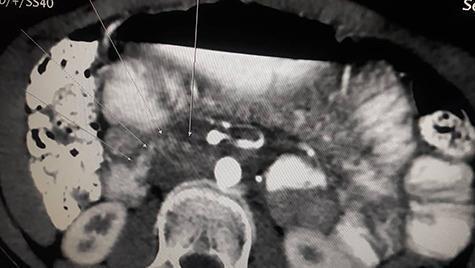
This diagnostic axial contrast-enhanced CT scan of the abdomen demonstrates classic findings of Superior Mesenteric Artery (SMA) Syndrome. The image shows significant proximal duodenal dilation transitioning into an abrupt narrowing (transition zone) as the duodenum passes through the angle between the abdominal aorta (AA) and the superior mesenteric artery (SMA). The third part of the duodenum is visibly compressed in the narrow aortomesenteric space. The scan highlights a reduced aortomesenteric distance and angle, leading to proximal gastrointestinal obstruction. Annotations indicate the key vascular landmarks (SMA and aorta) and the site of duodenal compression. This imaging is clinically significant for diagnosing Wilkie's syndrome in patients presenting with symptoms of high intestinal obstruction. It serves as an educational example of vascular compression of the gastrointestinal tract and the radiological evaluation of the aortomesenteric region.
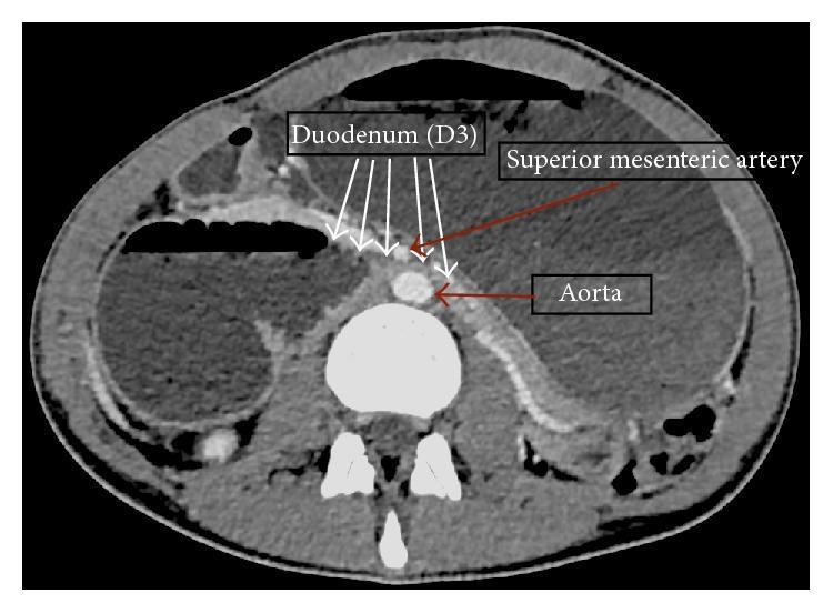
An axial contrast-enhanced CT scan of the abdomen demonstrating the hallmark anatomical findings of Superior Mesenteric Artery (SMA) syndrome. The image highlights the compression of the third part of the duodenum (D3) as it passes through the narrow aortomesenteric angle, formed between the abdominal aorta posteriorly and the superior mesenteric artery anteriorly. Key landmarks include the hyperdense, contrast-filled circular lumen of the aorta and the smaller SMA originating from its anterior wall. The D3 segment is visibly narrowed (beaked) at the midline. Secondary signs of high-grade proximal obstruction are evident, including a markedly distended stomach and dilated proximal duodenum. The scan also provides visualization of the left renal vein in close proximity, relevant for the 'Nutcracker phenomenon' associated with this vascular geometry. This diagnostic image is intended for educational use in gastroenterology and radiology to illustrate the mechanism of vascular compression leading to duodenal obstruction.
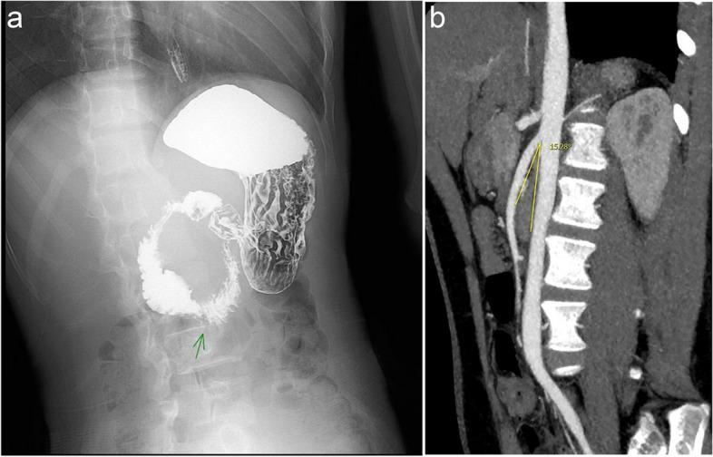
This composite diagnostic image illustrates the radiological findings of Superior Mesenteric Artery Syndrome (SMAS). Panel a presents an abdominal X-ray from a barium meal study, showing significant gastric distension and dilation of the first and second portions of the duodenum. A characteristic abrupt narrowing is visible at the third duodenal portion (indicated by a green arrow), known as the 'penholder sign,' representing extrinsic compression. Panel b shows a sagittal CT angiography (CTA) reconstruction of the abdominal aorta and the superior mesenteric artery (SMA). Annotated yellow lines measure the aortomesenteric angle at 15.28 degrees, which is significantly below the normal range (typically 25-60 degrees). This reduced angle demonstrates the mechanical compression of the duodenum as it passes between the SMA and the aorta. These imaging modalities collectively demonstrate the classic diagnostic criteria for SMAS: proximal duodenal obstruction and a narrowed vascular angle.
Nutcracker Syndrome & SMA Syndrome
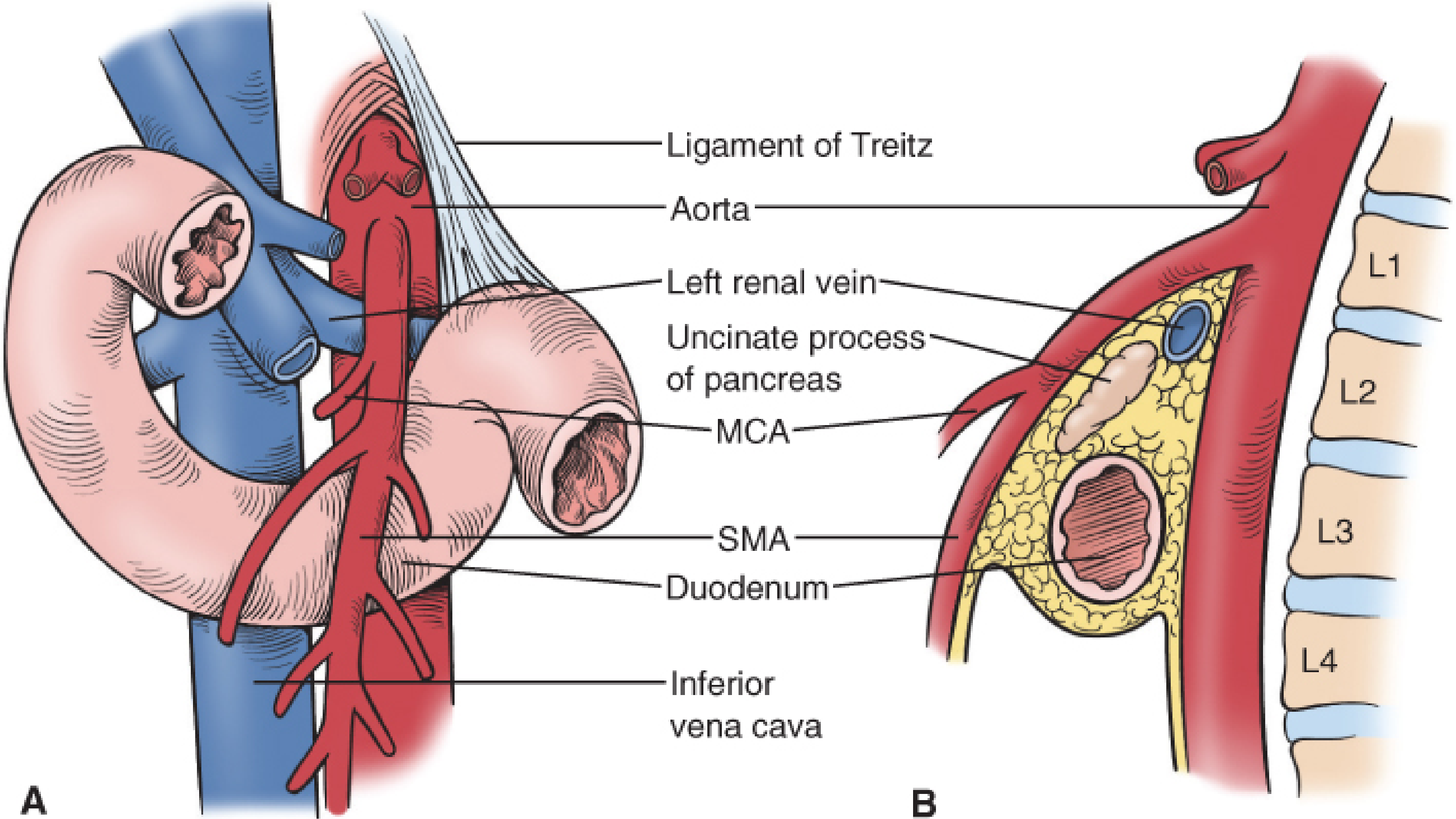
Nutcracker Syndrome (Renal Vein Entrapment Syndrome)
What gets compressed?
Pathophysiology
Clinical Features
- Gross or microscopic hematuria (most common presenting feature) - typically left-sided on cystoscopy
- Left flank / loin pain
- Left-sided varicocele in males (from gonadal vein reflux)
- Pelvic congestion syndrome in females (dyspareunia, dysmenorrhea, chronic pelvic pain) - from Berek & Novak's Gynecology
- Orthostatic proteinuria in children
Imaging
- Color Doppler ultrasound: dilated LRV hilar segment vs. compressed aortomesenteric segment (ratio >4:1 is suggestive); increased peak velocity in the compressed segment
- CT/CTA: shows the compressed LRV with pre-stenotic dilatation; measures the aortomesenteric angle
Management
- Conservative: watchful waiting in young patients (especially children), as spontaneous resolution can occur with growth and weight gain
- Surgical:
- Left renal vein transposition (repositioning behind the aorta)
- SMA transposition
- Nephrectomy (last resort)
- Endovascular: LRV stenting - less invasive, increasingly reported - Campbell-Walsh Wein Urology
SMA Syndrome (Wilkie's Syndrome / Vascular Compression of the Duodenum)
Other names
What gets compressed?
Why does fat matter?
Predisposing factors
| Category | Examples |
|---|---|
| Rapid weight loss | Anorexia nervosa, post-surgical |
| Immobilization | Spinal surgery, body casts ("cast syndrome"), prolonged supine positioning |
| Anatomic variants | Low SMA origin, high/foreshortened ligament of Treitz, excessive lumbar lordosis |
| Post-surgical | Scoliosis surgery, proctocolectomy, aortic aneurysm repair |
| Rapid growth | Tall, thin adolescents |
Clinical Features
- Nausea and vomiting (bilious)
- Postprandial epigastric pain (intermittent to constant depending on severity)
- Early satiety, abdominal distension
- Weight loss (often precedes other symptoms)
- Young, thin/asthenic females more commonly affected - Sabiston Textbook of Surgery, Sleisenger & Fordtran's
Diagnosis
- Barium upper GI series: abrupt cutoff at D3 ("to and fro" peristalsis) with proximal dilatation; a "penholder sign" / extrinsic compression is seen, especially with patient supine
- CT/CTA: measures the aortomesenteric angle and distance; confirms duodenal compression
Management
- Nutritional rehabilitation - high-calorie enteral or parenteral nutrition to rebuild mesenteric fat
- Postural maneuvers (left lateral decubitus, prone, knee-chest position - relieves compression)
- Nasojejunal tube feeding past the obstruction
- Metoclopramide (prokinetic)
- Duodenojejunostomy (most common - bypasses the obstructed segment; can be done laparoscopically)
- Gastrojejunostomy
- Strong procedure (duodenal derotation - mobilizes the duodenum and divides the ligament of Treitz)
- Sleisenger & Fordtran's Gastrointestinal and Liver Disease; Sabiston Textbook of Surgery
Side-by-Side Comparison
| Feature | Nutcracker Syndrome | SMA Syndrome |
|---|---|---|
| Structure compressed | Left renal vein | 3rd part of duodenum |
| Compressing structures | Aorta + SMA | Aorta + SMA |
| Key symptom | Hematuria, pelvic pain | Nausea, vomiting, weight loss |
| Key precipitant | Thin body habitus, growth | Rapid weight loss, immobilization |
| Diagnostic test | Doppler US, CT venography | Barium meal, CT (aortomesenteric angle) |
| Diagnostic threshold | LRV ratio >4:1 | Angle ≤25°, distance ≤8 mm |
| Conservative Rx | Watchful waiting | Nutritional rehab, positioning |
| Surgery | LRV transposition / stenting | Duodenojejunostomy |
| Specialty | Urology / Vascular | GI / General surgery |