
Complications of third stage of labour
"Postpartum Hemorrhage"[MeSH Terms] AND "third stage of labor"
uterine inversion postpartum
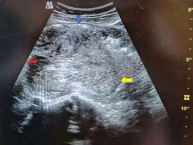
This bedside transabdominal ultrasound image demonstrates an acute uterine inversion in a postpartum patient. The diagnostic imaging displays a characteristic loss of normal uterine contour. A blue arrow identifies the serosal/plasma layer of the anterior uterine wall, which appears as a hypoechoic linear structure. A red arrow highlights the diagnostic 'cupping' or 'target' sign, indicating the indented area where the uterine wall has begun to fold inward. Yellow arrows point to the inverted uterine fundus, which has been displaced inferiorly into the uterine cavity and cervical region. The uterine tissue exhibits a heterogeneous echotexture with mixed echogenicity, reflecting tissue edema or retained products following delivery. This imaging is crucial for distinguishing uterine inversion from other postpartum complications like uterine fibroids or retained placenta. The visual evidence of the fundus within the lower uterine segment, rather than its normal superior position, confirms the anatomical diagnosis in an emergency obstetric context.
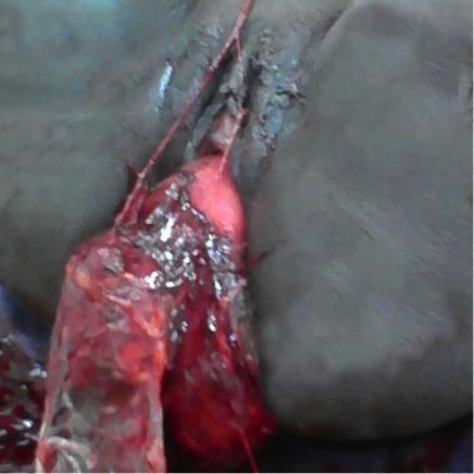
Clinical photograph demonstrating Stage III uterine inversion in a postpartum patient. The image shows the uterus completely exteriorized through the vaginal introitus. The visible uterine fundus exhibits a bright red, hemorrhagic, and fleshy appearance with an irregular, rough texture. Notably, the placenta remains firmly adhered to the uterine fundal surface, a hallmark feature that can contribute to this obstetric emergency. The cervical region and upper vaginal vault appear dark and congested with evidence of tissue disruption. Elongated vascular structures and remnants of the umbilical cord are visible extending from the mass. This visual represents a critical obstetric complication, typically associated with postpartum hemorrhage and cardiovascular collapse, requiring immediate manual or surgical repositioning (taxis) and management of placental attachment.
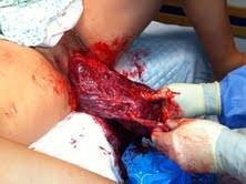
This clinical photograph illustrates a critical obstetric emergency: a complete acute uterine inversion following delivery. The image shows the uterine fundus and the still-attached placenta protruding entirely through the vaginal introitus. The exteriorized mass appears as a large, globular, reddish-purple structure with a rough, irregular texture, consistent with the maternal surface of the placenta and fetal membranes. A healthcare provider’s gloved hands are visible, performing an assessment or initial management of the exteriorized tissue. Significant blood staining is present on the surrounding perineal area and surgical drapes. This visual serves as a primary example of Stage 3 or 4 uterine inversion, where the uterine corpus has collapsed and descended outside the vaginal opening, often associated with fundal placental implantation and potentially life-threatening postpartum hemorrhage. It is a vital educational resource for identifying the physical presentation of obstetric shock and uterine prolapse/inversion in a labor and delivery setting.
Complications of the third stage of labour
1. Postpartum haemorrhage (the commonest and most dangerous)
- Tone: uterine atony is the commonest cause.
- Trauma: cervical, vaginal or uterine lacerations, episiotomy bleeding, and uterine rupture.
- Tissue: retained placenta or membranes, and placenta accreta.
- Thrombin: coagulopathy, either spontaneous or iatrogenic.
- Monitor vital signs frequently, set up large-bore IV access, give crystalloid and prepare for transfusion.
- Look for the source: atony, lacerations, coagulopathy, accreta and inversion. Ultrasound helps to find retained tissue or clots.
- Give oxytocin infusion, 20-30 units in 1000 mL at no more than 100 mU/min. Avoid bolus doses because they cause hypotension.
- Give methylergometrine 0.2 mg IM. Never give it IV, because of the risk of hypertension and vasospasm.
- Give carboprost (15-methyl PGF2α) 250 μg IM, repeated if needed. Use it with great caution in asthma or cardiovascular disease.
- Misoprostol is an option, especially where oxytocin is unavailable.
- If bleeding persists, use a Foley or Bakri balloon tamponade, uterine packing, or selective pelvic artery embolisation.
- The last resort is laparotomy with arterial ligation (including B-Lynch compression suture) and then hysterectomy.
2. Retained placenta
- If the placenta has not been delivered after 30 minutes of gentle cord traction and uterine massage, remove it manually. Use general anaesthesia, or IV tocolysis with adequate analgesia.
- Nitroglycerin is a useful tocolytic here: 400 μg sublingual spray (1-2 sprays, repeated every 3-5 minutes up to 3 doses) or 50-150 μg IV.
- Risk factors include maternal age over 30, extreme preterm delivery, and stillbirth.
- Placenta accreta is the rare cause. Chorionic villi attach directly to the myometrium because the decidua basalis is defective. It is associated with placenta praevia and prior caesarean. If manual removal fails, hysterectomy is usually needed to avoid catastrophic haemorrhage. Conservative management is possible in focal cases if fertility preservation is paramount.
- Retained fragments can also cause delayed PPH and infection.
3. Uterine inversion
- Mechanism: it is linked to fundal placental implantation, which thins the myometrium. Excessive cord traction or fundal pressure can then turn the uterus inside out. Connective tissue disorders and uterine anomalies also predispose.
- Complete inversion: the fundus protrudes past the cervix as a beefy-red mass at the introitus.
- Incomplete inversion: the fundus does not pass the external os. Suspect it when there is PPH and the fundus cannot be felt abdominally. Ultrasound helps with diagnosis.
- Presentation: pain, haemorrhage and neurogenic shock that is out of proportion to the blood loss.
- Management:
- Resuscitate and call the anaesthetist early.
- Reinvert immediately with firm, steady pressure on the fundus (Johnson manoeuvre). Tocolytics (magnesium sulfate, β-mimetics or nitroglycerin) or general anaesthesia may be needed.
- Reinversion is easier if the placenta is left in place until afterwards. Opinions differ on this.
- If this fails, go to laparotomy with the Huntington procedure (traction on the round ligaments), or the Haultain procedure (posterior incision of the fundus).
- Give uterotonics once the uterus is replaced.
- Delay allows cervical constriction and oedema, with a risk of necrosis and bladder or urethral injury.
4. Other complications
- Genital tract trauma: cervical, vaginal and perineal tears. Inspect and repair promptly after delivery.
- Uterine rupture: this is rare. Previous caesarean is the main risk factor. It presents with pain, bleeding and a palpable defect.
- Hypovolaemic shock and its sequelae: DIC, acute tubular necrosis, ARDS and Sheehan syndrome (pituitary necrosis). These are avoidable with careful observation and aggressive treatment of haemorrhage.
- Amniotic fluid embolism: this can present around delivery with collapse and DIC. It is not in the textbook passages I read, so this comes from my own knowledge.
- Prolonged third stage: this is itself a risk factor for PPH. A 2025 systematic review and meta-analysis (PMID 39032724) examined the length of the third stage and adverse maternal outcomes after vaginal birth. Its title questions whether "prolonged third stage" should be redefined. I did not read the full paper, so check it if you need the current thresholds.
- Complications of the interventions themselves:
- Oxytocin: water intoxication and hypotension.
- Ergometrine: hypertension and vasospasm.
- Carboprost: bronchospasm.
- Manual removal: pain and infection.
Quick summary
| Complication | Key feature | Key action |
|---|---|---|
| Atonic PPH | Soft, boggy uterus | Massage, oxytocin, ergometrine, carboprost, balloon, surgery |
| Retained placenta | No delivery after 30 minutes | Manual removal under anaesthesia or tocolysis |
| Accreta | Placenta will not separate | Hysterectomy or conservative surgery |
| Inversion | Shock, no palpable fundus | Immediate replacement, tocolysis or general anaesthesia |
| Lacerations | Bleeding with a firm uterus | Inspect and repair |