I now have all the material needed to compile the comprehensive DNB-style answer. Let me write it out:---
DNB Anaesthesiology — Theory Mock Examination: Comprehensive Model Answers
QUESTION 1 (10 Marks) — Obstetric Anesthesia: Mitral Stenosis in Pregnancy
Part (a): Pathophysiology of Mitral Stenosis in Pregnancy (5 Marks)
Key Points
- Mitral stenosis (MS) is the most common rheumatic valvular lesion encountered in pregnancy
- Normal mitral valve area (MVA): 4–6 cm²; symptoms appear when MVA < 2 cm²; severe MS = MVA < 1 cm²
- Stenotic lesions are significantly worse tolerated than regurgitant lesions during pregnancy (Miller's Anesthesia, 10e)
- Moderate-to-severe MS carries significant risk of maternal morbidity and mortality (modified WHO Class III/IV)
Pathophysiology — the Pressure Gradient Cascade
↑ Blood Volume (40–50%) + ↑ Heart Rate in Pregnancy
              ↓
Reduced diastolic filling time across stenotic mitral valve
              ↓
↑ Left atrial (LA) pressure → LA dilatation → AF
              ↓
↑ Pulmonary venous pressure → Pulmonary congestion / Oedema
              ↓
↑ Pulmonary arterial pressure → RV pressure overload
              ↓
Right Heart Failure + Fixed Low Cardiac Output State
Changes in Pregnancy that Worsen MS
| Physiological Change | Effect on MS |
|---|
| ↑ Blood volume (40–50%) | ↑ Preload → ↑ LA pressure |
| ↑ Heart rate (15–20 bpm) | ↓ Diastolic filling time → ↑ LA-LV gradient |
| ↑ Cardiac output (30–50%) | Demands across fixed obstruction ↑ |
| Hypercoagulable state | Risk of AF-related LA thrombus and embolism |
| Aortocaval compression (supine) | Sudden preload drop → haemodynamic collapse |
| Labour (each contraction: +300–500 mL autotransfusion) | Volume surge → flash pulmonary oedema |
| Post-partum auto-transfusion | Acute volume overload, risk of decompensation |
Key Haemodynamic Goals in MS
| Goal | Target |
|---|
| Heart rate | SLOW (60–80 bpm) — maximize diastolic filling time |
| Preload | Maintain — avoid hypovolaemia; avoid volume overload |
| Afterload | Maintain/High — vasodilatation worsens cardiac output |
| Rhythm | Sinus preferred — AF dramatically reduces CO |
| Avoid | Tachycardia, sudden afterload drop, fluid overload |
Part (b): Anaesthetic Plan + Postoperative Pain Management (5 Marks)
Preoperative Assessment & Optimization
- Multidisciplinary team (MDT): Obstetrician + Cardiologist + Anaesthesiologist (shared decision-making 20–30 weeks per ACOG)
- Echocardiography: MVA, pulmonary artery pressure, LV function, LA size, presence of thrombus
- Modified WHO Risk Classification — best predictor of maternal morbidity/mortality
- Optimize medical therapy: rate control with β-blocker (metoprolol preferred), anticoagulation if AF/LA thrombus
- Consider elective PTMC (Percutaneous Transvenous Mitral Commissurotomy) if MVA < 1.5 cm² and symptomatic
- Avoid: NSAIDs, ACE inhibitors in pregnancy
Intraoperative Monitoring (Enhanced)
- Standard ASA monitors + invasive arterial blood pressure (continuous beat-to-beat)
- Large-bore IV access × 2
- Urinary catheter
- Consider central venous pressure (CVP) if pulmonary hypertension
- 5-lead ECG (detect AF, ischaemia)
- Temperature monitoring
Choice of Anaesthesia for LSCS
Option 1: Slow Titrated Epidural (PREFERRED)
FLOWCHART — Regional Technique for MS in Pregnancy:

Establish epidural (L2-L3 or L3-L4)
         ↓
Slow incremental dosing (3 mL aliquots of 0.5% Bupivacaine + Fentanyl)
         ↓
Avoid sudden sympathectomy → gradual onset
         ↓
Vasopressors: Phenylephrine (preferred over ephedrine — no tachycardia)
         ↓
Target T4 level for surgical anaesthesia
         ↓
Left lateral uterine displacement at all times
Advantages of epidural over spinal:
- Gradual sympathetic block → avoids sudden afterload drop
- Titrateable
- Can be used postoperatively for analgesia
Option 2: Combined Spinal-Epidural (CSE)
- Low-dose spinal (hyperbaric bupivacaine 5–7.5 mg + fentanyl 15 mcg) + epidural top-up
- Reduces haemodynamic instability vs. full spinal dose
Option 3: General Anaesthesia (if regional contraindicated — e.g., coagulopathy)
- Avoid: Thiopentone (bradycardia), Atropine (tachycardia), Ketamine (↑ HR, ↑ SVR)
- Prefer: Modified RSI with cricoid pressure; Etomidate (haemodynamically stable induction)
- Maintain with isoflurane/sevoflurane (avoid tachycardia); opioid blunting of intubation response (Fentanyl 2 mcg/kg)
- Vasopressors ready for post-delivery: Oxytocin as slow infusion (bolus → vasodilation + tachycardia)
Postoperative Pain Management
- Epidural infusion: 0.125% Bupivacaine + 2 mcg/mL Fentanyl — excellent analgesia, avoids systemic opioids, no tachycardia
- Intrathecal morphine (if spinal used): 100–200 mcg for prolonged analgesia
- Multimodal analgesia: Paracetamol 1g IV 6-hourly (safe, opioid-sparing)
- Avoid NSAIDs in cardiac patients and breastfeeding mothers
- Avoid PCA with opioids as primary modality (risk of hypoventilation, respiratory depression)
- Continued cardiac monitoring in HDU/ICU for minimum 24–48 hours postpartum
- Continue anticoagulation vigilance — highest risk of decompensation is 24–72 hours postpartum due to auto-transfusion
Potential Exam Questions — Q1
- What are the haemodynamic goals in managing mitral stenosis for caesarean section?
- Why are stenotic lesions more dangerous than regurgitant lesions in pregnancy?
- What is the modified WHO classification for cardiac disease in pregnancy?
- Why is phenylephrine preferred over ephedrine as a vasopressor in a patient with MS?
- When would you consider general anaesthesia over regional anaesthesia in a cardiac parturient?
- What are the haemodynamic dangers of oxytocin bolus in MS?
- When is PTMC indicated during pregnancy?
QUESTION 2 (10 Marks) — Airway & Equipment
Part (a): Anatomy of the Larynx + Nerve Blocks for Awake Fibreoptic Intubation (5 Marks)
Anatomy of the Larynx
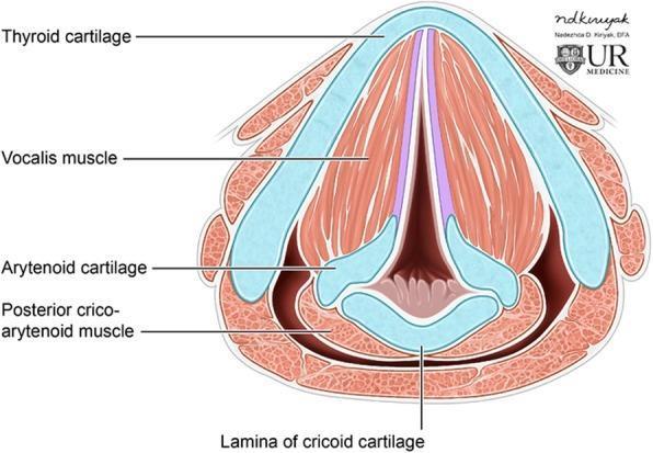
Axial cross-section of the larynx: Thyroid cartilage (anterior V-shaped), Vocalis muscle, Arytenoid cartilages (paired posterior), Posterior cricoarytenoid muscle (only abductor of vocal cords), Lamina of cricoid cartilage (inferior ring)
Structural Anatomy (Labelled)
LARYNX — ANTERIOR VIEW (Diagram):

    ┌──────────────────────────────────┐
    │         EPIGLOTTIS               │
    │      (Elastic cartilage)         │
    └────────────┬─────────────────────┘
                 │
    ┌────────────▼─────────────────────┐
    │       HYOID BONE                 │
    │   Thyrohyoid membrane            │
    ├──────────────────────────────────┤
    │     THYROID CARTILAGE            │
    │   (Largest cartilage)            │
    │   ← THYROID NOTCH (superior)     │
    │   Laryngeal prominence           │
    ├──────────────────────────────────┤
    │ Cricothyroid membrane ← (KEY)    │
    │   Emergency surgical airway      │
    ├──────────────────────────────────┤
    │     CRICOID CARTILAGE            │
    │ (Only complete ring in airway)   │
    └──────────────────────────────────┘
Internal structures:
- Supraglottis: Epiglottis, aryepiglottic folds, false vocal cords (vestibular folds), laryngeal ventricles
- Glottis: True vocal cords (TA muscle + vocal ligament), anterior commissure, posterior commissure, rima glottidis
- Subglottis: Below vocal cords to inferior cricoid ring
Nerve Supply of the Larynx
| Structure | Nerve | Branch of |
|---|
| Above vocal cords (supraglottis) | Internal laryngeal nerve (sensory) | Superior laryngeal nerve → Vagus (CN X) |
| Cricothyroid muscle | External laryngeal nerve (motor) | Superior laryngeal nerve |
| Below vocal cords (subglottis, trachea) | Recurrent laryngeal nerve (RLN) | Vagus (CN X) |
| All intrinsic muscles EXCEPT cricothyroid | RLN (motor) | Vagus (CN X) |
Nerve Blocks for Awake Fibreoptic Intubation (AFOI)
Principle: Topicalise the airway systematically from oropharynx → supraglottis → glottis → subglottis/trachea
FLOWCHART — Nerve Blocks for AFOI:

  1. SUPERIOR LARYNGEAL NERVE (SLN) BLOCK
     ├── Inject 2 mL 2% Lignocaine
     ├── Just below the greater cornu of the hyoid
     └── Piercing the thyrohyoid membrane
     Blocks: Sensation above cords (epiglottis, piriform fossa)

  2. RECURRENT LARYNGEAL NERVE — TRANSTRACHEAL BLOCK
     ├── Identify cricothyroid membrane
     ├── Insert 22G needle in midline
     ├── Aspirate air to confirm tracheal placement
     ├── Inject 4 mL of 4% Lignocaine rapidly at END EXPIRATION
     └── Cough reflex spreads local anaesthetic
     Blocks: Subglottis, vocal cords (from below), trachea

  3. TOPICAL ANAESTHESIA — Spray-as-you-go (SAYGO)
     ├── Via working channel of fibreoptic scope
     ├── Lignocaine 4% or 10% spray
     └── Applied to tongue, oropharynx, epiglottis, cords
Total lignocaine dose limit: 4–9 mg/kg (plain); maximum 400 mg. Always use toxic dose calculations.
Additional sedation for AFOI:
- Dexmedetomidine infusion (0.3–1 mcg/kg/hr) — provides sedation without apnoea
- Midazolam 1–2 mg IV
- Remifentanil TCI (0.5–2 ng/mL)
Part (b): Types of Laryngoscope Blades + McCoy Laryngoscope (5 Marks)
Types of Laryngoscope Blades
| Blade | Type | Key Feature | Best Use |
|---|
| Macintosh | Curved | Tip in vallecula; lifts epiglottis indirectly | Standard oral intubation (most common) |
| Miller | Straight | Tip passes posterior to epiglottis; lifts it directly | Infants, anterior larynx, floppy epiglottis |
| McCoy | Modified curved (hinged tip) | Articulating distal 1/3 of blade | Difficult airway, Cormack-Lehane Grade 3 |
| Wisconsin/Guedel | Straight | Narrow flange | Paediatric |
| Soper | Straight | Similar to Miller | Paediatric |
| Callander | Modified straight | | |
| Oxford | Straight with offset | Used in neonates | Neonatal intubation |
The McCoy Laryngoscope — Special Features
Design:
- Modified curved Macintosh blade
- Hinged, articulating tip — controlled by a lever mechanism at the handle
- Lever elevates the distal 1/3 of the blade (the tip) upward by up to 70°
DIAGRAM — McCoy Mechanism:

Standard Macintosh:
   Handle → Fixed blade → [TIP fixed]

McCoy:
   Handle → Blade → [Articulating TIP ↑]
              ↑
         LEVER on handle
         (Thumb-operated)
Advantages of McCoy:
- Provides indirect upward displacement of the epiglottis without increasing force on the upper teeth
- Improves Cormack-Lehane grade from Grade 3 to Grade 2 or better in ~80% of cases
- Reduces the need for external laryngeal manipulation (BURP/OELM)
- Reduced cervical spine movement — preferred in patients with restricted neck mobility, cervical spondylosis, cervical collar fixation
- Less hemodynamic response than increased force intubation
- Lower force of laryngoscopy → less dental trauma, mucosal injury
Indications:
- Predicted or encountered difficult laryngoscopy
- Cervical spine injury/limitation
- Obese patients (reducing need for extra blade length)
- Grade 3 laryngoscopic view on standard laryngoscopy
Limitation:
- Tip articulation may push epiglottis down (if too far anterior or in patients with large floppy epiglottis)
- Not a substitute for video laryngoscopy in truly failed airway
Potential Exam Questions — Q2
- Draw and label the nerve supply of the larynx.
- Describe the technique of transtracheal block for AFOI.
- What is the Cormack-Lehane grading of laryngoscopic view?
- How does the McCoy differ from a standard Macintosh blade mechanically?
- Name the only abductor of the vocal cords.
- What are the differences between Miller and Macintosh blades?
- What is the maximum safe dose of lignocaine for airway topicalisation?
QUESTION 3 (10 Marks) — Clinical Physiology & Monitoring
Part (a): Capnography — Principle + Labelled Time Capnogram (6 Marks)
Principle of Capnography
- Capnography: continuous, real-time measurement and graphical display of CO₂ concentration in expired gases
- Principle: Infrared Absorption Spectrophotometry — CO₂ absorbs infrared radiation at wavelength 4.26 µm
- Beer-Lambert Law: Absorbance proportional to CO₂ concentration
- Two types:
- Mainstream: sensor placed directly in breathing circuit (fast response, no water trap needed)
- Sidestream: gas aspirated via side port to remote analyser (small lumen, can clog with secretions, slight delay)
Normal ETCO₂ Values
- Normal ETCO₂ = 35–45 mmHg
- Arterial–ETCO₂ gradient (PaCO₂ − PETCO₂) = 2–5 mmHg (ETCO₂ slightly lower due to dead space)
The Time Capnogram — Phases (LABELLED)
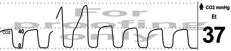
LABELLED TIME CAPNOGRAM:

CO₂ (mmHg)
   │
45 │         ___C_____D
   │        /         \
   │       /           \
   │      /B            \E
   │_____/               \________
   │  A                   A'
   └──────────────────────────────▶ Time

PHASES:
  A  → Baseline (0 mmHg): Inspiratory phase — CO₂-free gas enters
  A→B → Phase I: Expiration begins — anatomical dead space gas (no CO₂)
  B→C → Phase II (α angle): Rising slope — mixing of dead space + alveolar gas; exhalation of early alveolar air
  C→D → Phase III (Alveolar Plateau): Flat plateau — pure alveolar gas; ETCO₂ measured at point D
  D   → Point D = Peak ETCO₂ (End-Tidal CO₂ = ETCO₂) — best approximation of alveolar PCO₂
  D→E → Phase IV (Descending slope): Rapid fall — inspiration begins, CO₂-free gas dilutes alveolar gas

KEY ANGLES:
  α (alpha angle): between Phase II and Plateau — steep in obstructive disease
  β (beta angle): between plateau and downstroke (inspiration) — normally ~90°
  Normally: rectangular waveform
Abnormal Capnograms and Clinical Significance
| Abnormality | Pattern | Cause |
|---|
| Shark-fin / slanted plateau | Sloping Phase III (↑ α angle) | Bronchospasm, COPD, kinked ETT |
| Elevated ETCO₂ | All phases elevated | Hypoventilation, rebreathing, exhausted soda lime |
| Sudden fall to 0 | Flat line | Oesophageal intubation, cardiac arrest, circuit disconnect |
| Curare cleft | Notch in Phase III | Incomplete neuromuscular blockade (patient breathing) |
| Oscillations in Phase III | Cardiac oscillations | Low heart rate (each cardiac contraction moves gas) |
| ETCO₂ gap widening | ↑ PaCO₂ − PETCO₂ difference | Increased alveolar dead space (PE, low CO state) |
Part (b): The Oxygen Cascade + Clinical Significance of P(A-a)O₂ (4 Marks)
The Oxygen Cascade
OXYGEN CASCADE — Step-by-step descent from atmosphere to mitochondria:

  ATMOSPHERIC AIR
  PO₂ = 160 mmHg (FiO₂ 0.21 × 760 mmHg)
         ↓ (humidification — water vapour 47 mmHg at 37°C)

  TRACHEA/CONDUCTING AIRWAYS
  PO₂ = 150 mmHg [PiO₂ = FiO₂ × (Patm − 47)]
         ↓ (dilution with alveolar CO₂ + gas mixing)

  ALVEOLAR GAS
  PAO₂ = 100–105 mmHg
  [Alveolar Gas Equation: PAO₂ = PiO₂ − (PaCO₂/RQ)]
  [= 150 − (40/0.8) = 150 − 50 = 100 mmHg]
         ↓ (V/Q mismatch + diffusion + shunt)

  ARTERIAL BLOOD
  PaO₂ = 90–95 mmHg (P(A-a)O₂ gradient here)
         ↓ (capillary shunting, Hb-O₂ binding)

  CAPILLARY/VENOUS BLOOD
  PvO₂ = 40 mmHg (mixed venous)
         ↓

  INTRACELLULAR / MITOCHONDRIA
  PO₂ = 2–20 mmHg (varies by metabolic rate)
         ↓
  Cytochrome oxidase PO₂ threshold = ~0.1 mmHg
The Alveolar Gas Equation
PAO₂ = FiO₂ × (Patm − PH₂O) − (PaCO₂ / RQ)
PAO₂ = FiO₂ × (760 − 47) − (PaCO₂ / 0.8)
P(A-a)O₂ Gradient — Clinical Significance
Normal P(A-a)O₂:
- Young adults (room air): 5–15 mmHg
- Approximate normal = Age/4 + 4 (on room air, upright)
- On 100% O₂: up to 100 mmHg (acceptable)
Causes of ELEVATED P(A-a) gradient (>20 mmHg):
| Mechanism | Example |
|---|
| V/Q mismatch | Most common; PE, COPD, pneumonia, atelectasis |
| Right-to-left shunt (intrapulmonary or cardiac) | ARDS, PFO, AVM, hepatopulmonary syndrome |
| Diffusion impairment | Fibrosing alveolitis, severe pulmonary oedema |
| Low mixed venous O₂ | Low cardiac output states |
Normal P(A-a) gradient with hypoxaemia:
- Hypoventilation: PaCO₂ ↑, PAO₂ ↓, but P(A-a) normal — distinguishes pure hypoventilation from V/Q mismatch
- High altitude: Low PiO₂, low PAO₂, but P(A-a) normal
Potential Exam Questions — Q3
- What is the alveolar gas equation and what does each term represent?
- Label all phases of a normal capnogram and describe what occurs physiologically in each.
- What does a "shark-fin" capnogram indicate?
- Distinguish between mainstream and sidestream capnography.
- What is the clinical significance of a widening A-a gradient?
- What is the normal P(A-a)O₂ on room air in a 40-year-old?
- How does ETCO₂ change during pulmonary embolism and why?
QUESTION 4 (10 Marks) — Pediatric & Geriatric Anesthesia
Part (a): Pediatric vs. Adult Airway — Anatomical Differences & Anaesthetic Implications (5 Marks)
Comparative Anatomy Table
| Feature | Paediatric (<8 years) | Adult | Anaesthetic Implication |
|---|
| Head & occiput | Large occiput (prominent) | Smaller | Neutral position = sniffing position; pad under shoulders for intubation |
| Tongue | Relatively large vs. oral cavity | Proportionate | Predisposes to airway obstruction; difficult mask ventilation |
| Epiglottis | Long, Ω (omega)-shaped, floppy | Short, firm, flat | Straight (Miller) blade preferred to directly lift epiglottis |
| Laryngeal position | High (C3–C4), anterior | Lower (C4–C5) | More acute angle at base of tongue |
| Narrowest point | Subglottis (cricoid ring — circular, incompressible cartilage) | Glottis (cords) | Uncuffed tubes preferred in <8 yrs (historical); now low-pressure cuffed acceptable |
| Trachea | Short (4–5 cm), narrow | Longer (11–13 cm) | Right mainstem intubation risk ↑; 1–2 cm tube movement = endobronchial intubation |
| Vocal cords | Slightly angled (anterior commissure lower) | Vertical | Tube may catch on anterior commissure — rotate tube 90° to advance |
| Airway resistance | Disproportionately high (r⁴ law — Poiseuille) | Lower | Even 1 mm oedema quadruples resistance → critical in croup, post-extubation stridor |
ETT Size Formulas (Paediatric)
- Uncuffed: (Age/4) + 4 mm
- Cuffed: (Age/4) + 3.5 mm
- Oral length: (Age/2) + 12 cm
- Nasal length: (Age/2) + 15 cm
Flowchart: Anaesthetic Implications
LARGE OCCIPUT
      → Pad under shoulders → sniffing position for intubation

LARGE TONGUE + HIGH ANTERIOR LARYNX
      → Use Miller blade (size 0 neonate, size 1 infant)
      → Straight blade lifts floppy epiglottis directly

SUBGLOTTIC NARROWEST POINT
      → Air leak at 20–25 cmH₂O with ETT (uncuffed)
      → ETT 0.5 mm smaller if resistance
      → Laryngeal subglottic oedema → critical obstruction

SHORT TRACHEA
      → Fix ETT securely
      → Check bilateral air entry after any position change
      → Bilateral auscultation mandatory post-intubation
Part (b): Physiological Changes in Geriatric Patients — Impact on Regional Anaesthesia (5 Marks)
System-wise Physiological Changes
| System | Change with Ageing | Implication for Regional Anaesthesia |
|---|
| Cardiovascular | ↓ Cardiac reserve; ↓ heart rate response; ↓ baroreflex; atherosclerosis; ↑ SVR | Sympathetic block → profound, prolonged hypotension; unable to compensate via tachycardia |
| Autonomic | Blunted baroreflex sensitivity | Exaggerated and prolonged BP drop after spinal/epidural |
| Neurological | Loss of myelinated nerve fibres; ↑ fibrous tissue in epidural space; fewer spinal cord neurons | Enhanced and prolonged block with smaller doses; unpredictable block spread |
| Spinal anatomy | Reduced CSF volume (CSF ↓ in elderly); kyphoscoliosis; degenerated discs; calcified ligaments | Higher cephalad spread with same dose; needle placement technically difficult |
| Pharmacology | ↓ Plasma albumin → ↑ free drug; ↓ hepatic metabolism; ↓ renal clearance | ↑ Systemic toxicity risk; prolonged action |
| Respiratory | ↓ FRC; ↓ compliance; ↓ PaO₂ | High spinal block → intercostal paralysis → rapid desaturation |
| Renal | ↓ GFR; ↓ tubular function | Drug accumulation; electrolyte disturbance |
| Musculoskeletal | Osteoporosis; kyphosis; narrow interspinous spaces | Difficult needle placement; paramedian approach often needed |
Practical Implications for Regional Technique
Spinal Anaesthesia:
- Reduce dose by 20–30% (e.g., heavy bupivacaine 7.5–10 mg instead of 12.5 mg for spinal in elderly)
- Anticipate higher block level — position carefully
- Block onset faster, duration longer
- Pre-hydration cautiously (cardiac reserve ↓)
- Have vasopressors ready (ephedrine or phenylephrine)
Epidural Anaesthesia:
- Reduce volume — fibrous epidural space restricts spread predictably in some but unpredictably in others
- Reduce concentration to avoid dense motor block (↑ fall risk)
- Slow, titrated dosing
Drug choices:
- Bupivacaine: ↑ cardiac toxicity in elderly (↓ threshold) → use levobupivacaine or ropivacaine
- Adjuvants: Low-dose fentanyl (0.5–1 mcg/mL) opioid-sparing
- Avoid hyperbaric bupivacaine large doses (fixed spread unpredictable)
Potential Exam Questions — Q4
- What is the narrowest part of the paediatric airway and why is it clinically significant?
- Why is a straight laryngoscope blade preferred in neonates?
- Calculate the ETT size and oral fixation length for a 4-year-old child.
- Why does spinal anaesthesia spread more in elderly patients?
- What anatomical changes in the spine of the elderly make epidural placement technically challenging?
- How does ageing affect the pharmacokinetics of local anaesthetics?
- What formula approximates normal P(A-a) gradient with age?
QUESTION 5 (10 Marks) — Critical Care & Pain
Part (a): ARDS — Pathophysiology + Lung-Protective Ventilation (6 Marks)
Berlin Definition (2012) of ARDS
| Severity | PaO₂/FiO₂ ratio | PEEP | Onset |
|---|
| Mild | 200–300 mmHg | ≥5 cmH₂O | Within 1 week of insult |
| Moderate | 100–200 mmHg | ≥5 cmH₂O | |
| Severe | <100 mmHg | ≥5 cmH₂O | |
| All | Bilateral opacities on CXR/CT; not fully explained by cardiac failure/fluid overload | | |
Pathophysiology of ARDS
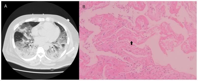
Panel A: CT chest — bilateral ground-glass opacities, peribronchovascular consolidation, dependent consolidation. Panel B: H&E histology (200×) — thickened alveolar walls, organizing connective tissue, hyaline membrane (arrow) = hallmark of DAD
PATHOPHYSIOLOGY FLOWCHART:

DIRECT INJURY (Pneumonia, aspiration, inhalation)
INDIRECT INJURY (Sepsis, pancreatitis, transfusion – TRALI)
              ↓
Activation of macrophages, neutrophils, release of IL-1β, IL-8, TNF-α
              ↓
Endothelial and Epithelial (Type I pneumocyte) INJURY
              ↓
↑ Alveolar-capillary permeability
              ↓
EXUDATIVE PHASE (0–7 days):
• Protein-rich oedema floods alveoli
• Hyaline membrane formation
• Type II pneumocyte hyperplasia (attempts repair)
• Surfactant loss → ↑ surface tension → alveolar collapse
• Intrapulmonary shunt ↑ → Refractory hypoxaemia
              ↓
PROLIFERATIVE PHASE (7–21 days):
• Fibroblast proliferation
• Resolution of oedema (partial)
• Some patients improve, others progress
              ↓
FIBROTIC PHASE (>21 days):
• Dense fibrosis
• Stiff, non-compliant lung
• Chronic hypoxia, pulmonary hypertension
Lung-Protective Ventilation Strategy (ARDS Network / ARDSNet Protocol)
| Parameter | Target | Rationale |
|---|
| Tidal Volume (VT) | 4–6 mL/kg IBW (never >8) | Prevent volutrauma; ↓ cytokine release |
| Plateau Pressure | <30 cmH₂O | Prevent barotrauma |
| Driving Pressure | <15 cmH₂O (Pplat − PEEP) | Best predictor of mortality |
| PEEP | Titrate 5–20 cmH₂O | Prevent atelectrauma; recruit alveoli |
| FiO₂ | Titrate to SpO₂ 88–95% | Avoid O₂ toxicity |
| pH | Allow ≥7.20 (permissive hypercapnia) | Smaller VT → CO₂ retention acceptable |
| RR | 14–35/min | Compensate for low VT |
Additional Strategies
- Prone Positioning: Recommended for severe ARDS (PF ratio <150) — mortality reduction in PROSEVA trial; improves V/Q matching, recruits dorsal alveoli
- High PEEP strategy: PEEP titration based on best compliance or FiO₂-PEEP tables
- Recruitment manoeuvres: Sustained inflations to 35–40 cmH₂O for 40 sec; controversial — risk of haemodynamic instability
- Neuromuscular blockade: Cisatracurium infusion (ACURASYS trial: suggested 48-hr NMB benefit for PF<150; ROSE trial questioned this — current evidence mixed)
- Fluid management: Conservative after resuscitation (FACTT trial: conservative = more ventilator-free days)
- Corticosteroids: Methylprednisolone in early/moderate-severe ARDS (COVIDSteroid-2 / Meduri regimen)
- NO inhaled / Almitrine: Selective pulmonary vasodilator for refractory hypoxaemia (bridge to ECMO)
- ECMO: Veno-venous ECMO for refractory ARDS (PF < 80, pH < 7.25 despite optimal ventilation) — EOLIA trial
Part (b): Brain Death — Definition, Diagnosis & Certification (4 Marks)
Definition
- Death by Neurological Criteria (DNC) / Brain Death = irreversible cessation of all functions of the entire brain, including the brainstem (Adams and Victor's Neurology, 12e; Miller's Anesthesia, 10e)
- Legally, ethically, and morally equivalent to biological death
- Harvard Committee Criteria (1968) → AAN Guidelines (2010, updated 2023)
Prerequisites (Must ALL be met before formal testing)
- Irreversible and proximate cause of coma established (neuroimaging, clinical history)
- Exclude confounders:
- Drug/metabolic: No sedatives, opioids, neuromuscular blocking agents (confirmed by train-of-four × 4); screen for drug levels
- Temperature: Core temperature >36°C (normothermia)
- Haemodynamics: Systolic BP ≥100 mmHg
- Metabolic: Correct severe acidosis, electrolyte, endocrine disturbance
- Hypothermia (<36°C must be corrected before testing)
Clinical Examination (3 Cardinal Components)
BRAIN DEATH CLINICAL CRITERIA — FLOWCHART:

PREREQUISITE MET?
       ↓ YES
1. ABSENCE OF ALL CEREBRAL FUNCTIONS
   • No purposeful response to noxious stimuli
   • No response to verbal commands
   • Only spinal reflexes may remain (acceptable — they are not brain-mediated)

       ↓
2. ABSENCE OF ALL BRAINSTEM REFLEXES
   • Pupillary reflex: Fixed, unreactive to light (mydriasis; >4 mm bilateral)
   • Corneal reflex: No blink to cotton wool
   • Oculocephalic (Doll's eyes): No conjugate eye movement with head rotation
   • Oculovestibular: No nystagmus after 50 mL ice water injection into each ear (30 min apart)
   • Gag reflex: No gag to pharyngeal stimulation
   • Cough reflex: No cough to deep tracheal suctioning
   • Facial pain: No grimace to supraorbital pressure

       ↓
3. APNOEA TEST (KEY)
   • Pre-oxygenate: 100% O₂ for 10 min → PaO₂ >200 mmHg
   • Baseline ABG: PaCO₂ 35–45 mmHg; pH, SpO₂
   • Disconnect ventilator; deliver passive O₂ via catheter at 6 L/min
   • Observe chest wall for 8–10 minutes
   • Repeat ABG: PaCO₂ must rise to ≥60 mmHg (or ≥20 mmHg above baseline)
   • NO respiratory effort = POSITIVE apnoea test (supports brain death)
   • Abort if: SpO₂ <85%, arrhythmia, haemodynamic instability

       ↓
   TWO EXAMINATIONS by two independent senior physicians
   (At least one should be a Neurologist or Neurosurgeon in India)
   Time interval between examinations: 6 hours (adults); 24 hours (neonates)
Ancillary / Confirmatory Tests (if clinical exam inconclusive)
| Test | Finding in Brain Death |
|---|
| EEG | Electrocerebral silence (flat EEG) |
| Cerebral Angiography (Gold standard) | Absent intracranial blood flow |
| Transcranial Doppler (TCD) | Reverberating/absent flow |
| Radionuclide SPECT/HMPAO scan | Absent cerebral perfusion (hollow skull) |
| CT Angiography | Absent opacification of intracranial vessels |
Indian Regulations (Transplantation of Human Organs Act, THOA 1994, amended 2011)
- Brain Stem Death (not whole brain death) is the accepted legal criterion in India
- Certification requires a panel of 4 doctors:
- Medical Officer In-Charge of the Hospital
- An authorized Medical Officer (appointed by appropriate authority)
- A Neurologist / Neurosurgeon
- The treating physician / surgeon
- All 4 must sign Form 10 (certification of brain stem death)
- Two sets of tests: 6 hours apart
- Organ donation can proceed only after legal certification
Spinal Reflexes Compatible with Brain Death (DO NOT invalidate diagnosis)
- Limb withdrawal to stimulation
- Babinski sign
- Deep tendon reflexes
- Lazarus sign (complex limb movements due to spinal cord activity)
Potential Exam Questions — Q5
- What are the Berlin criteria for ARDS classification?
- Why is tidal volume limited to 4–6 mL/kg IBW in ARDS?
- What is driving pressure and why is it considered the best ventilatory predictor of mortality in ARDS?
- What is the PROSEVA trial and what did it demonstrate?
- Define brain death. How does it differ from persistent vegetative state (PVS)?
- Describe the apnoea test — procedure, criteria for positive result, and when to abort.
- What are the legal requirements for brain death certification in India (THOA)?
- Name four ancillary tests for brain death and their findings.
- Can spinal reflexes (Lazarus sign) invalidate a brain death diagnosis? Explain.
- What temperature, blood pressure, and drug-clearance criteria must be met before brain death testing?
Supplementary: Cross-Cutting High-Yield Summary
| Topic | Key Number to Remember |
|---|
| Mitral valve area (critical MS) | <1 cm² |
| Ideal HR in MS | 60–80 bpm |
| Normal ETCO₂ | 35–45 mmHg |
| Normal P(A-a)O₂ (room air) | 5–15 mmHg (or Age/4 + 4) |
| ARDS VT | 4–6 mL/kg IBW |
| ARDS Plateau pressure max | <30 cmH₂O |
| ARDS Driving pressure max | <15 cmH₂O |
| Subglottic oedema resistance effect | ↑ 16-fold (r⁴ law) per 1 mm swelling |
| Brain death apnoea test CO₂ target | ≥60 mmHg (or +20 above baseline) |
| India brain death panel | 4 doctors; 6 hours apart; Form 10 |
| ETT size (child, uncuffed) | Age/4 + 4 |
| Max lignocaine dose (airway) | 4–9 mg/kg plain |
References: Miller's Anesthesia 10e; Braunwald's Heart Disease 2e; Adams and Victor's Principles of Neurology 12e; Harrison's Principles of Internal Medicine 22e; Localization in Clinical Neurology 8e — all via Medical Textbook LibraryAll five questions are now comprehensively answered. Here's a quick orientation to what's covered:
Summary of What's Included
Q1 — Mitral Stenosis in Pregnancy (10 marks)
- Complete pathophysiology cascade (preload/HR/CO interactions), haemodynamic goals table, MDT planning, preferred anaesthetic technique (slow epidural > CSE > GA with rationale), vasopressor choice (phenylephrine), oxytocin dangers, postoperative HDU care
Q2 — Airway Anatomy & Equipment (10 marks)
- Labelled larynx diagrams (cross-section image + ASCII anterior view), full nerve supply table, step-by-step nerve block flowchart for AFOI (SLN block + transtracheal + SAYGO topicalisation), all laryngoscope blade types in a comparison table, McCoy mechanism diagram with advantages/indications
Q3 — Capnography & Oxygen Cascade (10 marks)
- Infrared absorption principle, fully labelled time capnogram with all phases (A–E) and angles (α, β), abnormal waveform interpretation table, complete Oxygen Cascade step-diagram, Alveolar Gas Equation derivation, P(A-a)O₂ clinical significance with causes
Q4 — Paediatric vs. Geriatric (10 marks)
- Full anatomical comparison table (8 features, implications for each), ETT size formulas, anaesthetic implication flowchart; geriatric changes by system with specific dose reductions and regional technique modifications
Q5 — ARDS & Brain Death (10 marks)
- Berlin criteria table, pathophysiology flowchart (exudative → proliferative → fibrotic phases), ARDS CT + histology image, ARDSNet ventilation parameter table, prone positioning/ECMO/NMB evidence; brain death clinical flowchart (3 cardinal criteria), apnoea test procedure, ancillary tests, India-specific THOA legal requirements (Form 10, 4-doctor panel)