Tissues
What aspect of tissues are you interested in? · What level of detail do you need?
connective tissue types histology diagram
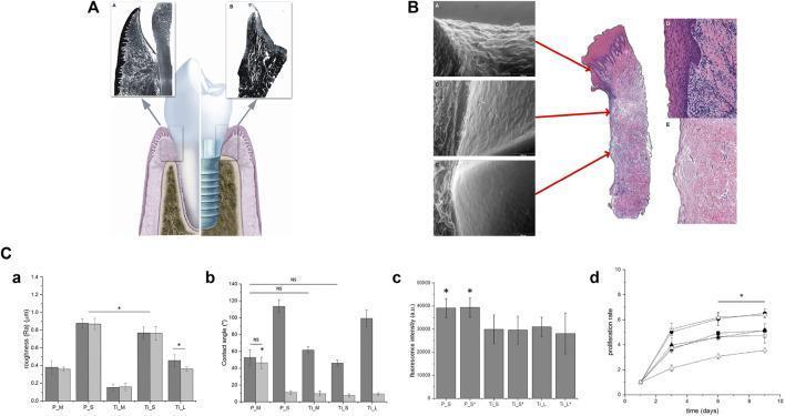
Educational figure illustrating dental implantology and periodontal histology. Panel A features an anatomical diagram comparing a natural tooth with a screw-type dental implant embedded in alveolar bone. It shows key interfaces: the periodontal ligament for the tooth and the biological seal (sulcular/junctional epithelium and connective tissue) for the implant. Histological insets highlight the peri-implant mucosa and gingival structure. Panel B presents a larger histological cross-section of peri-implant tissue with corresponding scanning electron microscope (SEM) images, detailing the surface morphology of the barrier epithelium and underlying fibrous connective tissue. Panel C provides quantitative data via four graphs (a-d) evaluating material properties and biological response. Graph (a) shows surface roughness (Ra) for titanium (Ti) and polyetheretherketone (PEEK) samples; (b) measures contact angle (wettability); (c) evaluates NIH-3T3 cell adhesion via fluorescence intensity; and (d) displays a 10-day proliferation rate curve. The data illustrates how surface modifications like air-plasma treatment affect biomaterial-cell interactions, emphasizing the clinical importance of soft tissue integration for implant success.
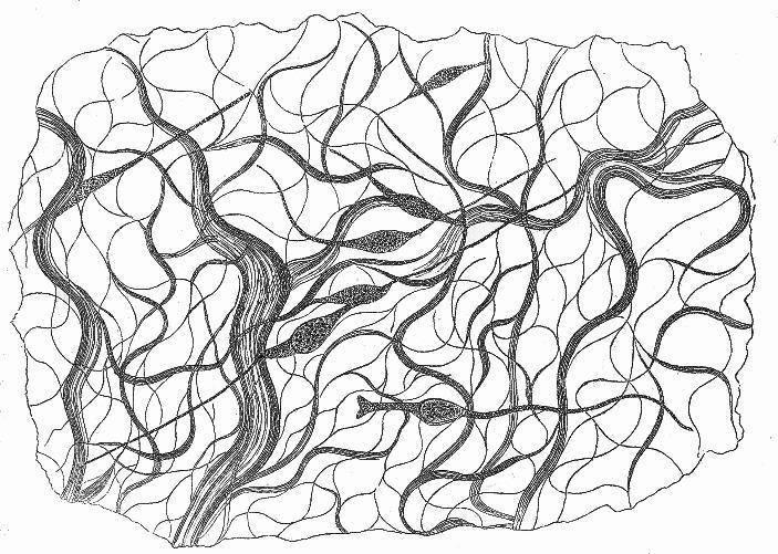
This historical anatomical diagram is a schematic illustration of embryonic connective tissue from a five-month-old human embryo's serous membrane. The illustration depicts the nucleated, spindle-shaped connective tissue corpuscles (Bindegewebskörperchen) as they develop into fibrous connective tissue. The visual content features a complex, branching network of elongated elements distributed throughout the field. Thinner, thread-like lines form a mesh-like pattern or lacunae, while thicker, cord-like structures show a striated or bundled texture, suggesting the formation of parallel fiber strands. Several darker, oval-shaped cellular bodies with visible internal texture (nuclei) are interconnected within this network, with some fiber cords directly emerging from or terminating at these cellular structures. This diagram serves as a pathophysiology and anatomical reference for the histological development of the extracellular matrix and collagenous metamorphosis from mesenchymal precursors in human embryology.
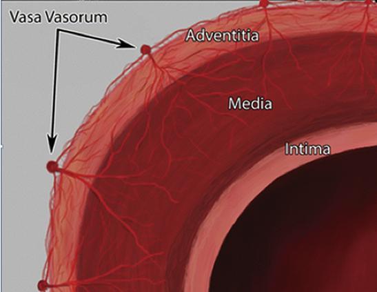
This anatomical diagram provides a cross-sectional view of a large systemic artery wall, illustrating the structural layers and the microvascular supply. The diagram identifies three distinct histological layers: the tunica intima (innermost lining), the tunica media (thick muscular middle layer), and the tunica adventitia (outermost connective tissue layer). A primary focus of the illustration is the 'vasa vasorum'—the 'vessels of the vessels.' The vasa vasorum are depicted as a branching network of small capillaries originating from larger nutrient vessels on the external surface of the adventitia. These microvessels penetrate through the adventitia and extend into the outer portions of the tunica media, highlighting the physiological mechanism for delivering oxygen and nutrients to thick-walled vessels where simple diffusion from the lumen is insufficient. The image serves as a pedagogical tool for understanding vascular histology and the pathophysiology of arterial wall maintenance in conditions such as atherosclerosis or aortic disease.
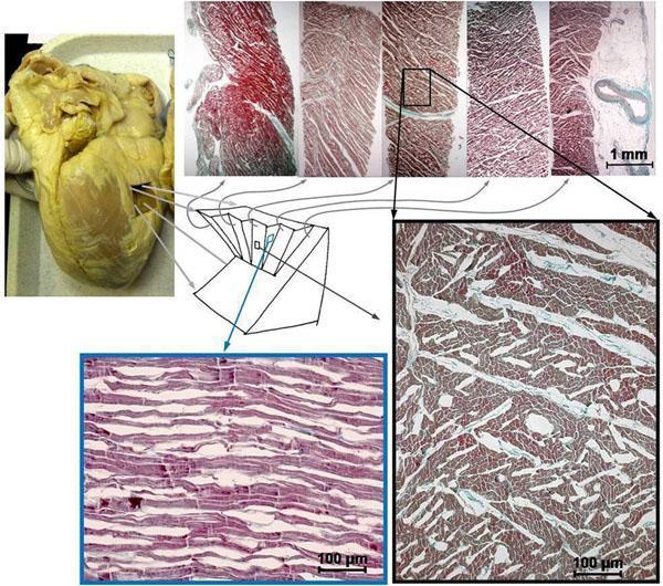
This composite educational image illustrates human myocardial histology and structural organization. At the top left, a gross clinical photograph shows a human heart with a highlighted mid-lateral block sampling site. Beside it, a series of five tangential histological slices, stained with Masson's trichrome or similar (red myocytes, blue-green collagen/connective tissue), demonstrates the laminar microstructure of the ventricular wall. These slices show varying orientations of muscle bundles and visible 'shear layers' or interstitial gaps, which may be accentuated post-mortem. A central diagram clarifies the three-dimensional planes of sectioning relative to the myocardial wall. Below, two high-magnification light microscopy frames provide a detailed comparison: the 'wall tangent section' (left) displays myocytes in longitudinal profile, revealing elongated, parallel cellular arrangements; the 'mid-wall cross-myocyte section' (right) shows myocytes in transverse cross-section, appearing as individual polygonal units separated by fine connective tissue. This figure is used in cardiology and pathology education to demonstrate the complex, anisotropic architecture of the myocardium and the transition between transmural orientations.
loose dense connective tissue fibroblast collagen histology microscopy
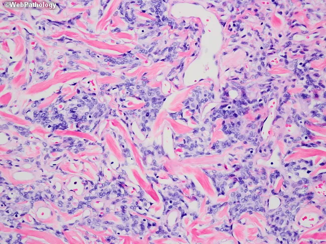
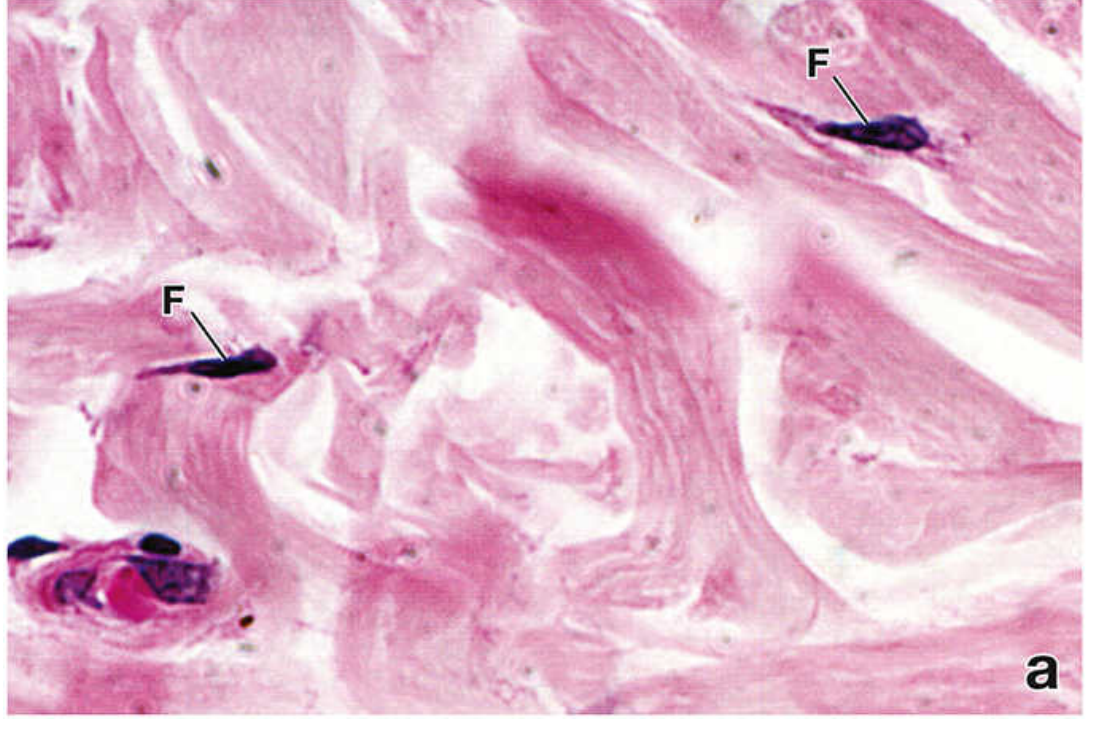
This histopathology image shows a hematoxylin and eosin (H&E) stained soft tissue section examined with bright-field microscopy. The tissue displays a dense, eosinophilic collagen-rich stroma with wavy, interlacing fibers forming an abundant fibrous matrix. Within this matrix are scattered spindle-shaped fibroblasts containing elongated basophilic nuclei. The cellularity is moderate and the vascular channels are present but not prominent. A mild inflammatory infiltrate composed of small lymphocytes and plasma cells is interspersed among the fibrous strands. No evident epithelial elements or malignant cytology are observed in this field, and there is no overt necrosis or high mitotic activity. The overall pattern suggests fibrous tissue proliferation, scar-like remodeling, or a desmoplastic reaction within soft tissue. The image highlights classic histologic features of connective tissue: collagen deposition, fibroblast morphology, and stromal architecture, along with minor inflammatory components. Clinically, these findings are nonspecific without clinical history and ancillary studies but are relevant to differential diagnoses including fibrosis, chronic inflammation, granulation tissue, or low-grade fibrous neoplasms. This slide is useful for teaching about connective tissue histology, pattern recognition for fibrous lesions, and correlating morphological observations with patient presentation and imaging findings to guide diagnosis and management.
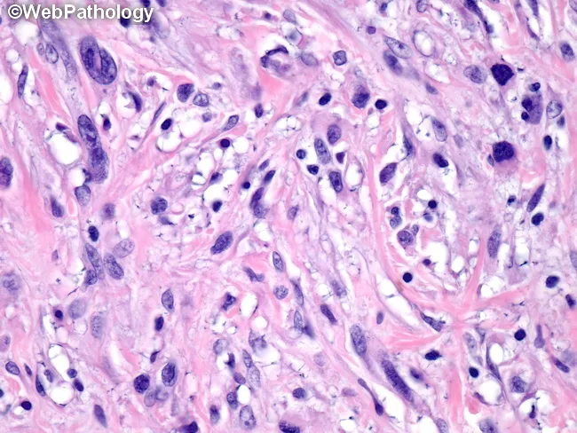
Brightfield light microscopy of an H&E-stained biopsy specimen reveals a loose to moderately dense fibrous stroma populated by scattered spindle-shaped fibroblast-like cells with elongated, oval nuclei and minimal cytoplasm. The cells are arranged in irregular, wavy cords within a pink eosinophilic collagen matrix, reflecting a desmoplastic-type reaction. Interspersed inflammatory elements include small lymphocytes and occasional plasma cells, consistent with a chronic or subacute process. In this field there is no obvious necrosis, and mitotic activity is not readily evident, arguing against high-grade malignancy in this limited view. Vascular channels are present but not markedly abnormal. The overall pattern suggests a fibroblastic or myofibroblastic proliferative process within connective tissue, with preserved tissue architecture and absence of overt differentiation toward epithelial, neural, or adipocytic lineages. Clinically, these features support differential diagnoses that include fibromatosis (desmoid-type), benign fibrous scar or granulation tissue, and low-grade fibrosarcoma; inflammatory myofibroblastic tumor remains a consideration, particularly in younger patients or when inflammatory infiltrate is prominent. Correlation with clinical history, imaging, and, if indicated, immunohistochemical staining (e.g., SMA, desmin, CD34, Ki-67) is recommended to refine diagnosis and guide management. Imaging-pathology concordance is essential for definitive classification and subsequent therapeutic planning, including surgical excision or observation, and multidisciplinary discussion.
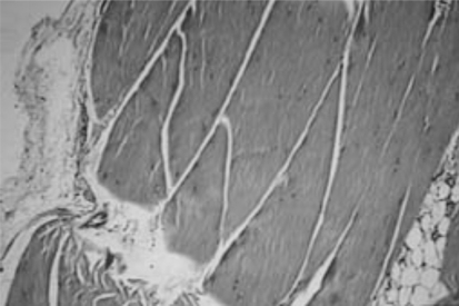
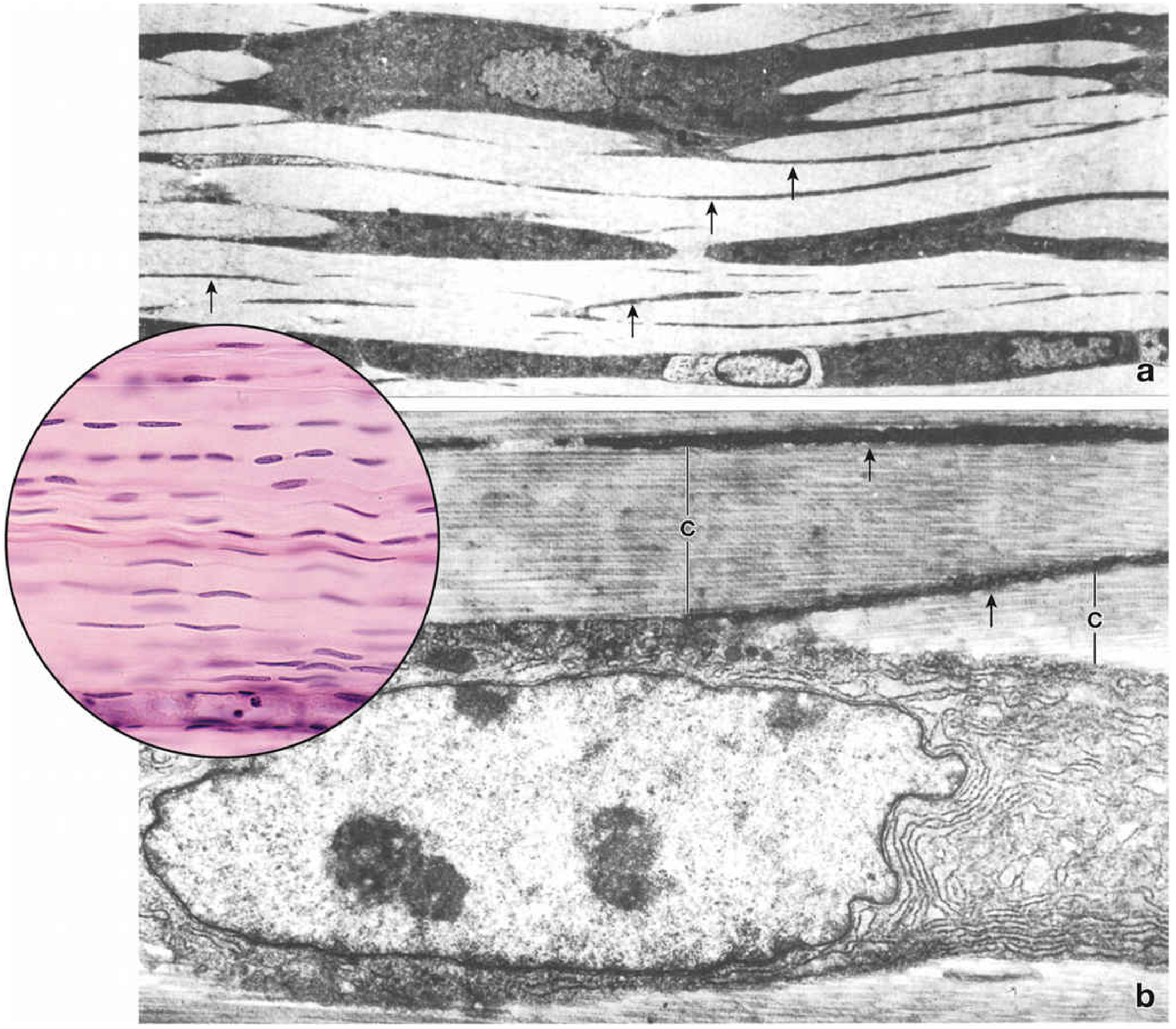
**Imaging Modality:** Histopathology (Light microscopy) **Stain:** Hematoxylin and Eosin (H&E) **Magnification:** 40x **Anatomical Region:** Tendon tissue **Description:** The micrograph displays a longitudinal section of dense regular connective tissue characteristic of a healthy tendon. The primary architectural feature is the parallel arrangement of eosinophilic type I collagen fiber bundles (fascicles). Interspersed between these bundles are elongated, flattened nuclei of tenocytes (fibrocytes), oriented parallel to the long axis of the collagen fibers. **Key Visual Features:** - **Fascicular Organization:** Distinct primary fiber bundles are visible, separated by thin endotenon layers. - **Cellularity:** Low cellular density with quiescent tenocytes exhibiting spindle-shaped morphology. - **Extracellular Matrix:** Dense, wavy (crimped) eosinophilic matrix indicating mechanical integrity. - **Associated Tissue:** Minimal peripheral loose connective tissue and adipocytes are visible at the margin, likely representing the paratenon or epitenon interface. **Diagnostic Relevance:** The image serves as a reference for normal tendon histology, showing absence of neovascularization, mucoid degeneration, or inflammatory infiltration, which are key markers for tendinopathy or chronic injury.
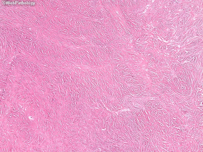
This is a histopathology slide prepared from a soft tissue/dermal biopsy, stained with Hematoxylin and Eosin and evaluated by light microscopy. The tissue shows dense eosinophilic collagen arranged in broad, intersecting fibrous bands with low cellularity. Spindle-shaped fibroblast-like cells are present along the collagenous stroma, exhibiting elongated nuclei and minimal cytologic atypia. Mitotic figures are rare or not evident in the field, and there is no overt necrosis or inflammatory infiltrate. The overall pattern may display a storiform or interlacing fascicular architecture, consistent with fibrous tissue proliferation. The epidermis overlying or surrounding the lesion is not clearly involved in this field, suggesting a dermal/soft tissue process rather than an overt epidermal neoplasm. The histologic impression favors a benign fibrous lesion such as scar tissue- or fibroma-type stroma; however, because desmoid-type fibromatosis and other fibrous tumors can mimic scar on routine sections, correlation with immunohistochemical stains (e.g., beta-catenin for desmoid; CD34 for DFSP) and clinical history is recommended. The diagnostic significance lies in distinguishing non-neoplastic scar from low-grade fibrous neoplasms, guiding management toward observation or surgical excision with clean margins. This image is educational for pathology trainees studying soft tissue histology and differential diagnosis of fibrous lesions.
Connective Tissue - In-Depth Study Notes
1. Overview and Defining Characteristics
2. Classification of Connective Tissue
Connective Tissue
├── Embryonic Connective Tissue
│   ├── Mesenchyme (found in embryo)
│   └── Mucous connective tissue (umbilical cord - Wharton's jelly)
│
├── Connective Tissue Proper
│   ├── Loose (areolar) connective tissue
│   └── Dense connective tissue
│       ├── Dense irregular
│       └── Dense regular
│
└── Specialized Connective Tissues
    ├── Cartilage
    ├── Bone
    ├── Adipose tissue
    └── Blood & lymphoid tissue
3. Connective Tissue Proper
3.1 Loose (Areolar) Connective Tissue
- Found in close association with most epithelia - most epithelia rest upon it
- ECM contains loosely arranged collagen fibers
- Contains numerous cells of several types (fibroblasts + many immune migrants)
- Highly cellular; nuclei vary in size and shape on histology
- Functions as a conduit for blood vessels, nerves; site of immune surveillance
3.2 Dense Connective Tissue
Dense Irregular Connective Tissue
- Abundant fibers, few cells; fibers run in multiple directions
- Cell type: almost exclusively fibroblasts
- Contains very little ground substance
- High proportion of collagen → stains more intensely blue with Mallory/Mazan stains
- Location: reticular (deep) layer of the dermis; submucosa of hollow organs (intestinal tract)
- Function: resists tearing from multidirectional stretching forces
Dense Regular Connective Tissue
- Fibers arranged in parallel, densely packed arrays
- Provides maximum tensile strength in one direction
- Found in: tendons (muscle-to-bone), ligaments (bone-to-bone), aponeuroses
- Tendons specifically:
- Parallel bundles of type I collagen fibers
- Cells between bundles = tendinocytes (specialized fibroblasts)
- Surrounded by thin capsule: epitendineum
- Subdivided into fascicles by endotendineum (carries vessels + nerves)
- In H&E sections: tendinocytes appear as rows of flattened basophilic nuclei between fibers
4. Components of the ECM
4.1 Fibers
| Fiber Type | Composition | Properties | Location |
|---|---|---|---|
| Collagen | Type I-XXVIII+ | Strong, inextensible; most abundant protein in body | Tendons, dermis, bone, everywhere |
| Reticular | Type III collagen | Thin, forms meshwork (argyrophilic - stains with silver) | Lymphoid organs, liver, bone marrow |
| Elastic | Elastin + fibrillin microfibrils | Stretch and recoil; stained with Verhoeff's or orcein | Skin, lung, aorta, ligamentum flavum |
4.2 Ground Substance
- Fills space between cells and fibers
- Composed of glycosaminoglycans (GAGs) - long repeating disaccharide chains (e.g., hyaluronic acid/hyaluronan, chondroitin sulfate, keratan sulfate, heparan sulfate)
- GAGs are largely attached to protein cores as proteoglycans (e.g., aggrecan in cartilage)
- Hyaluronan is unique: extremely long GAG chain, NOT covalently bound to a protein core
- Highly hydrophilic → binds large amounts of water → creates a hydrated gel that resists compression
- Also contains glycoproteins (fibronectin, laminin) that mediate cell-ECM adhesion
5. Cells of Connective Tissue
- Resident (fixed) cells - permanent inhabitants
- Transient (wandering) cells - migrants from the vascular system, associated with immunity
5.1 Fibroblasts (Resident - Primary Cell)
- The principal cell of connective tissue
- Responsible for synthesizing ALL ECM components: collagen fibers, elastic fibers, reticular fibers, and ground substance carbohydrates
- A single fibroblast is capable of producing all ECM components
- Histology:
- In routine H&E: only the nucleus is visible - elongated/disc-like, sometimes with nucleolus
- Thin pale cytoplasm blends with surrounding collagen fibers
- In activated states (wound healing/growth): cytoplasm more extensive, basophilic (due to increased rER for protein synthesis)
- Ultrastructure (EM): prominent rER + Golgi apparatus (active secretory cell)
- An inactive fibroblast is sometimes called a fibrocyte
5.2 Myofibroblasts
- Display properties of BOTH fibroblasts AND smooth muscle cells
- Express α-smooth muscle actin (α-SMA)
- Key role in wound contraction and fibrosis/scarring
- Differentiate from fibroblasts in response to TGF-β signaling
5.3 Macrophages (Resident - Histiocytes)
- Derived from blood monocytes (which arise from hematopoietic stem cells)
- Major phagocytic cells: engulf debris, pathogens, foreign particles
- Antigen-presenting cells - initiate adaptive immune responses
- Activated by interferon-γ (IFN-γ) and bacterial products
- Part of the mononuclear phagocyte system (MPS)
5.4 Mast Cells (Resident)
- Arise from pluripotent hematopoietic stem cells (HSC) in bone marrow
- Mast cell progenitors (MCPs) circulate as agranular monocyte-like cells → migrate to connective tissue → differentiate and produce granules there
- Distinguished from basophils (which differentiate and remain in circulation):
| Feature | Mast Cells | Basophils |
|---|---|---|
| Site of differentiation | Connective tissue | Bone marrow |
| Location | Tissue | Circulation |
| Size | 20-30 μm | 7-10 μm |
| Nucleus | Round | Segmented |
| Life span | Weeks to months | Days |
| Cell division | Yes (occasionally) | No |
- Surface has high-affinity Fc receptors (FcεRI) for IgE
- Activation: antigen binds surface IgE → receptor aggregation → degranulation
Mast Cell Granule Contents:
- Histamine - increases vascular permeability, edema, mucus production, bronchial smooth muscle contraction; blocked by antihistamines
- Heparin - sulfated GAG; anticoagulant; inhibits coagulation factors via antithrombin III
- Tryptase - mast cell-specific marker; released with histamine
- Chymase - generates angiotensin II; activates MMPs; induces apoptosis of vascular smooth muscle cells
- ECF (eosinophil chemotactic factor) + NCF (neutrophil chemotactic factor)
Newly Synthesized Mediators (lipid-derived):
- Leukotriene C4 (LTC4) → cleaved to LTD4 and LTE4
- Promote inflammation, increase vascular permeability
- Cause prolonged bronchospasm (slower onset but longer duration than histamine)
- Blocked by leukotriene receptor antagonists (montelukast) - used for asthma management
- Prostaglandins, PAF (platelet-activating factor)
5.5 Plasma Cells (Transient)
- Differentiated from B lymphocytes upon antigen stimulation
- Produce and secrete antibodies (immunoglobulins)
- Histology: clock-face chromatin pattern, eccentric nucleus, prominent perinuclear hof (pale Golgi zone)
- Rich rER → deeply basophilic cytoplasm
5.6 Other Transient Cells
- Lymphocytes - T and B cells; surveillance, adaptive immunity
- Eosinophils - arrive in response to ECF; counteract histamine and leukotriene effects; prominent in parasitic infections and allergic reactions
- Neutrophils - arrive early in acute inflammation; phagocytic
6. Specialized Connective Tissues (Overview)
| Type | Defining Feature |
|---|---|
| Bone | Mineralized ECM (calcium phosphate + collagen type I); provides rigid support |
| Cartilage | ECM rich in water-binding hyaluronan aggregates; no vessels; resists compression |
| Adipose tissue | Defined by adipocytes (not ECM); stores energy as intracellular lipid droplets; endocrine function (leptin, adiponectin) |
| Blood | ECM = plasma (protein-rich fluid); transports O2, nutrients; removes CO2, metabolites |
| Lymphoid tissue | Supports immune cell development and immune responses |
7. Key Histological Stains for Connective Tissue
| Stain | What it highlights |
|---|---|
| H&E | General structure; collagen = pink, nuclei = purple |
| Mallory/Mazan (Mallory-Azan) | Collagen = blue; good for distinguishing loose vs. dense CT |
| Masson's trichrome | Collagen = green or blue; muscle = red |
| Verhoeff's elastic stain / Orcein | Elastic fibers = black/dark brown |
| Silver impregnation (Gomori) | Reticular fibers = black (argyrophilic) |
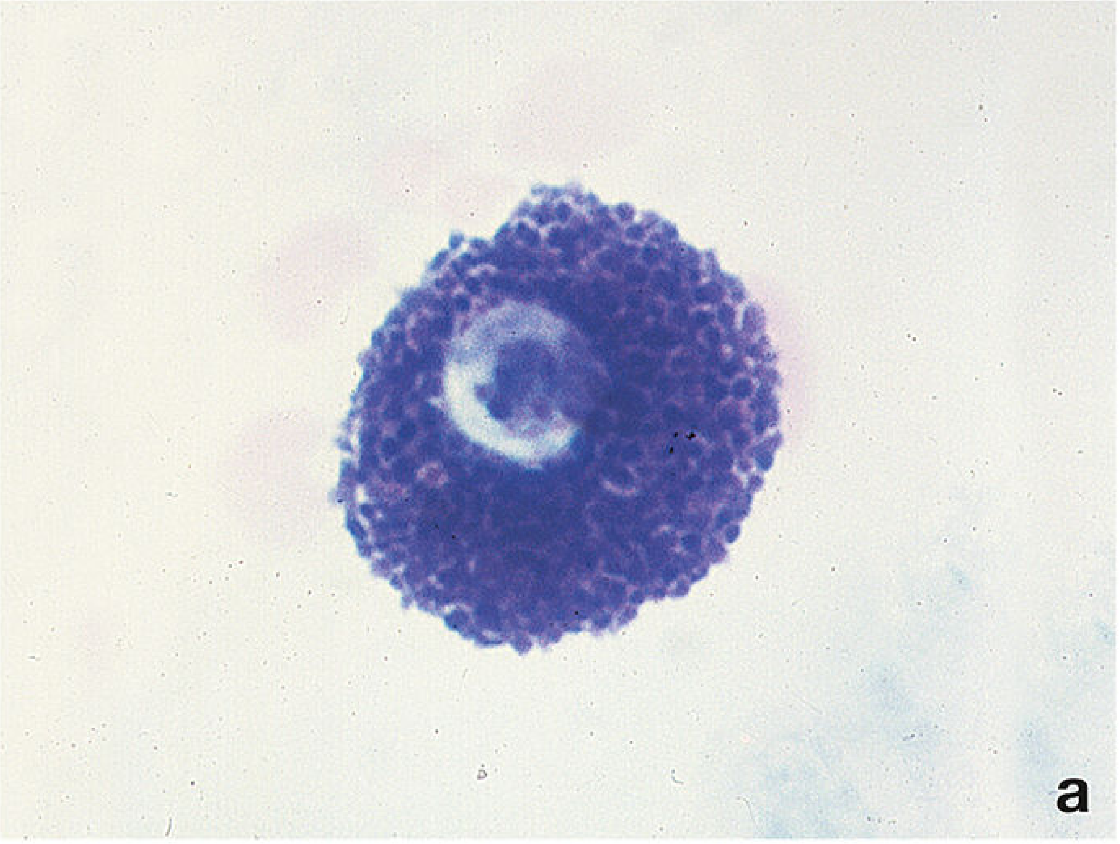
| Toluidine blue | Mast cell granules stain intensely (metachromasia) |
8. Key Histological Landmarks to Recognize
- Loose CT: many cells of varied nuclear shapes, loosely arranged pale fibers
- Dense irregular CT: thick collagen bundles running in multiple planes, sparse nuclei (dermis)
- Dense regular CT: parallel collagen bundles, rows of flattened nuclei between fibers (tendon)
- Fibroblast: elongated/spindle nucleus, cytoplasm blends with collagen
- Mast cell: round nucleus obscured by densely packed granules (toluidine blue)
- Plasma cell: clock-face nucleus, eccentric position, perinuclear hof
9. Clinical Connections
- Allergic reactions / Anaphylaxis: IgE-mediated mast cell degranulation → histamine and leukotrienes cause systemic vasodilation, bronchoconstriction (Type I hypersensitivity)
- Asthma: Leukotriene receptor antagonists (LTRAs) prevent and treat bronchospasm; antihistamines alone are insufficient
- Wound healing and fibrosis: Fibroblast → myofibroblast transition (driven by TGF-β) → wound contraction; excessive activity = pathological fibrosis (e.g., keloid, scleroderma)
- Ehlers-Danlos syndrome: Genetic defects in collagen synthesis → hyperextensible skin, joint hypermobility, fragile blood vessels
- Marfan syndrome: Fibrillin-1 mutation → defective elastic fibers → aortic dilation, lens dislocation
- Scurvy: Vitamin C deficiency → failed collagen hydroxylation → impaired collagen cross-linking → weak CT, bleeding gums