
Drug induced interestitial nephritis , clinical features,l diagnosis, management.
"acute interstitial nephritis" AND drug
drug induced acute interstitial nephritis diagnosis management clinical features authoritative guideline
https://pmc.ncbi.nlm.nih.gov/articles/PMC5718279
Drug-induced acute interstitial nephritis (DI-AIN)
- Antibiotics: beta-lactams, sulfonamides, rifampicin, fluoroquinolones, vancomycin
- NSAIDs
- Proton-pump inhibitors: omeprazole, pantoprazole, etc.
- Diuretics, allopurinol, anticonvulsants, antiretrovirals, and immune-checkpoint inhibitors
Clinical features
- Rising serum creatinine, sometimes oliguria
- Malaise, anorexia, nausea, vomiting
- Fever
- Maculopapular rash
- Arthralgia
- Peripheral eosinophilia
Diagnosis
- Review every prescribed, over-the-counter, and herbal drug, including recent courses.
- Measure serum creatinine, urea, electrolytes, complete blood count with differential, and urinalysis.
- Exclude common AKI causes: volume depletion, obstruction, sepsis, acute tubular injury, glomerulonephritis, and vasculitis.
- Sterile pyuria
- White blood cell casts
- Mild to moderate proteinuria
- Microscopic hematuria may occur
- Eosinophilia may occur
- Urinary eosinophils can support the diagnosis but have low diagnostic accuracy, so they cannot confirm or exclude AIN.
- Mainly to exclude urinary obstruction.
- Kidneys can be normal-sized or enlarged; small kidneys suggest chronic scarring and less likelihood of recovery.
- The diagnostic gold standard, especially if:
- diagnosis is uncertain,
- renal function is worsening or not improving after stopping suspected drugs,
- significant proteinuria or hematuria suggests another renal disorder,
- corticosteroid treatment is being considered.
Management
-
Stop the offending drug immediately
- Do not wait for biopsy if the suspected medication can be safely stopped.
- Record it clearly as a suspected drug allergy/adverse reaction and avoid re-exposure.
- Substitute treatment if the medicine is essential, such as choosing a non-nephrotoxic antibiotic.
-
Support AKI care
- Assess volume status and correct dehydration if present.
- Avoid further nephrotoxins, including NSAIDs, unnecessary contrast, and inappropriate doses of renally cleared medicines.
- Monitor creatinine, potassium, bicarbonate, urine output, and blood pressure.
- Treat hyperkalemia, acidosis, pulmonary edema, or uremic complications.
- Dialysis may be temporarily required for standard AKI indications.
-
Consider corticosteroids
- Best considered with nephrology input, preferably after biopsy confirmation when feasible.
- Observational evidence suggests more benefit when started early, particularly if renal function does not recover within about 7 to 14 days after drug withdrawal and biopsy shows limited interstitial fibrosis.
- A commonly used approach is prednisone equivalent 0.5 to 1 mg/kg/day, followed by reassessment in 1 to 3 weeks and a taper in responders. Regimens vary, and evidence is not from large definitive randomized trials.
- Avoid or use cautiously in uncontrolled infection, severe diabetes, active gastrointestinal bleeding, or marked chronic fibrosis on biopsy.
Prognosis
New drug exposure + AKI + sterile pyuria/WBC casts ± fever, rash, eosinophilia = suspect DI-AIN. Stop the drug, exclude other AKI causes, perform biopsy when uncertainty or steroid treatment is relevant, and consider early corticosteroids if recovery does not occur promptly.
Make a diagnostic and management flowchart
"acute interstitial nephritis" AND drug
drug-associated acute interstitial nephritis management algorithm biopsy corticosteroids
A clear educational clinical flowchart for drug-induced acute interstitial nephritis. Include: suspected DI-AIN with recent medication exposure and acute kidney injury; immediate actions to stop suspected culprit drug, assess AKI severity, urinalysis/CBC/creatinine/electrolytes, renal ultrasound to exclude obstruction, stop additional nephrotoxins and give supportive care; decision points for urgent dialysis indications and alternative cause of AKI; if diagnosis uncertain or kidney function does not improve, nephrology referral and renal biopsy; biopsy confirms AIN with limited fibrosis then consider early corticosteroids; marked chronic fibrosis or contraindication then supportive care; monitoring and follow-up with creatinine, electrolytes, urine output, steroid adverse effects, and avoidance of the culprit drug. Ensure arrows and decision diamonds are logically accurate.
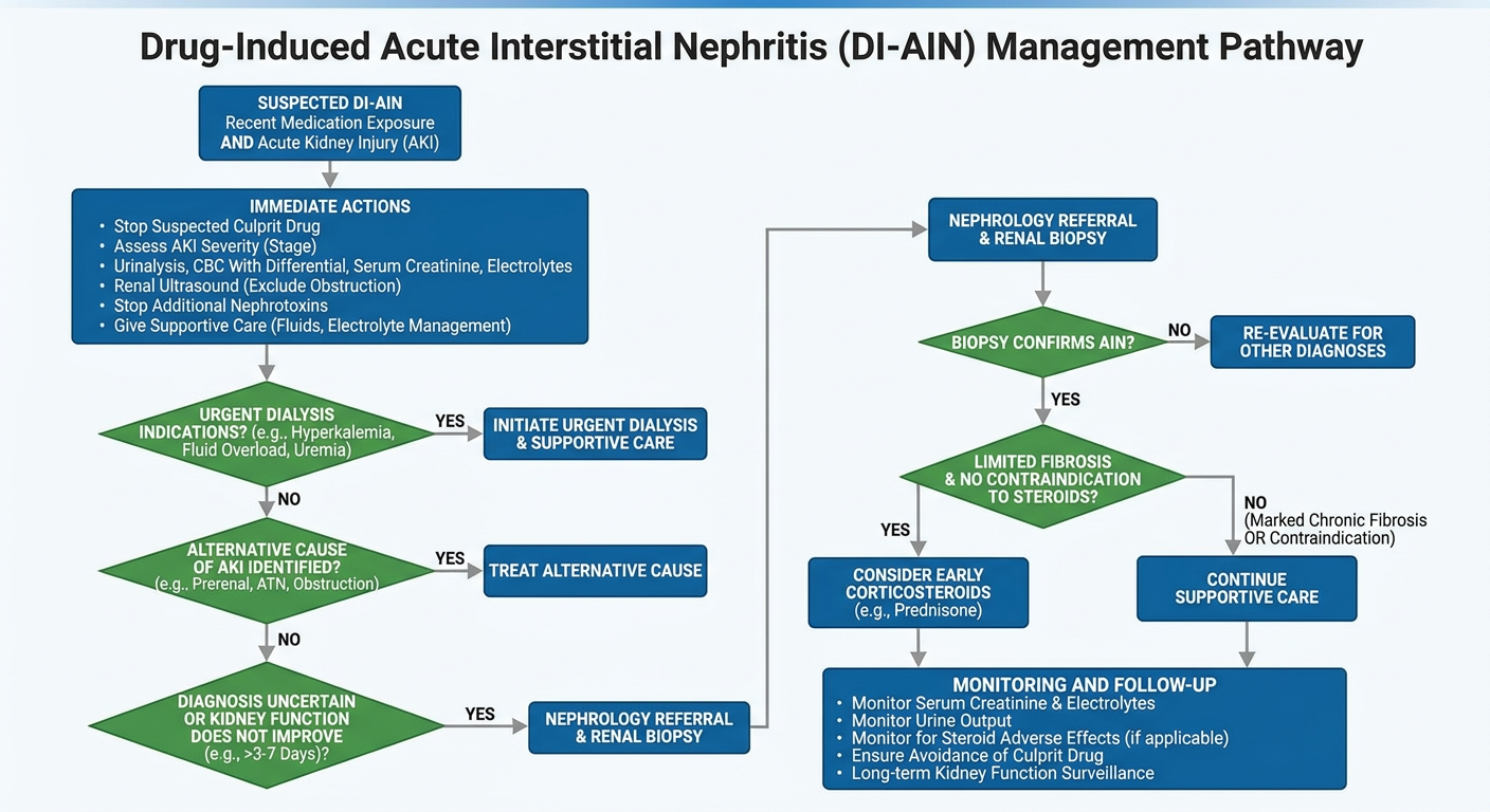
              SUSPECT DRUG-INDUCED AIN (DI-AIN)
       Recent drug exposure + unexplained AKI / rising creatinine
       ± fever, rash, eosinophilia, sterile pyuria, WBC casts
                              │
                              ▼
             IMMEDIATE ACTIONS: DO NOT DELAY
  • Stop suspected culprit drug(s): antibiotics, NSAIDs, PPIs, etc.
  • Document suspected adverse drug reaction and avoid re-exposure
  • Assess volume status, urine output, BP, and AKI severity
  • Stop other nephrotoxins; adjust doses of renally cleared drugs
                              │
                              ▼
                    INITIAL DIAGNOSTIC WORK-UP
  • Serum creatinine, urea, electrolytes, bicarbonate
  • CBC with differential: eosinophilia may be present
  • Urinalysis and microscopy: pyuria, WBC casts, mild proteinuria,
    microscopic hematuria
  • Urine protein quantification
  • Renal ultrasound: exclude obstruction; assess kidney size
  • Review all drugs, OTC NSAIDs, supplements, and recent exposures
                              │
                              ▼
      ┌─────────────────────────────────────────────────┐
      │ Are there urgent complications of AKI?           │
      │ Refractory hyperkalemia, acidosis, pulmonary     │
      │ edema/volume overload, uremic complications      │
      └─────────────────────────────────────────────────┘
                   │ Yes                        │ No
                   ▼                            ▼
      Urgent nephrology review,       Continue supportive AKI care
      treat complication, and         and evaluate for alternative causes
      initiate dialysis if indicated  of AKI
                   │                            │
                   └──────────────┬─────────────┘
                                  ▼
      ┌─────────────────────────────────────────────────┐
      │ Is DI-AIN diagnosis reasonably secure AND renal  │
      │ function improving after drug withdrawal?        │
      └─────────────────────────────────────────────────┘
                   │ Yes                        │ No / uncertain / worsening
                   ▼                            ▼
      Continue supportive care,           Nephrology referral
      monitor creatinine and              Consider renal biopsy promptly:
      electrolytes closely                • diagnosis uncertain
                                          • no renal recovery
                                          • active urine sediment/proteinuria
                                          • steroid treatment being considered
                                                    │
                                                    ▼
      ┌─────────────────────────────────────────────────┐
      │ Biopsy consistent with AIN and limited chronic   │
      │ fibrosis/tubular atrophy?                        │
      └─────────────────────────────────────────────────┘
                   │ Yes                        │ No / major chronic scarring
                   ▼                            ▼
       Consider corticosteroids,          Supportive care; steroids less likely
       ideally early and under            to benefit. Reconsider alternate
       nephrology supervision:            diagnoses and prognosis.
       prednisone equivalent
       0.5-1 mg/kg/day, then taper
       according to clinical response
                   │
                   ▼
                     FOLLOW-UP
  • Serial creatinine, potassium, bicarbonate, urine output, BP
  • Review recovery within 1-3 weeks
  • Monitor steroid adverse effects: glucose, infection, mood changes
  • Avoid the offending medicine permanently when clinically appropriate
  • Assess for incomplete recovery or chronic kidney disease