Give a detail explanation on anatomy, examination, investigations, rehabilitation exercises & add PROMS with treatment including ayurveda part

Asking for Preferences
Reading File
Finding Sources
Finding Sources
Finding Sources
Searching PubMed

"knee osteoarthritis"[MeSH Terms] AND "rehabilitation"[MeSH Terms]

Searching the Web

knee joint Ayurveda treatment Janu Basti Panchakarma osteoarthritis 2024

Reading File
Reading File
Finding Sources
Finding Sources
Searching PubMed

"knee"[MeSH Terms] AND "patient reported outcome"[MeSH Terms] AND "exercise"[MeSH Terms]

Reading File
Finding Sources
Searching PubMed

"knee osteoarthritis" AND "exercise therapy"

Searching the Web

KOOS WOMAC Oxford Knee Score patient reported outcome measures knee joint physiotherapy

Reading File
Reading File
Finding Sources
Reading File
Reading File
Reading Web Page

https://pmc.ncbi.nlm.nih.gov/articles/PMC12444219

Reading File
Reading File
Searching the Web

knee rehabilitation exercises phases ACL meniscal OA physiotherapy protocol 2024

Loading Image
Loading Image
Here is a comprehensive reference guide on the Knee Joint covering all the areas you asked about.

The Knee Joint - A Complete Clinical Reference


1. ANATOMY

1.1 Joint Type & Compartments

The knee is a complex synovial hinge joint - the largest in the body. It has three functional compartments:
  • Medial tibiofemoral compartment
  • Lateral tibiofemoral compartment
  • Patellofemoral compartment
The shape of the articular surfaces makes the joint inherently unstable; stability depends on both static and dynamic restraints. - Bailey & Love's Short Practice of Surgery, 28th Ed.

1.2 Osseous Structures

StructureKey Points
Distal FemurMedial and lateral condyles; intercondylar notch houses the cruciate ligaments
Proximal TibiaMedial and lateral tibial plateaus; tibial tuberosity (insertion of patellar tendon); Gerdy's tubercle (IT band insertion)
PatellaLargest sesamoid bone; acts as a pulley for quadriceps; has the thickest articular cartilage in the body (designed for loads up to 20x body weight)
Fibular HeadProvides attachment for the LCL and biceps femoris

1.3 Articular Cartilage

Hyaline cartilage covers all articulating surfaces. The medial femoral condyle is larger than the lateral, causing a combination of rolling, sliding, and tibial internal rotation during knee flexion - a pattern facilitated by the more mobile lateral meniscus.

1.4 Menisci

The menisci are C-shaped fibrocartilaginous structures that deepen the tibial plateau and distribute load.
FeatureMedial MeniscusLateral Meniscus
ShapeC-shaped (more open)O-shaped (more circular)
MobilityLess mobile (attached to MCL & capsule)More mobile
Blood supplyOuter 1/3 (red zone) onlyOuter 1/3
Injury frequencyMore commonly tornLess commonly torn

1.5 Ligaments

Cruciate Ligaments (Intracapsular, Extrasynovial):
  • ACL - runs from posterior lateral femoral condyle to anterior intercondylar area of tibia; primary restraint to anterior tibial displacement; also resists internal rotation
  • PCL - stronger than ACL; runs from anterior medial femoral condyle to posterior tibia; primary restraint to posterior tibial displacement
Collateral Ligaments:
  • MCL (Medial Collateral Ligament) - has superficial and deep components plus the posterior oblique ligament (POL); resists valgus and external rotation forces
  • LCL (Lateral Collateral Ligament) - resists varus forces; runs from lateral femoral epicondyle to fibular head
Posterolateral Corner (PLC):
  • Popliteus tendon, popliteofibular ligament, arcuate ligament - resist varus, external rotation, and posterior tibial translation

1.6 Joint Capsule & Bursae

The capsule is reinforced anteriorly by the quadriceps retinaculum. Key bursae include:
  • Suprapatellar bursa - communicates with joint cavity (fills with effusion)
  • Prepatellar bursa - "housemaid's knee" when inflamed
  • Infrapatellar bursa (superficial & deep)
  • Pes anserine bursa - medial, between MCL and pes anserine tendons
  • Baker's cyst (popliteal cyst) - posterior, communicates with joint via an opening between medial gastrocnemius and semimembranosus

1.7 Extensor Mechanism

Quadriceps (rectus femoris, vastus medialis, vastus lateralis, vastus intermedius) → quadriceps tendon → patella → patellar tendon → tibial tuberosity. The vastus medialis oblique (VMO) is critical for patellar tracking.

1.8 Dynamic Stabilizers (Muscles)

GroupMusclesFunction
ExtensorsQuadricepsKnee extension; stabilize patella
Flexors/RotatorsHamstrings (biceps femoris, semimembranosus, semitendinosus)Flexion; tibial rotation
Pes AnserinusSartorius, gracilis, semitendinosusMedial stabilizers; attach on proximal medial tibia
IT BandTFL + gluteus maximusLateral stabilizer via Gerdy's tubercle
PopliteusPopliteus"Unlocks" knee from full extension; resists posterolateral rotation

1.9 Biomechanics & Axes

Lower limb mechanical and anatomical axes diagram
The mechanical axis runs from the center of the femoral head through the intercondylar notch to the center of the ankle. The anatomical and mechanical axes of the femur diverge by 5-7° (valgus cut angle). Normal ROM: 5° hyperextension to 135° flexion. Loads across the patellofemoral joint can reach 20x body weight during jumping. - Bailey & Love's, 28th Ed.

2. CLINICAL EXAMINATION

A systematic LOOK - FEEL - MOVE approach is used. Always examine the patient standing, walking, and lying supine/prone.

2.1 Look (Inspection)

Standing (weight-bearing):
  • Alignment: Varus (bow-legs) or valgus (knock-knees) deformity
    • Measure intermalleolar distance for valgus; intermédial knee distance for varus
  • Muscle bulk: Quadriceps wasting (compare sides)
  • Side view: Fixed flexion deformity or recurvatum (hyperextension)
  • Back: Popliteal swelling, hamstring bulk
Gait analysis:
  • Antalgic gait - shortened stance phase on painful limb (OA)
  • Varus thrust - dynamic collapse into varus during stance (medial compartment OA)
  • High-stepping gait - foot drop
Supine:
  • Skin, scars, sinuses
  • Swelling (prepatellar, suprapatellar, popliteal)
  • Deformity

2.2 Feel (Palpation)

  • Temperature: Use dorsum of hand; warmth = inflammation/infection
  • Effusion: Patellar tap test (large effusion) / Bulge/sweep test (small effusion)
  • Joint lines: Medial and lateral joint line tenderness (meniscal pathology, OA)
  • Bony landmarks: Tibial tuberosity (Osgood-Schlatter), fibular head, femoral condyles, Gerdy's tubercle
  • Soft tissues: Quadriceps tendon, patellar tendon, collateral ligaments, popliteal fossa (Baker's cyst)
  • Muscle: Hamstrings, calf

2.3 Move (Range of Motion)

  • Active ROM: Ask patient to fully flex and extend
  • Passive ROM: Assessor moves joint; note end-feel (firm = normal, springy = meniscal block, empty = pain)
  • Normal: 0° extension (some hyperextension normal) to 135° flexion
  • Fixed flexion test: Sit patient upright with knees hanging over edge; if flexion deformity persists, it is in the knee (not hip)

2.4 Special Tests

Collateral Ligaments

  • Valgus stress test at 30° flexion - tests MCL (at 0° full extension, also tests posterior capsule/PCL)
  • Varus stress test at 10-30° flexion - tests LCL

Cruciate Ligaments

  • Lachman test (most sensitive for ACL) - 20-30° flexion, anterior tibial drawer; firm vs. soft end-feel
  • Anterior drawer test - 90° flexion, anterior tibial pull
  • Pivot shift test (most specific for ACL functional instability) - internal rotation + valgus stress while extending; a clunk = positive
  • Posterior drawer test - 90° flexion, posterior tibial push (PCL)
  • Posterior sag sign (Godfrey's test) - PCL rupture causes tibia to sag posteriorly with hip and knee at 90°

Meniscal Tests

  • McMurray's test - flex knee fully, then extend with external rotation (medial meniscus) or internal rotation (lateral meniscus); click = positive
  • Thessaly test - single-leg stance at 20° flexion, rotate body; joint line pain = positive (reported higher sensitivity than McMurray's)
  • Apley's grind test (compression/distraction) - prone, knee at 90°; compression + rotation tests meniscus; distraction tests ligaments

Patellofemoral Tests

  • Patellar tap test - large effusion
  • Patellar grind (Clarke's test) - compress patella into trochlea, patient contracts quads; pain = chondromalacia
  • Patellar apprehension (Fairbank's test) - lateral patellar displacement at 30° flexion; apprehension/contraction = instability
  • Patellar tracking ('J' sign) - sit, legs hanging at 90°, slowly extend; lateral jump near full extension = maltracking

Other

  • Ober's test - IT band tightness
  • Thomas test - hip flexor tightness
  • Trendelenburg test - hip abductor weakness
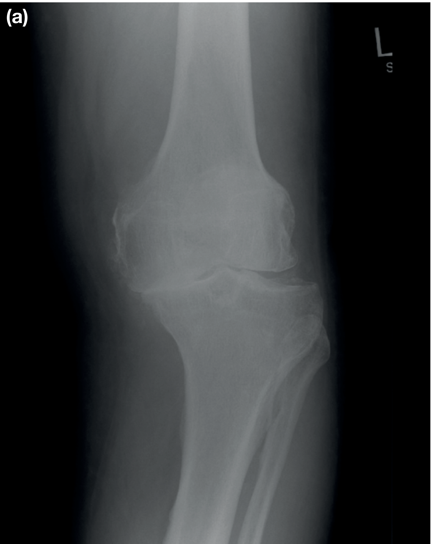
3. INVESTIGATIONS

3.1 Imaging

InvestigationIndicationsKey Findings
Plain X-ray (weight-bearing AP, lateral, skyline)First-line for OA, fractures, alignmentOA: joint space narrowing, subchondral sclerosis, osteophytes, subchondral cysts; Always do weight-bearing to show true joint space
MRISoft tissue pathology - first choice for meniscal tears, ligament injuries, cartilage defects, bone marrow oedemaNot routinely needed pre-TKR; most sensitive for ACL, meniscal, chondral assessment
CT scanComplex intra-articular fractures, tibial plateau fractures, rotational alignment, pre-op planningBony detail; CT arthrogram when MRI contraindicated
UltrasoundSoft tissue - Baker's cyst, popliteal vessels, tendon assessment; guided injectionsDynamic assessment of tendon; effusion aspiration guidance
Bone scan (SPECT/CT)Occult fractures, stress fractures, osteonecrosis, infectionIncreased uptake in active bone pathology

3.2 X-Ray Views

  • AP (weight-bearing): Kellgren-Lawrence grading of OA; alignment
  • Lateral (30° flexion): Patella height (Insall-Salvati ratio), posterior condyle, tibial slope
  • Skyline (Merchant/axial): Patellofemoral compartment, trochlear dysplasia
  • Long-leg alignment (scanogram): Mechanical axis; varus/valgus correction planning
  • Rosenberg view (posteroanterior at 45°): Most sensitive plain X-ray view for joint space in early OA

3.3 Kellgren-Lawrence Grading (OA)

GradeDescription
0Normal
1Doubtful narrowing; possible osteophyte
2Definite osteophyte; possible joint space narrowing
3Multiple osteophytes; definite narrowing; some sclerosis
4Large osteophytes; marked narrowing; severe sclerosis; possible bony deformity

3.4 Blood Investigations

TestPurpose
FBC, CRP, ESRInfection, inflammatory arthritis
Rheumatoid factor, anti-CCPRheumatoid arthritis
Uric acidGout (though normal during acute attack)
ANA, dsDNASLE
HLA-B27Seronegative spondyloarthropathy
Blood culturesSeptic arthritis
CoagulationPre-operative

3.5 Synovial Fluid Analysis (Aspiration)

ParameterNormalOAInflammatorySeptic
AppearanceClearYellow, clearYellow, turbidPurulent, opaque
WBC/mm³< 200< 2,0002,000-50,000> 50,000
PMN%< 25%< 25%> 50%> 75%
Glucose= serum= serum<< serum
CultureNegativeNegativeNegativePositive
CrystalsNoneNoneGout (urate), CPPD (pyrophosphate)None

3.6 Arthroscopy (Diagnostic)

Indicated when clinical + imaging inconclusive; allows direct visualization and simultaneous treatment of meniscal tears, loose bodies, synovitis, cartilage defects.

4. COMMON CONDITIONS, DIAGNOSIS & TREATMENT

4.1 Knee Osteoarthritis (OA)

Prevalence: ~10% in men and 13% in women aged 60+. Can be primary (idiopathic) or secondary (post-trauma, meniscectomy, ligament injury). - Bailey & Love's, 28th Ed.
Clinical features: Pain worse with loading and activity; stiffness; swelling; antalgic gait; varus deformity common; joint effusion; crepitus; reduced ROM.
Investigations: Weight-bearing AP X-ray (Kellgren-Lawrence grading); MRI not routine pre-TKR.
Anteroposterior X-ray of knee OA showing medial joint space narrowing, subchondral sclerosis, and osteophytes
Non-operative treatment (first line):
  • Weight loss, exercise, patient education, self-management
  • Analgesics: topical NSAIDs (first-line), oral NSAIDs, paracetamol
  • Walking aids, offloader brace, orthotics
  • Intra-articular corticosteroid injection (short-term relief in flares; not long-term)
  • Hyaluronic acid - not recommended by AAOS
  • Oral narcotics - not recommended
Surgical treatment:
  • High tibial osteotomy (HTO) - younger patients with unicompartmental OA + malalignment; realigns mechanical axis to offload diseased compartment
  • Unicompartmental knee replacement (UKR) - single compartment disease; shorter recovery
  • Total knee replacement (TKR) - end-stage disease; traditional goal = restore mechanical axis to neutral
  • Arthrodesis - last resort; young patient with failed TKR or infection
Per AAOS 2023 Evidence-Based Guidelines: Exercise (supervised/unsupervised/aquatic), patient education, topical & oral NSAIDs, and weight loss carry the strongest (4-star) recommendations. - Miller's Review of Orthopaedics, 9th Ed.

4.2 ACL Injury

Mechanism: Twisting or landing injury in pivoting sport; often audible 'pop' + immediate swelling; higher risk in females (smaller ligaments, narrower notch, different landing mechanics).
Examination: Positive Lachman test + pivot shift test.
Management:
  • Conservative: Physiotherapy, bracing - for low-demand, older, sedentary patients
  • Surgical (ACL reconstruction): Recommended for young, active patients; pivot sport athletes; associated meniscal/chondral injuries. Graft options: patellar tendon (BTB), hamstring (gracilis + semitendinosus), quadriceps tendon, allograft

4.3 Meniscal Tears

Types: Circumferential (bucket-handle), radial, horizontal, flap, degenerative
Symptoms: Joint line pain, locking (bucket-handle), giving way, swelling
Management:
  • Conservative: Degenerative tears in older patients without mechanical symptoms
  • Arthroscopic partial meniscectomy: Unstable tears with mechanical symptoms
  • Meniscal repair: Younger patients, early presentation, vascular outer 1/3, stable tear pattern, concomitant ACL reconstruction - Bailey & Love's, 28th Ed.

4.4 Patellofemoral Pain Syndrome (PFPS)

Anterior knee pain, worse on stairs and rising from sitting. Maltracking, VMO weakness, tight lateral retinaculum, trochlear dysplasia.
Treatment: VMO strengthening, taping/bracing, footwear, avoid aggravating activities. Surgery (realignment) rarely required.

4.5 Septic Arthritis

Orthopaedic emergency. Hot, swollen, extremely painful joint; fever; raised WBC, CRP, ESR. Needs urgent aspiration + culture → IV antibiotics → joint washout (arthroscopic or open).

5. REHABILITATION EXERCISES

Rehabilitation is phased and condition-specific. The following covers both OA management and post-surgical (ACL/meniscal) rehabilitation.

5.1 General Principles

A 2024 Cochrane systematic review confirmed that exercise is effective for knee OA - reducing pain and improving function with no increased risk of harm (Lawford et al., Cochrane 2024). The 2025 BMJ network meta-analysis (Yan et al.) found aquatic exercise and combined aerobic + strengthening provided the greatest benefit.
EULAR 2023 updated guidelines recommend exercise as the non-pharmacological cornerstone of hip and knee OA management.

5.2 Phase-Based Rehabilitation (OA & General Knee)

Phase 1 - Acute/Early Phase (Week 1-2)

Goals: Pain & swelling control, maintain ROM, prevent muscle wasting
ExerciseDescriptionSets/Reps
Ankle pumpsFlex/extend ankle; promote circulation3 × 20
Quad sets (isometric)Press back of knee to bed, contract quads3 × 10, hold 5s
Inner range quadsKnee supported at 30° under roll, extend actively3 × 10
Straight leg raise (SLR)Leg raised to 45°, quads contracted; all 4 planes3 × 10
Heel slidesSlide heel to bend knee (flexion ROM)3 × 10
Calf raises (seated)Improve venous return3 × 15
Ice + elevation20 minutes, 4-6 times/day-

Phase 2 - Sub-acute Phase (Week 3-6)

Goals: Restore ROM, begin weight-bearing strengthening, neuromuscular control
ExerciseDescriptionSets/Reps
Mini squats (0-30°)Partial weight-bearing squat, hands on support3 × 15
Step-ups (forward & lateral)Low step 5-10 cm; progress height gradually3 × 10 each side
Terminal knee extension (TKE)Resistance band behind knee, extend from 30° to 0°3 × 15
Hamstring curls (prone)Flex knee against gravity/resistance3 × 12
Hip abduction (side-lying)Strengthens glute medius; reduces knee valgus3 × 15
Static cyclingLow resistance; ROM & cardiovascular benefit15-20 min
VMO emphasis squatsFeet slightly externally rotated3 × 12

Phase 3 - Strengthening Phase (Week 6-12)

Goals: Progressive strength, balance, functional activity
ExerciseDescriptionSets/Reps
Full squats (0-90°)Bodyweight to loaded; watch for valgus collapse3 × 12
Leg pressControlled full ROM; start low resistance3 × 12
Lunges (forward/lateral)Functional multi-plane movement3 × 10 each leg
Wall squats (isometric)Sustained quad + VMO activation3 × 30-60s
Single-leg balanceEyes open → eyes closed → unstable surface3 × 30s each
Nordic hamstring curlsEccentric hamstring strengthening3 × 8
Aquatic exercisesPool walking, pool squats; offloads joint20-30 min

Phase 4 - Functional/Return-to-Sport Phase (Week 12+)

Goals: Return to full activity; sport-specific training (ACL patients)
ExerciseDescription
Bulgarian split squatsSingle-leg loaded squat
Box jumps / PlyometricsACL patients only after strength testing clears
Lateral band walksHip abductor strengthening in functional position
Agility ladder drillsNeuromuscular coordination
Running progressionWalk-jog intervals → continuous jog → sprint
Sport-specific drillsCutting, pivoting, deceleration
Criteria to progress to return-to-sport: Limb Symmetry Index (LSI) >90% on hop testing, KOOS-sports >90%, hamstring/quad ratio ≥66%, pain-free full ROM. - Ref: MGH Sports Medicine ACL Protocol 2024

5.3 Aquatic Exercise (Hydrotherapy)

Especially useful in OA, post-TKR, and obese patients where weight-bearing land exercise is painful. Water buoyancy reduces effective body weight by up to 90% (neck depth). Includes: pool walking, aqua cycling, water aerobics, and resistance exercises with floats.

6. PATIENT-REPORTED OUTCOME MEASURES (PROMs)

PROMs are standardized questionnaires completed by the patient to assess function, pain, and quality of life. They are essential for clinical audit, surgical decision-making, and monitoring rehabilitation progress.

6.1 Summary Table of Key Knee PROMs

PROMFull NameItemsSubscalesBest Used ForScore RangeInterpretation
KOOSKnee Injury & Osteoarthritis Outcome Score42Pain, Symptoms, ADL, Sport/Rec, QoLOA, ACL, Meniscal injury, TKR0-100 per subscaleHigher = better
KOOS-PSKOOS Physical Function Short Form7Physical functionQuick functional screen0-100Higher = better
WOMACWestern Ontario & McMaster Universities OA Index24Pain (5), Stiffness (2), Function (17)Knee & hip OA0-96 (Likert) or 0-100 (VAS)Lower = better
Oxford Knee Score (OKS)Oxford Knee Score12Single scoreTKR pre/post-op assessment0-48Higher = better
Lysholm Knee ScaleLysholm Knee Scoring Scale8Limp, locking, instability, pain, swelling, stair climbing, squatACL, ligament injuries0-100Higher = better
IKDCInt'l Knee Documentation Committee Subjective Knee Form18Symptoms, sport activity, functionLigament/meniscal injury, ACL0-100Higher = better
Tegner Activity ScaleTegner Activity Level Scale1Activity levelACL, sports-related injury0-10 (0=sick leave, 10=competitive sport)Higher = more active
VAS / NRSVisual Analogue / Numeric Rating Scale1Pain intensityAny knee condition0-10Lower = less pain
SF-36 / SF-12Short Form Health Survey36/128 domains, PCS + MCSGeneral health-related QoL0-100Higher = better
PROMISPatient-Reported Outcomes Measurement Information SystemAdaptivePain, function, fatigue, mental healthComprehensive; computer-adaptiveT-score (50 = norm)Condition dependent
KSSKnee Society ScoreDualKnee score + Function scoreTKR outcomes0-100 eachHigher = better
ACL-RSIACL Return to Sport Index12Psychological readinessPost-ACL reconstruction0-100≥90 = cleared psychologically

6.2 Detailed Notes on Key PROMs

KOOS (Knee Injury and Osteoarthritis Outcome Score):
  • 42 items across 5 subscales: Pain (9), Symptoms (7), ADL (17), Sport/Recreation (5), QoL (4)
  • Each item scored 0-4; transformed to 0-100 (0 = extreme problems, 100 = no problems)
  • Reliability ICC >0.8 on most subscales
  • Validated for ACL reconstruction, meniscal tears, OA; extends and is compatible with WOMAC
  • The 2024 KOOS review study confirms it as the most comprehensive functional PROM for the knee
WOMAC:
  • 24-item self-report; excellent reliability (ICC 0.90 for pain); high concurrent validity with SF-36
  • Primarily validated for OA; less sensitive for younger/more active patients
Oxford Knee Score (OKS):
  • 12-item questionnaire; 5-point Likert scale; score 0-48
  • Excellent reliability and validity especially for TKR
  • MCID (Minimum Clinically Important Difference) = 5 points
Lysholm Scale:
  • 8-item; clinician + patient scored; used widely for ligament injuries
  • Categories: Excellent (95-100), Good (84-94), Fair (65-83), Poor (<65)
Tegner Activity Scale:
  • Single item, 0-10 ordinal scale of activity
  • Pre-injury vs. post-treatment comparison important for return-to-sport decisions
ACL-RSI (Psychological Readiness):
  • Often overlooked but critical: psychological readiness is a predictor of re-injury
  • Score ≥90 on ACL-RSI is recommended before return to cutting/pivoting sport
When to use which PROM:
Clinical ScenarioRecommended PROM(s)
Knee OA (conservative management)KOOS or WOMAC + VAS
ACL reconstructionKOOS + IKDC + Lysholm + Tegner + ACL-RSI
Meniscal injuryKOOS + Lysholm
TKR/UKROxford Knee Score (OKS) + KOOS + KSS
Patellofemoral painKOOS + VAS (WOMAC less specific for PFPS)
Research / generalPROMIS (computer-adaptive, efficient)

7. TREATMENT SUMMARY

7.1 Pharmacological Treatment

Drug ClassExamplesUse
Topical NSAIDsDiclofenac gel, KetoprofenFirst-line OA; fewer GI/renal side effects
Oral NSAIDsIbuprofen, Naproxen, CelecoxibModerate OA, acute flares
Paracetamol (Acetaminophen)Paracetamol 1g QIDMild-moderate OA
OpioidsTramadol (weak opioid)Not recommended by AAOS for knee OA
Intra-articular corticosteroidsTriamcinolone, MethylprednisoloneShort-term flare control; max 3-4/year
Hyaluronic acid (viscosupplementation)Synvisc, OstenilEvidence conflicting; not recommended by AAOS
Duloxetine60-120 mg/dayChronic pain with central sensitization component
SupplementsGlucosamine, ChondroitinLimited evidence (AAOS: limited recommendation only)

7.2 Physical & Allied Health Interventions

  • Physiotherapy (strengthening, manual therapy, taping)
  • Occupational therapy (joint protection, aids)
  • Weight loss (each 1 kg lost reduces knee load by ~4 kg)
  • Orthotics (medial wedge for lateral OA; lateral wedge NOT recommended by AAOS)
  • Offloader brace (valgus brace for medial compartment OA)
  • TENS, acupuncture (limited evidence; AAOS 2-star)
  • PRP (Platelet-rich plasma) - AAOS: limited, may reduce pain (2-star)

7.3 Surgical Treatment

ProcedureIndication
Arthroscopic debridement/washoutNOT recommended for OA (AAOS strong against)
Arthroscopic meniscectomySymptomatic meniscal tear with mechanical symptoms
Meniscal repairYoung patient, vascular zone tear, repairable
ACL reconstructionActive patients with functional instability
Chondrocyte transplant (MACI/ACI)Young patient, full-thickness cartilage defect
Osteotomy (HTO/DFO)Young patient, unicompartmental OA + malalignment
Unicompartmental knee replacement (UKR)Single compartment OA, intact cruciate ligaments
Total Knee Replacement (TKR)End-stage, multi-compartment OA; failed conservative/other surgery
ArthrodesisFailed TKR, chronic septic arthritis

8. AYURVEDA & INTEGRATIVE APPROACH TO THE KNEE

8.1 Ayurvedic Understanding

In Ayurveda, the knee joint is called "Janu Sandhi". Knee pain is primarily linked to Vata dosha imbalance - specifically a condition called "Sandhigatavata" (degeneration of joints, analogous to OA). When Vata increases, it dries synovial lubrication (Sleshaka Kapha), causing friction, stiffness, crackling sounds, and pain.
Secondary pathology includes accumulation of Ama (toxins from incomplete digestion), which causes inflammatory changes resembling rheumatoid arthritis (Amavata).

8.2 Panchakarma Procedures for the Knee

Janu Basti (Knee Oil Retention Therapy)

  • A dough ring (made of black gram flour) is placed over the knee to create a well
  • Warm medicated oil (Dhanwantara Taila, Ksheerabala Taila, or Mahanarayan Taila) is poured into the well and retained for 30-45 minutes at therapeutic temperature
  • The warmth allows the oil to penetrate deeply into ligaments, cartilage, and synovial membrane
  • Benefits: Reduces pain, stiffness, and swelling; nourishes joint tissues; improves ROM
  • Indications: OA knee, chronic knee pain, Sandhigatavata
  • Contraindications: Acute infection, open wounds, severe acute trauma

Matra Basti (Therapeutic Oil Enema)

  • Small-volume medicated oil enema (30-60 mL) using Ksheerabala Taila
  • Considered the prime treatment for Vatavyadhi (musculoskeletal/degenerative disorders)
  • Rectal absorption provides high bioavailability; lipid-soluble drugs cross rectal mucosa rapidly
  • A 2025 RCT protocol (Rai et al., JMIR Protocols 2025) is evaluating Matra Basti + Janu Basti combined with Laksha Guggulu for primary knee OA

Abhyanga (Therapeutic Oil Massage)

  • Full-body or localized warm medicated oil massage
  • Oils used: Mahanarayan Taila (anti-inflammatory, rejuvenative), Ksheerabala Taila
  • Improves circulation, reduces Vata, nourishes joint tissues
  • Done before Swedana (fomentation)

Swedana (Heat Fomentation)

  • Steam therapy or local fomentation after Abhyanga
  • Relieves stiffness; opens channels (Srotas); prepares joint for deeper treatments
  • Types: Nadi Sweda (steam pipe), Patra Pinda Sweda (herbal leaf bolus), Jambira Pinda Sweda (lemon bolus)

Upanaha Sweda (Medicated Poultice)

  • Herbal paste applied warm over the knee joint overnight
  • Herbs: Eranda (castor), sesame, salt, vinegar
  • Used for chronic stiffness and degeneration

Mridu Samshodhana (Mild Purgation)

  • Mild virechana (purgation) to eliminate Ama
  • Indicated when inflammatory/toxic component is present

Lepa (External Medicated Paste)

  • Herbal paste applied topically
  • Guggulu-based formulations (Yogaraja Guggulu, Trayodashanga Guggulu)

8.3 Ayurvedic Internal Medicines

FormulationKey IngredientsAction
Yogaraja GugguluGuggul, triphala, trikatu, sesame oilAnti-inflammatory, anti-arthritic; Vata pacifying
Trayodashanga GugguluGuggul + 13 herbs incl. ashwagandhaAnalgesic, anti-rheumatic; Vata-pacifying
Laksha GugguluLaksha (lac), guggul, ashwagandha, nagabalaBone and cartilage regeneration; fracture healing
Muktashukti BhasmaCalcined pearl oyster shellCalcium supplementation; anti-inflammatory
Ashwagandha (Withania somnifera)Alkaloids, withanolidesAdaptogen; muscle and ligament strengthening; reduces stiffness
Shallaki / Boswellia serrataBoswellic acidsWell-evidenced anti-inflammatory; inhibits 5-LOX; reduces knee OA pain
Guggulu (Commiphora mukul)GuggulsteronesAnti-inflammatory; balances Vata & Kapha
TriphalaAmalaki, bibhitaki, haritakiAntioxidant; Ama removal; GI support for drug absorption
Shatavari (Asparagus racemosus)Steroidal saponinsRejuvenative; anti-inflammatory
Punarnava (Boerhavia diffusa)PunarnavineAnti-oedematous; reduces joint swelling

8.4 Dietary Guidelines (Ahara) in Ayurveda

  • Avoid: Cold, dry, stale food; excessive raw salads; cold drinks; refined flour; nightshades (in Vata conditions)
  • Include: Warm, unctuous, easily digestible food; ghee, sesame oil; root vegetables; ginger, turmeric, black pepper; warm soups
  • Turmeric (Haridra): Curcumin has documented anti-inflammatory effects; combine with black pepper for absorption

8.5 Evidence for Ayurveda in Knee OA

A 2024 prospective study (Kshirsagar et al., Int J Res Orthop 2024) using Panchakarma therapy in OA patients demonstrated statistically significant improvements in KOOS subscores (pain, sports, function, QoL) over 90 days (p<0.001). The study concluded Ayurvedic treatment is effective in reducing arthritis severity and improving KOOS scores.
Boswellia serrata (Shallaki) has multiple RCTs demonstrating superiority over placebo for knee OA pain reduction, with effects comparable to NSAIDs but with fewer GI side effects.

9. INTEGRATIVE TREATMENT ALGORITHM

KNEE PAIN PRESENTATION
         |
    ─────────────────
    |                |
ACUTE INJURY        CHRONIC / GRADUAL ONSET
(trauma, sport)     (OA, overuse, degeneration)
    |                |
Imaging (X-ray,     Weight-bearing X-ray
MRI) + Special      K-L grading
tests               |
    |                |
DIAGNOSE:           Grade I-II OA:              Grade III-IV OA:
- ACL/PCL           Non-operative:              Surgical evaluation
- Meniscus          Exercise + PT               HTO / UKR / TKR
- Ligament          NSAIDs (topical)
- Fracture          Weight loss
                    Ayurveda + Physio
    |               (Janu Basti +
    |               Strengthening exercises)
Surgical or              |
conservative        PROM monitoring:
decision            KOOS / WOMAC / OKS
    |
Rehabilitation
(Phased - Phase 1 to 4)
PROMS at baseline, 6 weeks, 3 months, 6 months, 1 year

Key References

  • Bailey & Love's Short Practice of Surgery, 28th Edition - Knee anatomy, examination, OA, ACL, meniscal injuries
  • Miller's Review of Orthopaedics, 9th Edition - AAOS evidence-based guidelines for knee OA
  • Rockwood & Green's Fractures in Adults, 10th Ed. - Medial and lateral knee anatomy
  • Lawford et al., Cochrane 2024 - Exercise for knee OA: systematic review
  • Yan et al., BMJ 2025 - Comparative efficacy of exercise modalities in knee OA (network meta-analysis)
  • EULAR 2023 Updated Guidelines - Non-pharmacological management of hip and knee OA
  • Rai et al., JMIR Protocols 2025 - Multimodal Ayurveda regimen (Janu Basti + Matra Basti + Laksha Guggulu) RCT protocol for primary knee OA
  • Kshirsagar et al., Int J Res Orthop 2024 - Panchakarma therapy improving KOOS in OA
  • Physio-pedia KOOS - KOOS subscale details

Generate a rehabilitation protocol PDF with exercise progression charts

Reading File
Reading File
Running Command

mkdir -p /home/daytona/workspace/knee-rehab-pdf && ls /home/daytona/workspace/kn…

Writing File

~/knee-rehab-pdf/generate_pdf.py

#!/usr/bin/env python3
"""
Knee Rehabilitation Protocol PDF Generator
Uses reportlab for PDF creation and matplotlib for charts.
"""

import matplotlib
matplotlib.use('Agg')
import matplotlib.pyplot as plt
import matplotlib.patches as mpatches
import numpy as np
import os
import io

from reportlab.lib.pagesizes import A4
from reportlab.lib import colors
from reportlab.lib.units import mm, cm
from reportlab.lib.styles import getSampleStyleSheet, ParagraphStyle
from reportlab.lib.enums import TA_CENTER, TA_LEFT, TA_RIGHT, TA_JUSTIFY
from reportlab.platypus import (
    SimpleDocTemplate, Paragraph, Spacer, Table, TableStyle,
    PageBreak, HRFlowable, Image, KeepTogether
)
from reportlab.platypus.flowables import Flowable
from reportlab.lib.colors import HexColor, white, black

# ── Colour palette ──────────────────────────────────────────────────────────
C_DARK_BLUE   = HexColor('#1A3A5C')
C_MID_BLUE    = HexColor('#2B6CB0')
C_LIGHT_BLUE  = HexColor('#EBF4FF')
C_ACCENT      = HexColor('#E53E3E')
C_TEAL        = HexColor('#2C7A7B')
C_GREEN       = HexColor('#276749')
C_LIGHT_GREEN = HexColor('#F0FFF4')
C_AMBER       = HexColor('#C05621')
C_LIGHT_AMBER = HexColor('#FFFAF0')
C_PURPLE      = HexColor('#553C9A')
C_LIGHT_GREY  = HexColor('#F7FAFC')
C_MID_GREY    = HexColor('#718096')
C_BORDER      = HexColor('#CBD5E0')

OUTPUT_DIR = '/home/daytona/workspace/knee-rehab-pdf'
os.makedirs(OUTPUT_DIR, exist_ok=True)

# ── Custom styles ────────────────────────────────────────────────────────────
def build_styles():
    base = getSampleStyleSheet()
    styles = {}

    styles['cover_title'] = ParagraphStyle(
        'cover_title', parent=base['Title'],
        fontSize=32, textColor=white, leading=40,
        spaceAfter=10, alignment=TA_CENTER, fontName='Helvetica-Bold'
    )
    styles['cover_sub'] = ParagraphStyle(
        'cover_sub', parent=base['Normal'],
        fontSize=14, textColor=HexColor('#BEE3F8'), leading=20,
        alignment=TA_CENTER, fontName='Helvetica'
    )
    styles['cover_footer'] = ParagraphStyle(
        'cover_footer', parent=base['Normal'],
        fontSize=10, textColor=HexColor('#90CDF4'),
        alignment=TA_CENTER, fontName='Helvetica-Oblique'
    )
    styles['section_header'] = ParagraphStyle(
        'section_header', parent=base['Heading1'],
        fontSize=16, textColor=white, leading=22,
        spaceBefore=6, spaceAfter=8,
        fontName='Helvetica-Bold', alignment=TA_LEFT
    )
    styles['subsection'] = ParagraphStyle(
        'subsection', parent=base['Heading2'],
        fontSize=12, textColor=C_DARK_BLUE, leading=16,
        spaceBefore=10, spaceAfter=4,
        fontName='Helvetica-Bold'
    )
    styles['body'] = ParagraphStyle(
        'body', parent=base['Normal'],
        fontSize=9.5, textColor=HexColor('#2D3748'), leading=14,
        spaceAfter=4, fontName='Helvetica', alignment=TA_JUSTIFY
    )
    styles['body_bold'] = ParagraphStyle(
        'body_bold', parent=base['Normal'],
        fontSize=9.5, textColor=C_DARK_BLUE, leading=14,
        spaceAfter=4, fontName='Helvetica-Bold'
    )
    styles['bullet'] = ParagraphStyle(
        'bullet', parent=base['Normal'],
        fontSize=9.5, textColor=HexColor('#2D3748'), leading=14,
        spaceAfter=3, leftIndent=14, firstLineIndent=-10,
        fontName='Helvetica'
    )
    styles['table_header'] = ParagraphStyle(
        'table_header', parent=base['Normal'],
        fontSize=9, textColor=white, leading=12,
        fontName='Helvetica-Bold', alignment=TA_CENTER
    )
    styles['table_cell'] = ParagraphStyle(
        'table_cell', parent=base['Normal'],
        fontSize=8.5, textColor=HexColor('#2D3748'), leading=12,
        fontName='Helvetica'
    )
    styles['table_cell_bold'] = ParagraphStyle(
        'table_cell_bold', parent=base['Normal'],
        fontSize=8.5, textColor=C_DARK_BLUE, leading=12,
        fontName='Helvetica-Bold'
    )
    styles['phase_label'] = ParagraphStyle(
        'phase_label', parent=base['Normal'],
        fontSize=11, textColor=white, leading=14,
        fontName='Helvetica-Bold', alignment=TA_CENTER
    )
    styles['caption'] = ParagraphStyle(
        'caption', parent=base['Normal'],
        fontSize=8, textColor=C_MID_GREY, leading=11,
        fontName='Helvetica-Oblique', alignment=TA_CENTER
    )
    styles['prom_name'] = ParagraphStyle(
        'prom_name', parent=base['Normal'],
        fontSize=9, textColor=C_DARK_BLUE, leading=12,
        fontName='Helvetica-Bold'
    )
    styles['footer_text'] = ParagraphStyle(
        'footer_text', parent=base['Normal'],
        fontSize=8, textColor=C_MID_GREY, leading=10,
        fontName='Helvetica', alignment=TA_CENTER
    )
    return styles

STYLES = build_styles()

# ── Helper: coloured section banner ─────────────────────────────────────────
def section_banner(title, bg_color=C_DARK_BLUE):
    data = [[Paragraph(title, STYLES['section_header'])]]
    t = Table(data, colWidths=[170*mm])
    t.setStyle(TableStyle([
        ('BACKGROUND', (0,0), (-1,-1), bg_color),
        ('LEFTPADDING',  (0,0), (-1,-1), 10),
        ('RIGHTPADDING', (0,0), (-1,-1), 10),
        ('TOPPADDING',   (0,0), (-1,-1), 7),
        ('BOTTOMPADDING',(0,0), (-1,-1), 7),
        ('ROUNDEDCORNERS', [4,4,4,4]),
    ]))
    return t

# ── Helper: phase badge ──────────────────────────────────────────────────────
def phase_badge(text, color):
    data = [[Paragraph(text, STYLES['phase_label'])]]
    t = Table(data, colWidths=[170*mm])
    t.setStyle(TableStyle([
        ('BACKGROUND', (0,0), (-1,-1), color),
        ('LEFTPADDING',  (0,0), (-1,-1), 8),
        ('RIGHTPADDING', (0,0), (-1,-1), 8),
        ('TOPPADDING',   (0,0), (-1,-1), 5),
        ('BOTTOMPADDING',(0,0), (-1,-1), 5),
    ]))
    return t

# ── Helper: exercise table ───────────────────────────────────────────────────
def exercise_table(headers, rows, col_widths, row_colors=None):
    header_row = [Paragraph(h, STYLES['table_header']) for h in headers]
    data = [header_row]
    for i, row in enumerate(rows):
        styled = []
        for j, cell in enumerate(row):
            if j == 0:
                styled.append(Paragraph(str(cell), STYLES['table_cell_bold']))
            else:
                styled.append(Paragraph(str(cell), STYLES['table_cell']))
        data.append(styled)

    t = Table(data, colWidths=col_widths, repeatRows=1)
    style_cmds = [
        ('BACKGROUND',   (0,0), (-1,0), C_DARK_BLUE),
        ('ROWBACKGROUNDS',(0,1),(-1,-1), [C_LIGHT_GREY, white]),
        ('GRID',         (0,0), (-1,-1), 0.4, C_BORDER),
        ('VALIGN',       (0,0), (-1,-1), 'MIDDLE'),
        ('LEFTPADDING',  (0,0), (-1,-1), 6),
        ('RIGHTPADDING', (0,0), (-1,-1), 6),
        ('TOPPADDING',   (0,0), (-1,-1), 4),
        ('BOTTOMPADDING',(0,0), (-1,-1), 4),
        ('LINEABOVE',    (0,1), (-1,1), 1, C_MID_BLUE),
    ]
    t.setStyle(TableStyle(style_cmds))
    return t

# ── CHART 1: Exercise Progression Overview (Gantt-style) ───────────────────
def make_progression_chart():
    fig, ax = plt.subplots(figsize=(11, 5.5))
    fig.patch.set_facecolor('#FAFAFA')
    ax.set_facecolor('#FAFAFA')

    phases = [
        ('Phase 1: Acute\n(Weeks 1-2)',      0,  2,  '#C53030'),
        ('Phase 2: Sub-acute\n(Weeks 3-6)',   2,  6,  '#C05621'),
        ('Phase 3: Strengthening\n(Wks 7-12)',6, 12, '#276749'),
        ('Phase 4: Functional\n(Weeks 12+)', 12, 20, '#1A3A5C'),
    ]

    exercises = [
        ('Ankle Pumps / Isometrics',    0, 6,   '#FC8181'),
        ('Straight Leg Raises',         0, 8,   '#FC8181'),
        ('Heel Slides / ROM exercises', 0, 12,  '#F6AD55'),
        ('Mini Squats (0-30 deg)',       2, 12,  '#F6AD55'),
        ('Step-ups / TKE',              4, 16,  '#68D391'),
        ('Stationary Cycling',          2, 20,  '#68D391'),
        ('Leg Press / Full Squats',     6, 20,  '#4299E1'),
        ('Single-Leg Balance',          6, 20,  '#4299E1'),
        ('Nordic Hamstring Curls',      8, 20,  '#9F7AEA'),
        ('Lunges / Bulgarian Splits',   8, 20,  '#9F7AEA'),
        ('Plyometrics / Agility Drills',12, 20, '#1A3A5C'),
        ('Return-to-Sport Drills',      14, 20, '#1A3A5C'),
    ]

    n = len(exercises)
    bar_height = 0.52

    # Phase background shading
    phase_colors_bg = ['#FFF5F5', '#FFFAF0', '#F0FFF4', '#EBF4FF']
    phase_bounds = [(0,2), (2,6), (6,12), (12,20)]
    for (start, end), bg in zip(phase_bounds, phase_colors_bg):
        ax.axvspan(start, end, alpha=0.45, color=bg, zorder=0)

    # Exercise bars
    for i, (name, start, end, color) in enumerate(exercises):
        y = n - 1 - i
        ax.barh(y, end - start, left=start, height=bar_height,
                color=color, alpha=0.88, edgecolor='white', linewidth=0.8, zorder=2)
        # Label inside bar if wide enough
        bar_len = end - start
        if bar_len >= 3:
            ax.text(start + bar_len/2, y, name,
                    va='center', ha='center', fontsize=7.2,
                    color='white', fontweight='bold', zorder=3)
        else:
            ax.text(end + 0.1, y, name,
                    va='center', ha='left', fontsize=7.2,
                    color='#2D3748', zorder=3)

    # Phase header bars at top
    for (name, start, end, color) in phases:
        width = end - start
        ax.barh(n + 0.5, width, left=start, height=0.7,
                color=color, alpha=0.92, edgecolor='white', linewidth=1, zorder=2)
        ax.text(start + width/2, n + 0.5, name,
                va='center', ha='center', fontsize=7.5,
                color='white', fontweight='bold', zorder=3)

    # Vertical week markers
    for w in range(0, 21, 2):
        ax.axvline(x=w, color='#CBD5E0', linewidth=0.5, linestyle='--', zorder=1)

    ax.set_xlim(0, 20)
    ax.set_ylim(-0.7, n + 1.2)
    ax.set_xlabel('Weeks', fontsize=10, color='#2D3748', fontweight='bold')
    ax.set_xticks(range(0, 21, 2))
    ax.set_xticklabels([f'Wk {w}' for w in range(0, 21, 2)], fontsize=8)
    ax.set_yticks([])
    ax.set_title('Knee Rehabilitation - Exercise Progression Timeline', fontsize=13,
                 fontweight='bold', color='#1A3A5C', pad=10)
    ax.spines['top'].set_visible(False)
    ax.spines['right'].set_visible(False)
    ax.spines['left'].set_visible(False)
    ax.tick_params(axis='x', colors='#4A5568')

    plt.tight_layout()
    buf = io.BytesIO()
    plt.savefig(buf, format='png', dpi=160, bbox_inches='tight',
                facecolor='#FAFAFA')
    plt.close(fig)
    buf.seek(0)
    return buf

# ── CHART 2: PROM Score Progression ──────────────────────────────────────────
def make_prom_chart():
    fig, axes = plt.subplots(1, 2, figsize=(11, 4.5))
    fig.patch.set_facecolor('#FAFAFA')

    weeks = [0, 6, 12, 24, 52]
    labels = ['Baseline', 'Wk 6', 'Wk 12', 'Wk 24\n(6 mo)', 'Wk 52\n(1 yr)']

    # Chart A - KOOS Subscales
    ax = axes[0]
    ax.set_facecolor('#FAFAFA')
    koos_data = {
        'Pain':       [35, 52, 65, 78, 85],
        'Symptoms':   [38, 50, 62, 74, 80],
        'ADL':        [40, 55, 68, 80, 88],
        'Sport/Rec':  [20, 32, 48, 64, 75],
        'QoL':        [18, 30, 45, 62, 72],
    }
    palette = ['#E53E3E','#DD6B20','#38A169','#3182CE','#805AD5']
    for (label, vals), color in zip(koos_data.items(), palette):
        ax.plot(weeks, vals, marker='o', markersize=5, linewidth=2,
                color=color, label=label)
        ax.fill_between(weeks, vals, alpha=0.08, color=color)

    ax.axhline(y=70, color='#718096', linewidth=1, linestyle=':', alpha=0.7)
    ax.text(50.5, 71, 'Good outcome\nthreshold (70)', fontsize=7,
            color='#718096', va='bottom')
    ax.set_title('KOOS Subscale Progression', fontsize=11,
                 fontweight='bold', color='#1A3A5C')
    ax.set_xlabel('Weeks post-treatment', fontsize=9)
    ax.set_ylabel('Score (0-100, higher = better)', fontsize=9)
    ax.set_xlim(-2, 55)
    ax.set_ylim(0, 105)
    ax.set_xticks(weeks)
    ax.set_xticklabels(labels, fontsize=8)
    ax.legend(fontsize=7.5, loc='upper left', framealpha=0.85)
    ax.spines['top'].set_visible(False)
    ax.spines['right'].set_visible(False)
    ax.grid(axis='y', linestyle='--', alpha=0.4)

    # Chart B - Strength Recovery (LSI %)
    ax2 = axes[1]
    ax2.set_facecolor('#FAFAFA')
    weeks2 = [0, 6, 12, 18, 24, 36]
    labels2 = ['Pre-op','Wk 6','Wk 12','Wk 18','Wk 24','Wk 36']
    quad_lsi  = [100, 55, 65, 74, 82, 90]
    hamst_lsi = [100, 65, 72, 80, 87, 92]
    thresh = [90] * len(weeks2)

    ax2.plot(weeks2, quad_lsi,  marker='s', markersize=5, linewidth=2,
             color='#3182CE', label='Quadriceps LSI %')
    ax2.plot(weeks2, hamst_lsi, marker='^', markersize=5, linewidth=2,
             color='#38A169', label='Hamstring LSI %')
    ax2.plot(weeks2, thresh, linewidth=1.5, linestyle='--',
             color='#E53E3E', label='Return-to-sport threshold (90%)')
    ax2.fill_between(weeks2, quad_lsi, 90,
                     where=[q < 90 for q in quad_lsi],
                     alpha=0.12, color='#3182CE')

    ax2.set_title('Limb Symmetry Index (LSI) - ACL Rehab', fontsize=11,
                  fontweight='bold', color='#1A3A5C')
    ax2.set_xlabel('Weeks post-reconstruction', fontsize=9)
    ax2.set_ylabel('LSI % vs. contralateral limb', fontsize=9)
    ax2.set_xlim(-2, 38)
    ax2.set_ylim(40, 105)
    ax2.set_xticks(weeks2)
    ax2.set_xticklabels(labels2, fontsize=8)
    ax2.legend(fontsize=7.5, loc='lower right', framealpha=0.85)
    ax2.spines['top'].set_visible(False)
    ax2.spines['right'].set_visible(False)
    ax2.grid(axis='y', linestyle='--', alpha=0.4)

    plt.tight_layout(pad=2)
    buf = io.BytesIO()
    plt.savefig(buf, format='png', dpi=160, bbox_inches='tight',
                facecolor='#FAFAFA')
    plt.close(fig)
    buf.seek(0)
    return buf

# ── CHART 3: Load Progression Pyramid ────────────────────────────────────────
def make_load_pyramid():
    fig, ax = plt.subplots(figsize=(7, 5))
    fig.patch.set_facecolor('#FAFAFA')
    ax.set_facecolor('#FAFAFA')
    ax.set_xlim(0, 10)
    ax.set_ylim(0, 10)
    ax.axis('off')
    ax.set_title('Exercise Load Progression Pyramid', fontsize=12,
                 fontweight='bold', color='#1A3A5C', pad=8)

    levels = [
        (1.0, 8.0, 8.0, '#E53E3E', 'RETURN TO SPORT\nAgilty, Plyometrics, Cutting drills'),
        (1.5, 6.8, 7.0, '#DD6B20', 'FUNCTIONAL TRAINING\nSingle-leg press, Lunges, Step-downs'),
        (2.0, 5.6, 6.0, '#D69E2E', 'STRENGTHENING\nSquats, Leg press, Nordic curls'),
        (2.5, 4.4, 5.0, '#38A169', 'NEUROMUSCULAR CONTROL\nBalance, TKE, Mini-squats'),
        (3.0, 3.2, 4.0, '#3182CE', 'EARLY MOBILITY\nHeel slides, Cycling, Quad sets'),
        (3.5, 2.0, 3.0, '#1A3A5C', 'FOUNDATION\nIsometrics, SLR, Ankle pumps, Ice/Elevation'),
    ]

    for (left, y, width, color, text) in levels:
        bar_w = width
        rect = mpatches.FancyBboxPatch(
            (5 - bar_w/2, y - 0.55), bar_w, 0.95,
            boxstyle='round,pad=0.05', linewidth=0,
            facecolor=color, alpha=0.9
        )
        ax.add_patch(rect)
        lines = text.split('\n')
        ax.text(5, y, lines[0], ha='center', va='center',
                fontsize=7.5, color='white', fontweight='bold')
        if len(lines) > 1:
            ax.text(5, y - 0.25, lines[1], ha='center', va='center',
                    fontsize=6.5, color='#E2E8F0')

    # Arrow
    ax.annotate('', xy=(0.5, 9.2), xytext=(0.5, 1.5),
                arrowprops=dict(arrowstyle='->', color='#718096', lw=1.5))
    ax.text(0.1, 5.5, 'PROGRESSION', fontsize=7, color='#718096',
            rotation=90, va='center', ha='center')

    plt.tight_layout()
    buf = io.BytesIO()
    plt.savefig(buf, format='png', dpi=160, bbox_inches='tight',
                facecolor='#FAFAFA')
    plt.close(fig)
    buf.seek(0)
    return buf

# ── CHART 4: PROM Selection Matrix (heatmap) ─────────────────────────────────
def make_prom_matrix():
    conditions = ['OA\nConservative', 'Post-\nTKR/UKR', 'ACL\nRehab',
                  'Meniscal\nInjury', 'PFPS']
    proms = ['KOOS', 'WOMAC', 'Oxford KS', 'Lysholm', 'IKDC', 'Tegner',
             'VAS/NRS', 'ACL-RSI', 'SF-36']

    # 1=Recommended, 0.5=Useful, 0=Not applicable
    matrix = np.array([
        [1,   1,   0.5, 1,   0.5],   # KOOS
        [1,   0.5, 0,   0,   0.5],   # WOMAC
        [0.5, 1,   0,   0,   0  ],   # Oxford KS
        [0,   0,   1,   1,   0.5],   # Lysholm
        [0,   0,   1,   1,   1  ],   # IKDC
        [0,   0,   1,   0.5, 0.5],   # Tegner
        [1,   1,   1,   1,   1  ],   # VAS/NRS
        [0,   0,   1,   0,   0  ],   # ACL-RSI
        [0.5, 1,   0.5, 0.5, 0.5],  # SF-36
    ])

    fig, ax = plt.subplots(figsize=(8, 5.5))
    fig.patch.set_facecolor('#FAFAFA')

    from matplotlib.colors import LinearSegmentedColormap
    cmap = LinearSegmentedColormap.from_list('rehab',
        ['#FFFFFF', '#BEE3F8', '#2B6CB0'])
    im = ax.imshow(matrix, cmap=cmap, aspect='auto', vmin=0, vmax=1)

    ax.set_xticks(range(len(conditions)))
    ax.set_xticklabels(conditions, fontsize=9)
    ax.set_yticks(range(len(proms)))
    ax.set_yticklabels(proms, fontsize=9, fontweight='bold')

    for i in range(len(proms)):
        for j in range(len(conditions)):
            val = matrix[i, j]
            if val == 1:
                txt, col = 'REC', 'white'
            elif val == 0.5:
                txt, col = 'USE', '#1A3A5C'
            else:
                txt, col = '-', '#CBD5E0'
            ax.text(j, i, txt, ha='center', va='center',
                    fontsize=8, fontweight='bold', color=col)

    # Legend patches
    from matplotlib.patches import Patch
    legend_elements = [
        Patch(facecolor='#2B6CB0', label='Recommended (REC)'),
        Patch(facecolor='#BEE3F8', label='Useful (USE)'),
        Patch(facecolor='white', edgecolor='#CBD5E0', label='Not Applicable (-)'),
    ]
    ax.legend(handles=legend_elements, loc='upper right', bbox_to_anchor=(1.38, 1.02),
              fontsize=8, framealpha=0.9)

    ax.set_title('PROM Selection Matrix by Clinical Condition', fontsize=11,
                 fontweight='bold', color='#1A3A5C', pad=8)
    ax.tick_params(top=True, bottom=False, labeltop=True, labelbottom=False)
    ax.tick_params(axis='x', which='both', length=0)
    ax.tick_params(axis='y', which='both', length=0)
    for spine in ax.spines.values():
        spine.set_visible(False)
    ax.set_xticks(np.arange(len(conditions)) - 0.5, minor=True)
    ax.set_yticks(np.arange(len(proms)) - 0.5, minor=True)
    ax.grid(which='minor', color='#E2E8F0', linewidth=0.8)

    plt.tight_layout()
    buf = io.BytesIO()
    plt.savefig(buf, format='png', dpi=160, bbox_inches='tight',
                facecolor='#FAFAFA')
    plt.close(fig)
    buf.seek(0)
    return buf

# ══════════════════════════════════════════════════════════════════════════════
# COVER PAGE builder
# ══════════════════════════════════════════════════════════════════════════════
class CoverBackground(Flowable):
    def draw(self):
        w, h = A4
        c = self.canv
        # Deep blue gradient background
        c.setFillColor(C_DARK_BLUE)
        c.rect(0, 0, w, h, fill=1, stroke=0)
        # Teal accent band
        c.setFillColor(C_MID_BLUE)
        c.rect(0, h*0.30, w, h*0.42, fill=1, stroke=0)
        # Bottom dark band
        c.setFillColor(HexColor('#0F2336'))
        c.rect(0, 0, w, h*0.15, fill=1, stroke=0)
        # Decorative circles
        c.setFillColor(HexColor('#2C5282'))
        c.circle(w*0.85, h*0.85, 80, fill=1, stroke=0)
        c.setFillColor(HexColor('#2A4365'))
        c.circle(w*0.1, h*0.2, 60, fill=1, stroke=0)
        c.circle(w*0.9, h*0.1, 40, fill=1, stroke=0)

# ══════════════════════════════════════════════════════════════════════════════
# PAGE TEMPLATE with header/footer
# ══════════════════════════════════════════════════════════════════════════════
def on_page(canvas, doc):
    canvas.saveState()
    w, h = A4
    page_num = doc.page

    if page_num > 1:
        # Header
        canvas.setFillColor(C_DARK_BLUE)
        canvas.rect(0, h - 22*mm, w, 22*mm, fill=1, stroke=0)
        canvas.setFillColor(white)
        canvas.setFont('Helvetica-Bold', 10)
        canvas.drawString(15*mm, h - 14*mm,
                          'KNEE REHABILITATION PROTOCOL')
        canvas.setFont('Helvetica', 9)
        canvas.drawRightString(w - 15*mm, h - 14*mm,
                               'Clinical Reference Guide 2026')

        # Footer
        canvas.setFillColor(C_LIGHT_GREY)
        canvas.rect(0, 0, w, 12*mm, fill=1, stroke=0)
        canvas.setFillColor(C_DARK_BLUE)
        canvas.rect(0, 12*mm, w, 0.5*mm, fill=1, stroke=0)
        canvas.setFont('Helvetica', 8)
        canvas.setFillColor(C_MID_GREY)
        canvas.drawString(15*mm, 4*mm,
            'Evidence-based protocol | EULAR 2024 | AAOS Guidelines | Ayurveda integrated')
        canvas.drawRightString(w - 15*mm, 4*mm, f'Page {page_num}')

    canvas.restoreState()

# ══════════════════════════════════════════════════════════════════════════════
# MAIN BUILD FUNCTION
# ══════════════════════════════════════════════════════════════════════════════
def build_pdf():
    output_path = os.path.join(OUTPUT_DIR, 'Knee_Rehabilitation_Protocol.pdf')

    doc = SimpleDocTemplate(
        output_path,
        pagesize=A4,
        leftMargin=18*mm,
        rightMargin=18*mm,
        topMargin=28*mm,
        bottomMargin=18*mm,
        title='Knee Rehabilitation Protocol',
        author='Orris Clinical Reference',
        subject='Knee Rehabilitation, PROMs, Ayurveda'
    )

    story = []
    PW = 174*mm  # usable page width

    # ── PAGE 1: COVER ─────────────────────────────────────────────────────────
    story.append(Spacer(1, 55*mm))

    cover_title_data = [[Paragraph(
        'KNEE JOINT<br/>REHABILITATION PROTOCOL',
        STYLES['cover_title']
    )]]
    ct = Table(cover_title_data, colWidths=[PW])
    ct.setStyle(TableStyle([('BACKGROUND',(0,0),(-1,-1),C_DARK_BLUE)]))
    story.append(ct)
    story.append(Spacer(1, 8*mm))

    story.append(Paragraph(
        'Comprehensive Clinical Reference: Anatomy | Examination | Investigations<br/>'
        'Rehabilitation Exercises | PROMs | Treatment | Ayurveda Integration',
        STYLES['cover_sub']
    ))
    story.append(Spacer(1, 18*mm))

    # Info boxes on cover
    info_rows = [[
        Paragraph('<b>Evidence Base</b><br/>AAOS 2023 | EULAR 2024<br/>Cochrane 2024', STYLES['table_cell']),
        Paragraph('<b>Target Conditions</b><br/>OA | ACL | Meniscal<br/>PFPS | TKR/UKR', STYLES['table_cell']),
        Paragraph('<b>Includes</b><br/>KOOS | WOMAC | OKS<br/>Lysholm | IKDC | ACL-RSI', STYLES['table_cell']),
        Paragraph('<b>Ayurveda</b><br/>Janu Basti | Matra Basti<br/>Herbs | Panchakarma', STYLES['table_cell']),
    ]]
    info_t = Table(info_rows, colWidths=[PW/4]*4)
    info_t.setStyle(TableStyle([
        ('BACKGROUND', (0,0), (-1,-1), HexColor('#EBF4FF')),
        ('BOX',        (0,0), (-1,-1), 1, C_MID_BLUE),
        ('INNERGRID',  (0,0), (-1,-1), 0.5, C_BORDER),
        ('TOPPADDING', (0,0), (-1,-1), 8),
        ('BOTTOMPADDING',(0,0),(-1,-1), 8),
        ('ALIGN',      (0,0), (-1,-1), 'CENTER'),
        ('VALIGN',     (0,0), (-1,-1), 'MIDDLE'),
    ]))
    story.append(info_t)
    story.append(Spacer(1, 10*mm))

    story.append(Paragraph('Generated July 2026 | Orris Clinical Reference System', STYLES['cover_footer']))
    story.append(Spacer(1, 6*mm))
    story.append(Paragraph(
        'For clinical use by qualified healthcare professionals. Not a substitute for individual patient assessment.',
        STYLES['caption']
    ))
    story.append(PageBreak())

    # ── PAGE 2-3: PHASE OVERVIEW + PROGRESSION CHART ─────────────────────────
    story.append(section_banner('REHABILITATION PHASES - OVERVIEW'))
    story.append(Spacer(1, 4*mm))

    # Phase cards
    phases_info = [
        (C_ACCENT,   'PHASE 1: ACUTE / PROTECTION',    'Weeks 1-2',
         ['Control pain and swelling (RICE principle)',
          'Restore early range of motion (ROM)',
          'Prevent quadriceps atrophy',
          'Minimize effusion through elevation + ice (20 min, 4-6x/day)',
          'Weight-bearing: partial (crutches post-op) or as tolerated (OA)']),
        (C_AMBER,    'PHASE 2: SUB-ACUTE / MOBILITY',  'Weeks 3-6',
         ['Restore full passive ROM (0-135 deg)',
          'Begin closed-chain weight-bearing exercises',
          'Normalize gait pattern',
          'Initiate proprioception and neuromuscular training',
          'Stationary cycling: low resistance for ROM']),
        (C_GREEN,    'PHASE 3: STRENGTHENING',          'Weeks 7-12',
         ['Progressive resistance training for quads, hamstrings, glutes',
          'Closed and open kinetic chain exercises',
          'Advanced balance and proprioception (unstable surfaces)',
          'Aquatic exercise (especially OA/TKR patients)',
          'Nordic hamstring curls, leg press, lunges introduced']),
        (C_DARK_BLUE,'PHASE 4: FUNCTIONAL / RETURN',   'Week 12+',
         ['Sport-specific training and agility drills (ACL patients)',
          'Plyometrics when LSI >85% and pain-free',
          'Criteria-based return-to-sport (LSI >90%, KOOS-sports >90%)',
          'Psychological readiness assessed (ACL-RSI >/= 90)',
          'OA patients: maintain exercise as lifestyle activity']),
    ]

    for color, title, timing, bullets in phases_info:
        header_data = [[
            Paragraph(title, STYLES['phase_label']),
            Paragraph(timing, ParagraphStyle('timing', parent=STYLES['phase_label'],
                                              alignment=TA_RIGHT))
        ]]
        ht = Table(header_data, colWidths=[PW*0.7, PW*0.3])
        ht.setStyle(TableStyle([
            ('BACKGROUND', (0,0), (-1,-1), color),
            ('LEFTPADDING',  (0,0), (-1,-1), 8),
            ('RIGHTPADDING', (0,0), (-1,-1), 8),
            ('TOPPADDING',   (0,0), (-1,-1), 5),
            ('BOTTOMPADDING',(0,0), (-1,-1), 5),
        ]))
        bullet_paras = [Paragraph(f'<bullet>\u2022</bullet> {b}', STYLES['bullet']) for b in bullets]
        content_data = [[bullet_paras]]
        ct2 = Table([[ht], bullet_paras[0:2] + bullet_paras[2:]], colWidths=[PW])
        # Build as stacked
        for bp in bullet_paras:
            pass

        story.append(KeepTogether([
            ht,
            Table([[Paragraph(f'  \u2022  {b}', STYLES['bullet'])] for b in bullets],
                  colWidths=[PW],
                  style=TableStyle([
                      ('BACKGROUND', (0,0), (-1,-1), C_LIGHT_GREY),
                      ('LEFTPADDING',  (0,0), (-1,-1), 14),
                      ('RIGHTPADDING', (0,0), (-1,-1), 8),
                      ('TOPPADDING',   (0,0), (-1,-1), 2),
                      ('BOTTOMPADDING',(0,0), (-1,-1), 2),
                  ])),
            Spacer(1, 3*mm),
        ]))

    story.append(Spacer(1, 4*mm))
    story.append(Paragraph('Exercise Progression Timeline Chart', STYLES['subsection']))

    chart1_buf = make_progression_chart()
    img1 = Image(chart1_buf, width=PW, height=PW*0.45)
    story.append(img1)
    story.append(Paragraph(
        'Figure 1. Gantt-style exercise progression chart showing introduction and continuation of exercises across rehabilitation phases.',
        STYLES['caption']
    ))
    story.append(PageBreak())

    # ── PAGE 4: PHASE 1 & 2 EXERCISE TABLES ───────────────────────────────────
    story.append(section_banner('PHASE 1 & 2 - DETAILED EXERCISE PRESCRIPTION', C_ACCENT))
    story.append(Spacer(1, 3*mm))

    story.append(phase_badge('PHASE 1: ACUTE / PROTECTION  |  Weeks 1-2', C_ACCENT))
    story.append(Spacer(1, 2*mm))

    p1_headers = ['Exercise', 'Technique', 'Sets x Reps', 'Frequency', 'Precaution']
    p1_rows = [
        ['Ankle Pumps', 'Flex/point foot rhythmically; supine', '3 x 20', 'Hourly', 'None'],
        ['Quad Sets (Isometric)', 'Press back of knee to bed; tighten quads; hold 5s', '3 x 10', '4-6x/day', 'Pain-free only'],
        ['Inner Range Quads', 'Towel roll under knee (30 deg); actively extend', '3 x 10', '3x/day', 'Avoid if swollen'],
        ['Straight Leg Raise (SLR)', 'Quads contracted; lift to 45 deg; 4 planes', '3 x 10', '3x/day', 'No lag allowed'],
        ['Heel Slides', 'Supine; slide heel toward buttock; ROM only', '3 x 10', '3x/day', 'No forcing'],
        ['Supine Hip Abduction', 'Straight leg; abduct to 40 deg; side-lying alt.', '3 x 12', '2x/day', 'None'],
        ['Terminal Knee Extension (TKE)', 'Band behind knee; extend from 30 to 0 deg', '3 x 15', '3x/day', 'ACL: avoid early post-op'],
        ['Patella Mobilization', 'Passive superior-inferior + medial-lateral glides', '2 min', '2x/day', 'Post-op only after wound healed'],
        ['Cryotherapy', 'Ice pack over towel; knee elevated', '20 min', '4-6x/day', 'No direct skin contact'],
    ]
    p1_cols = [38*mm, 50*mm, 25*mm, 25*mm, 32*mm]
    story.append(exercise_table(p1_headers, p1_rows, p1_cols))
    story.append(Spacer(1, 4*mm))

    story.append(phase_badge('PHASE 2: SUB-ACUTE / MOBILITY  |  Weeks 3-6', C_AMBER))
    story.append(Spacer(1, 2*mm))

    p2_rows = [
        ['Mini Squats (0-30 deg)', 'Bilateral; hands on surface for balance; progress to unsupported', '3 x 15', 'Daily', 'No valgus collapse'],
        ['Step-Ups (Forward)', 'Low step (5 cm); controlled ascent/descent; progress height', '3 x 10 each', 'Daily', 'Pain < 3/10'],
        ['Stationary Cycling', 'Low resistance; saddle high; pedal smoothly', '15-20 min', '5x/week', 'Stop if sharp pain'],
        ['Calf Raises (Standing)', 'Bilateral; progress to single-leg', '3 x 15', 'Daily', 'None'],
        ['Hip Abduction (Resistance Band)', 'Standing; band at ankle; lift laterally', '3 x 15', '3x/week', 'None'],
        ['VMO Emphasis Squat', 'Feet 30 deg external rotation; drop into squat focusing medial quad', '3 x 12', 'Daily', 'Patella tracking'],
        ['Hamstring Curls (Prone)', 'Gravity or light resistance; full ROM', '3 x 12', '3x/week', 'ACL: check protocol'],
        ['Single-Leg Balance', 'Eyes open; 30s; progress to eyes closed / foam pad', '3 x 30s', 'Daily', 'Stand near wall'],
        ['Prone Hip Extension', 'Squeeze glutes; lift leg 15 cm; hold 3s', '3 x 12', '3x/week', 'None'],
    ]
    story.append(exercise_table(p1_headers, p2_rows, p1_cols))
    story.append(Spacer(1, 3*mm))

    # Load pyramid
    pyramid_buf = make_load_pyramid()
    img_pyr = Image(pyramid_buf, width=PW*0.55, height=PW*0.40)
    pyr_table = Table([[img_pyr]], colWidths=[PW])
    pyr_table.setStyle(TableStyle([('ALIGN',(0,0),(-1,-1),'CENTER')]))
    story.append(pyr_table)
    story.append(Paragraph(
        'Figure 2. Exercise load progression pyramid - build from the base upwards.',
        STYLES['caption']
    ))
    story.append(PageBreak())

    # ── PAGE 5: PHASE 3 & 4 EXERCISE TABLES ───────────────────────────────────
    story.append(section_banner('PHASE 3 & 4 - STRENGTHENING & FUNCTIONAL RETURN', C_GREEN))
    story.append(Spacer(1, 3*mm))

    story.append(phase_badge('PHASE 3: STRENGTHENING  |  Weeks 7-12', C_GREEN))
    story.append(Spacer(1, 2*mm))

    p3_rows = [
        ['Full Squats (0-90 deg)', 'Bodyweight to loaded; feet shoulder-width; neutral spine', '3-4 x 12', '3x/week', 'Valgus watch'],
        ['Leg Press', 'Low seat; full ROM; progressive load increase', '3-4 x 12', '3x/week', 'No locking out'],
        ['Lunges (Forward/Lateral)', 'Controlled descent; knee tracks over 2nd toe', '3 x 10 each', '3x/week', 'Pain-free'],
        ['Wall Squats (Isometric)', 'Sustained hold at 60-90 deg; VMO focused', '3 x 30-60s', 'Daily', 'Bilateral only initially'],
        ['Nordic Hamstring Curls', 'Eccentric; partner holds ankles; slow lowering', '3 x 6-8', '2x/week', 'High intensity - progress carefully'],
        ['Aquatic Exercises', 'Pool walking; pool squats; resistance with floats', '20-30 min', '3x/week', 'Wound healed (post-op)'],
        ['Lateral Band Walks', 'Band at ankles; side-step maintaining hip neutral', '3 x 15 each', '3x/week', 'None'],
        ['Step-Downs (Eccentric)', '20 cm step; single-leg controlled descent; hip neutral', '3 x 8-10 each', '3x/week', 'Pain < 3/10'],
        ['Open Chain Knee Extension', '90 to 40 deg only (protect ACL graft); resistance machine', '3 x 12', '3x/week', 'ACL: no full extension'],
    ]
    story.append(exercise_table(p1_headers, p3_rows, p1_cols))
    story.append(Spacer(1, 4*mm))

    story.append(phase_badge('PHASE 4: FUNCTIONAL / RETURN TO SPORT  |  Week 12+', C_DARK_BLUE))
    story.append(Spacer(1, 2*mm))

    p4_rows = [
        ['Bulgarian Split Squats', 'Rear foot elevated; loaded; single-leg strength', '3 x 8 each', '3x/week', 'LSI >80% before starting'],
        ['Box Jumps', 'Two-foot takeoff and landing; soft knees; progress height', '3 x 8', '2x/week', 'ACL: >12 weeks post-op'],
        ['Lateral Bounding', 'Single-leg lateral hops; stick landing; 3-4 bounds', '3 x 5 each side', '2x/week', 'Land mechanics'],
        ['Agility Ladder Drills', 'In-out, lateral shuffle, carioca; speed progression', '3 x 30s', '3x/week', 'Pain-free only'],
        ['Change of Direction Drills', 'T-test, 5-10-5 shuttle; progress to unplanned', '3 x 3', '2x/week', 'ACL: 16+ weeks'],
        ['Running Progression', 'Walk 5 min -> jog 2 min alternating -> continuous jog', 'Protocol', '3x/week', 'No limp; no pain'],
        ['Perturbation Training', 'Therapist-applied unexpected perturbations on unstable surface', '10-15 min', '2x/week', 'Therapist supervised'],
        ['Sport-Specific Drills', 'Position-specific movements; reactive activities', 'As tolerated', '3x/week', 'Match intensity to sport'],
    ]
    p4_cols = [42*mm, 50*mm, 23*mm, 22*mm, 33*mm]
    story.append(exercise_table(p1_headers, p4_rows, p4_cols))
    story.append(Spacer(1, 4*mm))

    # Return-to-sport criteria box
    rts_data = [[
        Paragraph('<b>RETURN-TO-SPORT CRITERIA (ACL)</b>', STYLES['body_bold']),
    ],[
        Paragraph(
            u'\u2022 LSI \u2265 90% on single-leg hop tests (single, triple, crossover, 6-metre timed)\n'
            u'\u2022 Hamstring:Quadriceps ratio \u2265 66%\n'
            u'\u2022 Full pain-free ROM and no effusion\n'
            u'\u2022 KOOS-Sports subscale \u2265 90\n'
            u'\u2022 IKDC score \u2265 93\n'
            u'\u2022 ACL-RSI score \u2265 90 (psychological readiness)\n'
            u'\u2022 Minimum 9 months post-reconstruction (reduces re-rupture risk)',
            STYLES['body']
        )
    ]]
    rts_t = Table(rts_data, colWidths=[PW])
    rts_t.setStyle(TableStyle([
        ('BACKGROUND', (0,0), (0,0), C_DARK_BLUE),
        ('BACKGROUND', (0,1), (0,1), C_LIGHT_BLUE),
        ('LEFTPADDING',  (0,0), (-1,-1), 10),
        ('RIGHTPADDING', (0,0), (-1,-1), 10),
        ('TOPPADDING',   (0,0), (-1,-1), 6),
        ('BOTTOMPADDING',(0,0), (-1,-1), 6),
        ('BOX', (0,0), (-1,-1), 1, C_MID_BLUE),
    ]))
    story.append(rts_t)
    story.append(PageBreak())

    # ── PAGE 6: PROMs ─────────────────────────────────────────────────────────
    story.append(section_banner('PATIENT-REPORTED OUTCOME MEASURES (PROMs)', C_TEAL))
    story.append(Spacer(1, 3*mm))

    story.append(Paragraph(
        'PROMs are standardized, patient-completed questionnaires that capture the patient\'s perspective on pain, '
        'function, and quality of life. They are essential for monitoring rehabilitation progress, surgical decision-making, '
        'and clinical audit. Administer at: Baseline, 6 weeks, 3 months, 6 months, and 12 months.',
        STYLES['body']
    ))
    story.append(Spacer(1, 3*mm))

    prom_headers = ['PROM', 'Items', 'Subscales / Score Range', 'Best Use', 'MCID']
    prom_rows = [
        ['KOOS', '42', '5 subscales; 0-100 each\n(higher = better)', 'OA, ACL, Meniscal, TKR', '8-10 pts'],
        ['KOOS-PS', '7', 'Physical function; 0-100', 'Quick functional screen', '8 pts'],
        ['WOMAC', '24', 'Pain/Stiffness/Function; 0-96', 'OA (hip & knee)', '10-15 pts'],
        ['Oxford Knee Score', '12', '0-48 (higher = better)', 'TKR/UKR pre & post-op', '5 pts'],
        ['Lysholm Scale', '8', '0-100; Excellent:95-100', 'Ligament / ACL injuries', '10 pts'],
        ['IKDC Subjective', '18', '0-100 (higher = better)', 'ACL, meniscal, ligament', '9-11 pts'],
        ['Tegner Activity', '1', '0-10 ordinal activity scale', 'Return-to-sport baseline', '1 level'],
        ['VAS / NRS Pain', '1', 'VAS 0-100mm / NRS 0-10', 'All conditions; daily monitoring', '1.5-2 pts'],
        ['ACL-RSI', '12', '0-100 psychological readiness', 'Post-ACL reconstruction', '17 pts'],
        ['SF-36 / SF-12', '36/12', '8 domains; PCS + MCS', 'General health-related QoL', '5-10 pts'],
        ['PROMIS', 'Adaptive', 'T-score (50 = population norm)', 'Research; multi-domain', 'Domain-specific'],
        ['KSS (Knee Society)', 'Dual', 'Knee score + Function score (0-100 each)', 'TKR outcomes', '10 pts'],
    ]
    prom_cols = [32*mm, 12*mm, 48*mm, 42*mm, 20*mm]
    story.append(exercise_table(prom_headers, prom_rows, prom_cols))
    story.append(Spacer(1, 4*mm))

    # PROM charts
    story.append(Paragraph('PROM Outcome Tracking & PROM Selection Matrix', STYLES['subsection']))
    prom_chart_buf = make_prom_chart()
    img_prom = Image(prom_chart_buf, width=PW, height=PW*0.38)
    story.append(img_prom)
    story.append(Paragraph(
        'Figure 3. Left: Expected KOOS subscale trajectory in OA rehabilitation over 12 months. '
        'Right: ACL reconstruction - Limb Symmetry Index (LSI) recovery curve for quad and hamstring strength.',
        STYLES['caption']
    ))
    story.append(Spacer(1, 4*mm))

    prom_matrix_buf = make_prom_matrix()
    img_matrix = Image(prom_matrix_buf, width=PW*0.70, height=PW*0.42)
    matrix_wrap = Table([[img_matrix]], colWidths=[PW])
    matrix_wrap.setStyle(TableStyle([('ALIGN',(0,0),(-1,-1),'CENTER')]))
    story.append(matrix_wrap)
    story.append(Paragraph(
        'Figure 4. PROM selection matrix - recommended instruments by clinical condition.',
        STYLES['caption']
    ))
    story.append(PageBreak())

    # ── PAGE 7: TREATMENT INCLUDING AYURVEDA ──────────────────────────────────
    story.append(section_banner('TREATMENT - CONVENTIONAL & AYURVEDA INTEGRATION', C_PURPLE))
    story.append(Spacer(1, 3*mm))

    # Conventional treatment table
    story.append(Paragraph('Conventional Treatment by Evidence Level (AAOS 2023)', STYLES['subsection']))

    conv_headers = ['Treatment', 'AAOS Strength', 'Recommendation']
    conv_rows = [
        ['Patient Education Programs', '4-Star (Strong)', 'Recommended - first-line'],
        ['Exercise (supervised / unsupervised / aquatic)', '4-Star (Strong)', 'Recommended - cornerstone of management'],
        ['Self-Management Programs', '4-Star (Strong)', 'Recommended'],
        ['Topical NSAIDs (e.g., Diclofenac gel)', '4-Star (Strong)', 'Recommended - preferred over oral for local OA'],
        ['Oral NSAIDs (e.g., Ibuprofen, Naproxen)', '4-Star (Strong)', 'Recommended - with GI protection'],
        ['Sustained Weight Loss', '3-Star (Moderate)', 'Recommended - each 1 kg loss reduces load by ~4 kg'],
        ['Brace Treatment / Walking Aids', '3-Star (Moderate)', 'Recommended - offloader for medial OA'],
        ['Intraarticular Corticosteroids', '3-Star (Moderate)', 'Short-term relief only; max 3-4x/year'],
        ['PRP (Platelet-rich Plasma)', '2-Star (Limited)', 'May reduce pain; not standard care'],
        ['Acupuncture / TENS', '2-Star (Limited)', 'May improve pain; adjunct use'],
        ['Hyaluronic Acid (Viscosupplementation)', '3-Star AGAINST', 'NOT Recommended (AAOS)'],
        ['Arthroscopy with lavage/debridement', '4-Star AGAINST', 'NOT Recommended for OA'],
    ]
    conv_cols = [58*mm, 32*mm, 80*mm]
    story.append(exercise_table(conv_headers, conv_rows, conv_cols))
    story.append(Spacer(1, 4*mm))

    # Ayurveda section
    story.append(section_banner('AYURVEDA INTEGRATION - JANU SANDHI (KNEE JOINT)', HexColor('#553C9A')))
    story.append(Spacer(1, 3*mm))

    story.append(Paragraph(
        'In Ayurveda, knee conditions are classified as <b>Sandhigatavata</b> (degenerative, Vata-dominant) '
        'or <b>Amavata</b> (inflammatory, with Ama toxins). Treatment is individualized based on Dosha and disease stage.',
        STYLES['body']
    ))
    story.append(Spacer(1, 3*mm))

    # Panchakarma table
    story.append(Paragraph('Panchakarma Procedures for Janu Sandhi', STYLES['subsection']))
    pk_headers = ['Procedure', 'Description', 'Oils / Materials', 'Duration', 'Evidence Level']
    pk_rows = [
        ['Janu Basti', 'Warm medicated oil retained in dough ring over knee joint',
         'Dhanwantara Taila, Ksheerabala Taila, Mahanarayan Taila',
         '30-45 min; 7-14 days', 'Clinical studies; RCT in progress (2025)'],
        ['Matra Basti', 'Small-volume medicated oil enema (30-60 mL)',
         'Ksheerabala Taila', 'Daily x 7-14 days', 'Promising RCTs; prime Vatavyadhi Rx'],
        ['Abhyanga', 'Therapeutic full or localized warm oil massage',
         'Mahanarayan Taila, Ksheerabala Taila', '45-60 min', 'Standard pre-Panchakarma'],
        ['Swedana', 'Heat fomentation / herbal steam after Abhyanga',
         'Herbal decoction steam', '15-20 min', 'Standard; relieves stiffness'],
        ['Patra Pinda Sweda', 'Herbal leaf bolus massage (hot compress)',
         'Eranda, Nirgundi leaves, sesame oil', '30-45 min', 'Case series evidence'],
        ['Upanaha Sweda', 'Medicated poultice applied overnight to knee',
         'Eranda, salt, sesame, vinegar paste', 'Overnight', 'Traditional; widely used'],
        ['Lepa (External paste)', 'Herbal anti-inflammatory paste applied topically',
         'Guggulu-based formulations', '30-60 min', 'Traditional use'],
    ]
    pk_cols = [28*mm, 40*mm, 40*mm, 24*mm, 38*mm]
    story.append(exercise_table(pk_headers, pk_rows, pk_cols))
    story.append(Spacer(1, 3*mm))

    story.append(Paragraph('Internal Ayurvedic Medicines', STYLES['subsection']))
    herb_headers = ['Formulation / Herb', 'Key Ingredients', 'Primary Action', 'Indication']
    herb_rows = [
        ['Yogaraja Guggulu', 'Guggul, triphala, trikatu, sesame', 'Anti-inflammatory; Vata-pacifying', 'OA, joint pain, stiffness'],
        ['Trayodashanga Guggulu', 'Guggul + 13 herbs incl. ashwagandha', 'Analgesic; anti-rheumatic', 'Sandhigatavata'],
        ['Laksha Guggulu', 'Lac, guggul, ashwagandha, nagabala', 'Bone & cartilage regeneration', 'OA with bone loss; fractures'],
        ['Muktashukti Bhasma', 'Calcined pearl oyster shell', 'Calcium; anti-inflammatory', 'OA; osteoporosis component'],
        ['Shallaki (Boswellia serrata)', 'Boswellic acids (AKBA)', 'Inhibits 5-LOX; reduces cartilage breakdown', 'Strong RCT evidence for knee OA'],
        ['Ashwagandha (Withania somnifera)', 'Withanolides', 'Adaptogen; muscle/ligament strengthening', 'Stiffness, weakness, degeneration'],
        ['Guggulu (Commiphora mukul)', 'Guggulsterones', 'Anti-inflammatory; lipid-lowering', 'OA, rheumatoid arthritis'],
        ['Punarnava (Boerhavia diffusa)', 'Punarnavine', 'Anti-oedematous; renal support', 'Knee swelling/effusion'],
    ]
    herb_cols = [42*mm, 40*mm, 42*mm, 46*mm]
    story.append(exercise_table(herb_headers, herb_rows, herb_cols))
    story.append(Spacer(1, 3*mm))

    # Evidence note
    evidence_data = [[
        Paragraph('<b>Evidence Note:</b> A 2024 prospective study (Kshirsagar et al., Int J Res Orthop 2024) demonstrated '
                  'statistically significant improvement in <b>KOOS subscores</b> (pain, sport, function, QoL) over 90 days '
                  'with Panchakarma therapy (p&lt;0.001). Boswellia serrata has multiple RCTs showing pain reduction in '
                  'knee OA comparable to NSAIDs. A multimodal RCT (Rai et al., JMIR Protocols 2025) is evaluating '
                  'Matra Basti + Janu Basti + Laksha Guggulu for primary knee OA.',
                  STYLES['body'])
    ]]
    ev_t = Table(evidence_data, colWidths=[PW])
    ev_t.setStyle(TableStyle([
        ('BACKGROUND', (0,0), (-1,-1), HexColor('#FAF5FF')),
        ('BOX', (0,0), (-1,-1), 1, C_PURPLE),
        ('LEFTPADDING',  (0,0), (-1,-1), 10),
        ('RIGHTPADDING', (0,0), (-1,-1), 10),
        ('TOPPADDING',   (0,0), (-1,-1), 7),
        ('BOTTOMPADDING',(0,0), (-1,-1), 7),
    ]))
    story.append(ev_t)
    story.append(PageBreak())

    # ── PAGE 8: MONITORING & REFERENCES ──────────────────────────────────────
    story.append(section_banner('MONITORING, ASSESSMENT SCHEDULE & REFERENCES'))
    story.append(Spacer(1, 3*mm))

    story.append(Paragraph('Recommended PROM Assessment Schedule', STYLES['subsection']))
    sched_headers = ['Timepoint', 'PROM(s)', 'Clinical Assessment', 'Notes']
    sched_rows = [
        ['Baseline (pre-treatment)', 'KOOS / WOMAC / OKS\nVAS / Tegner', 'Full examination\nX-ray (weight-bearing)', 'Set goals; document deformity'],
        ['6 Weeks', 'KOOS / VAS / Lysholm', 'ROM; Effusion; Strength\nGait assessment', 'Phase 2 progression check'],
        ['3 Months (12 weeks)', 'KOOS / IKDC / VAS\nACL-RSI (ACL patients)', 'Single-leg squat; Hop tests\nLSI measurement', 'Phase 3-4 criteria check'],
        ['6 Months', 'KOOS / IKDC / OKS\nSF-12 / ACL-RSI', 'Functional assessment\nReturn-to-sport battery', 'Surgical review if indicated'],
        ['12 Months (1 Year)', 'All relevant PROMs\n+ Patient Satisfaction', 'Full functional review', 'Annual thereafter for OA'],
    ]
    sched_cols = [32*mm, 42*mm, 50*mm, 46*mm]
    story.append(exercise_table(sched_headers, sched_rows, sched_cols))
    story.append(Spacer(1, 4*mm))

    # Red flags
    story.append(Paragraph('Red Flags - When to Stop Exercise & Seek Urgent Review', STYLES['subsection']))
    rf_data = [
        [Paragraph(u'\u26a0 RED FLAGS - STOP EXERCISE AND REFER URGENTLY', ParagraphStyle(
            'rf', parent=STYLES['body_bold'], textColor=white, fontSize=10))],
        [Paragraph(
            u'\u2022  Sudden severe pain unrelated to exertion\n'
            u'\u2022  Significant new swelling / hot red joint (consider septic arthritis / DVT)\n'
            u'\u2022  Inability to weight-bear that was not present before\n'
            u'\u2022  New neurological symptoms (numbness, tingling, weakness in foot)\n'
            u'\u2022  Locking of the knee that cannot be resolved with gentle movement\n'
            u'\u2022  Post-op: wound dehiscence, discharge, fever > 38.5 C\n'
            u'\u2022  Chest pain, calf swelling or breathlessness (possible PE/DVT)',
            STYLES['body']
        )]
    ]
    rf_t = Table(rf_data, colWidths=[PW])
    rf_t.setStyle(TableStyle([
        ('BACKGROUND', (0,0), (0,0), C_ACCENT),
        ('BACKGROUND', (0,1), (0,1), HexColor('#FFF5F5')),
        ('BOX', (0,0), (-1,-1), 1, C_ACCENT),
        ('LEFTPADDING',  (0,0), (-1,-1), 10),
        ('RIGHTPADDING', (0,0), (-1,-1), 10),
        ('TOPPADDING',   (0,0), (-1,-1), 6),
        ('BOTTOMPADDING',(0,0), (-1,-1), 6),
    ]))
    story.append(rf_t)
    story.append(Spacer(1, 4*mm))

    # References
    story.append(Paragraph('Key References', STYLES['subsection']))
    refs = [
        '1. Bailey & Love\'s Short Practice of Surgery, 28th Edition - Knee anatomy, examination, OA, ACL.',
        '2. Miller\'s Review of Orthopaedics, 9th Edition - AAOS evidence-based guidelines for knee OA.',
        '3. Rockwood & Green\'s Fractures in Adults, 10th Ed. - Medial and lateral knee anatomy.',
        '4. Lawford BJ et al. Exercise for osteoarthritis of the knee. Cochrane Database Syst Rev. 2024 Dec.',
        '5. Yan L et al. Comparative efficacy and safety of exercise modalities in knee OA. BMJ. 2025 Oct.',
        '6. Moseng T et al. EULAR recommendations for non-pharmacological management of hip/knee OA: 2023 update. Ann Rheum Dis. 2024.',
        '7. Rai AK et al. Efficacy of a Multimodal Ayurveda Regimen in knee OA. JMIR Protocols. 2025.',
        '8. Kshirsagar J et al. Impact of Ayurvedic Panchakarma therapy on osteoarthritis (KOOS). Int J Res Orthop. 2024;10(6).',
        '9. Collins NJ et al. Measures of knee function (KOOS, WOMAC, OKS, IKDC, Lysholm, Tegner). Arthritis Care Res. 2011.',
        '10. AAOS. Management of Osteoarthritis of the Knee (Non-Arthroplasty) Evidence-Based CPG. 2023.',
    ]
    for ref in refs:
        story.append(Paragraph(ref, STYLES['bullet']))
    story.append(Spacer(1, 4*mm))

    # Disclaimer
    disc_data = [[Paragraph(
        '<b>Disclaimer:</b> This protocol is a clinical reference guide for use by qualified healthcare professionals. '
        'All rehabilitation programs should be individualized to the patient\'s specific condition, fitness level, '
        'and surgical status. This document does not replace clinical judgement or direct patient examination. '
        'Ayurvedic treatments should be administered by qualified Ayurvedic practitioners.',
        STYLES['caption']
    )]]
    disc_t = Table(disc_data, colWidths=[PW])
    disc_t.setStyle(TableStyle([
        ('BACKGROUND', (0,0), (-1,-1), C_LIGHT_GREY),
        ('BOX', (0,0), (-1,-1), 0.5, C_BORDER),
        ('LEFTPADDING',  (0,0), (-1,-1), 8),
        ('RIGHTPADDING', (0,0), (-1,-1), 8),
        ('TOPPADDING',   (0,0), (-1,-1), 6),
        ('BOTTOMPADDING',(0,0), (-1,-1), 6),
    ]))
    story.append(disc_t)

    # Build
    doc.build(story, onFirstPage=on_page, onLaterPages=on_page)
    print(f"PDF generated: {output_path}")
    return output_path

if __name__ == '__main__':
    path = build_pdf()
    print(f"SUCCESS: {path}")
Running Command

cd /home/daytona/workspace/knee-rehab-pdf && python3 generate_pdf.py 2>&1

Running Command

ls -lh /home/daytona/workspace/knee-rehab-pdf/Knee_Rehabilitation_Protocol.pdf &…

Your PDF is ready! Here's the download link:

Knee Rehabilitation Protocol

PDF Document · PDF


What's inside the PDF (13 pages, 420 KB)

Page(s)Content
1Cover page with condition overview, evidence base summary, and Ayurveda note
2-34-phase rehabilitation overview cards + Figure 1: Gantt-style exercise progression timeline (12 exercises mapped across 20 weeks)
4Phase 1 & 2 detailed exercise prescription tables (sets/reps/frequency/precautions) + Figure 2: Load Progression Pyramid
5Phase 3 & 4 exercise tables + Return-to-Sport criteria box (LSI, KOOS, ACL-RSI thresholds)
6Full PROM reference table (12 PROMs) + Figure 3: KOOS subscale trajectory chart & LSI recovery curve + Figure 4: PROM selection matrix heatmap
7Conventional treatment table (AAOS evidence star ratings) + Panchakarma procedures table + Ayurvedic internal medicines table + evidence note
8PROM assessment schedule, Red Flag warnings, 10 key references, disclaimer

Charts included

  1. Exercise Progression Timeline - Gantt chart showing when each exercise is introduced across Weeks 1-20
  2. Load Progression Pyramid - Visual pyramid from foundation exercises up to return-to-sport
  3. KOOS Subscale Trajectory - Expected improvement curve over 12 months
  4. Limb Symmetry Index (LSI) Chart - ACL rehab quadriceps and hamstring recovery with 90% return-to-sport threshold line
  5. PROM Selection Matrix - Heatmap showing recommended/useful/not-applicable for each PROM across OA, TKR, ACL, Meniscal, and PFPS

make a pdf together for both the above generate one & add images for the examination part

Finding Sources
Reading File
Finding Sources
Reading File
Loading Image
Loading Image
Loading Image

give it in a textform

Here is the complete combined reference in text form:

KNEE JOINT — COMPLETE CLINICAL REFERENCE

Anatomy | Examination | Investigations | Conditions | Rehabilitation | PROMs | Treatment | Ayurveda


PART 1: ANATOMY

1.1 Joint Type & Compartments

The knee is the largest and most complex synovial hinge joint in the body. It is inherently unstable by design — the shape of the articular surfaces provides little bony stability, so the joint depends almost entirely on its soft tissue restraints. It has three functional compartments: the medial tibiofemoral compartment, the lateral tibiofemoral compartment, and the patellofemoral compartment.
Stability is divided into:
  • Static stabilizers: joint capsule, menisci, cruciate ligaments, collateral ligaments
  • Dynamic stabilizers: quadriceps, hamstrings, popliteus, IT band, pes anserine muscles

1.2 Osseous Structures

Distal Femur: The medial and lateral femoral condyles articulate with the tibial plateaus. The medial condyle is larger than the lateral, which determines the rolling-gliding-rotation kinematics of the knee during flexion. The intercondylar notch lies between the condyles and houses the cruciate ligaments.
Proximal Tibia: The medial tibial plateau is slightly concave; the lateral plateau is convex. The tibial tuberosity on the anterior surface is the insertion point of the patellar tendon. Gerdy's tubercle on the anterolateral surface is where the iliotibial (IT) band inserts.
Patella: The largest sesamoid bone in the body. It is embedded within the quadriceps tendon. Its posterior surface bears the thickest articular cartilage in the entire human body — designed to withstand loads of up to 20 times body weight during jumping. It functions as a pulley, increasing the mechanical advantage of the quadriceps by lengthening their lever arm.
Fibula: The fibular head lies at the posterolateral corner of the knee and serves as an attachment for the lateral collateral ligament and biceps femoris tendon.

1.3 Articular Cartilage

All articulating surfaces are covered by hyaline cartilage. Cartilage is avascular and aneural — damage heals poorly. The medial femoral condyle being larger than the lateral one causes the tibia to internally rotate during knee flexion, a motion facilitated by the more mobile lateral meniscus and the relatively lax lateral ligaments in flexion.

1.4 Menisci

The menisci are C-shaped fibrocartilaginous wedges that deepen the tibial plateau, distribute load, absorb shock, provide secondary stability, and lubricate the joint. They are essential structures — total meniscectomy dramatically accelerates the development of osteoarthritis.
Medial Meniscus: More open, C-shaped. Less mobile because it is firmly attached to the deep MCL and the joint capsule. This makes it more vulnerable to injury — it cannot escape a compressive force as easily as the lateral meniscus.
Lateral Meniscus: Nearly circular (O-shaped). More mobile — it has fewer capsular attachments, allowing it to move more freely during knee motion. It covers a greater percentage of the lateral tibial plateau than the medial meniscus covers of the medial plateau.
Blood Supply: Only the outer one-third (red zone) has a blood supply sufficient for healing. Tears in the inner two-thirds (white zone) do not heal and may require debridement if symptomatic.

1.5 Ligaments

Cruciate Ligaments (Intracapsular, Extrasynovial)

Anterior Cruciate Ligament (ACL): Runs from the posterior aspect of the lateral femoral condyle to the anterior intercondylar area of the tibia. Its primary function is to resist anterior tibial displacement; it also resists tibial internal rotation. The ACL is the most commonly seriously injured knee ligament. It is the weaker of the two cruciates.
Posterior Cruciate Ligament (PCL): Runs from the anterior aspect of the medial femoral condyle to the posterior intercondylar area of the tibia. It is the primary restraint to posterior tibial displacement and is approximately twice as strong as the ACL. It is the central stabilizing axis of the knee.

Collateral Ligaments

Medial Collateral Ligament (MCL): Has three components — the superficial MCL (the main structure), the deep MCL (a thickening of the capsule with meniscofemoral and meniscotibial parts), and the posterior oblique ligament (POL). Together these form the medial complex. It resists valgus stress and external rotation.
Lateral Collateral Ligament (LCL): A cord-like structure running from the lateral femoral epicondyle to the fibular head. Unlike the MCL, it has no direct attachment to the lateral meniscus. It resists varus stress. It works in combination with the posterolateral corner structures.

Posterolateral Corner (PLC)

The PLC consists of the popliteus tendon, popliteofibular ligament, and arcuate ligament complex. Together they resist varus stress, external tibial rotation, and posterior tibial translation in lower degrees of flexion. Isolated PLC injuries are rare — they are usually combined with ACL or PCL injuries.

1.6 Joint Capsule and Bursae

The joint capsule is a fibrous sleeve reinforced anteriorly by the quadriceps retinaculum. The posterior capsule is reinforced by the oblique popliteal ligament (an extension of the semimembranosus tendon).
Key bursae around the knee:
  • Suprapatellar bursa: Located between the quadriceps tendon and femur; communicates freely with the joint cavity and fills with fluid in effusions.
  • Prepatellar bursa: Lies over the anterior patella. Inflamed in "housemaid's knee" (chronic kneeling).
  • Superficial infrapatellar bursa: Over the patellar tendon insertion. Inflamed in "clergyman's knee."
  • Deep infrapatellar bursa: Between patellar tendon and tibia.
  • Pes anserine bursa: Between the MCL and the pes anserine tendons on the medial tibia. Often inflamed in OA, especially obese patients.
  • Baker's cyst (popliteal cyst): A posterior outpouching of the joint capsule, typically between the medial gastrocnemius and semimembranosus tendons. It communicates with the joint and fills with synovial fluid in the presence of chronic effusion or OA. May rupture, mimicking a DVT.

1.7 Extensor Mechanism

The extensor mechanism is a continuous functional unit: the quadriceps muscle group (rectus femoris, vastus medialis, vastus lateralis, vastus intermedius) converge into the quadriceps tendon, which inserts into the superior pole of the patella. The patellar tendon then connects the inferior pole of the patella to the tibial tuberosity. Disruption at any point causes loss of active knee extension.
The Vastus Medialis Oblique (VMO) is the most distal and obliquely oriented part of the vastus medialis. It is the only quadriceps component that pulls the patella medially, making it critical for proper patellar tracking. VMO atrophy or weakness leads to lateral patellar maltracking and patellofemoral pain.

1.8 Dynamic Muscle Stabilizers

Quadriceps (anterior): Primary knee extensors. Also the main dynamic anterior stabilizers of the knee — they prevent anterior tibial translation under load, supplementing the ACL.
Hamstrings (posterior): Biceps femoris (lateral), semitendinosus and semimembranosus (medial). Primary knee flexors and tibial rotators. The medial hamstrings contribute to the pes anserinus and provide dynamic medial support.
Pes Anserinus: The conjoined insertion of the sartorius, gracilis, and semitendinosus on the proximal medial tibia, just below the tibial plateau. They form a dynamic medial brace that resists valgus and rotatory stress.
IT Band / TFL: The iliotibial band runs from the tensor fascia lata and gluteus maximus down to Gerdy's tubercle. It provides lateral stabilization but when tight or inflamed, causes the IT band friction syndrome (lateral knee pain in runners).
Popliteus: A small but important muscle running from the posterior tibia to the lateral femoral condyle. It "unlocks" the knee from full extension (by internally rotating the tibia), and is a key component of the posterolateral corner.
Gastrocnemius: Crosses the knee posteriorly from the medial and lateral femoral condyles. Acts as a knee flexor and contributes to posterior capsule stability.

1.9 Biomechanics and Axes

The mechanical axis of the lower limb runs from the center of the femoral head through the intercondylar notch to the center of the ankle joint. The anatomical axis of the femur (defined by its medullary canal) diverges from the mechanical axis by 5-7 degrees (the valgus cut angle). In the tibia, the anatomical and mechanical axes coincide.
Normal knee range of motion is 5 degrees of hyperextension to 135 degrees of flexion. Knee motion is predominantly in the sagittal plane, but a limited degree of axial rotation also occurs — it increases as flexion increases. During knee flexion, a combination of rolling, sliding, and internal tibial rotation occurs (the "screw-home mechanism" reverses this in extension). Malalignment (varus or valgus) concentrates load on the respective compartment, accelerating cartilage degeneration.

PART 2: CLINICAL EXAMINATION

A systematic approach is used: Look, Feel, Move, Special Tests. Always examine the patient standing, walking, and lying supine and prone. Always compare both sides.

2.1 Look (Inspection)

Standing

Observe the patient from the front, side, and back.
Front view: Look for varus (bow-legged) or valgus (knock-kneed) deformity of the lower limbs. For valgus, measure the intermalleolar distance (normal = 0, i.e., malleoli touching). For varus, measure the distance between the medial aspects of the knees. Look at muscle bulk, especially quadriceps wasting, which indicates chronicity.
Side view: Look for fixed flexion deformity (a visible gap between the back of the knee and the couch when supine, or visible flexion when standing) or recurvatum (hyperextension). Also look for sagittal plane swelling.
From behind: Look for popliteal swelling (Baker's cyst), hamstring bulk asymmetry, and pelvic tilt.

Gait

Watch the patient walk several steps before they are aware you are examining.
  • Antalgic gait: Short stance phase on the painful limb; the patient spends minimal time loading it. Classic in knee OA.
  • Varus thrust: A dynamic collapse of the knee into greater varus during the stance phase as body weight is applied. Indicates medial compartment structural failure.
  • High-stepping gait: Suggests foot drop from peroneal nerve palsy — a potential complication of lateral knee injury or proximal fibula fracture.

Supine

Examine the skin for scars (arthroscopy portal scars are small; open surgery scars are larger). Look for swelling — prepatellar swelling indicates bursitis; medial or lateral swelling indicates a joint effusion or synovial hypertrophy; posterior swelling indicates a Baker's cyst. Assess bony alignment with the hip and ankle.

2.2 Feel (Palpation)

Begin away from the painful area and move toward it.
Temperature: Use the dorsum of your hand. Feel from the thigh down across the knee. Increased warmth indicates inflammation or infection (compare with the other knee and with the thigh above — the thigh is warmer than the knee in normal individuals; if the knee is warmer than the thigh, this is abnormal).
Effusion tests:
  • Bulge (sweep) test for small effusions: Stroke fluid from the medial side up into the suprapatellar pouch with your hand, then push it inferiorly while watching the medial gutter. A ripple or refilling of the medial gutter indicates a small effusion (positive bulge sign).
  • Patellar tap test for large effusions: Compress the suprapatellar pouch to push fluid under the patella, then tap the patella downward with one finger. A ballotable patella (bouncing sensation) confirms a tense large effusion.
Joint lines: Palpate both the medial and lateral joint lines with the knee in 90 degrees of flexion, placing your thumbs on either side of the patellar tendon. Medial joint line tenderness is the most reliable physical sign for medial meniscal pathology. Lateral joint line tenderness suggests lateral meniscal or LCL pathology.
Bony landmarks: Tibial tuberosity (tenderness in Osgood-Schlatter disease in adolescents), inferior pole of patella (patellar tendinopathy), patellar facets (chondromalacia), femoral condyles (osteochondral defects), fibular head (LCL / biceps femoris injury, peroneal nerve).
Soft tissue: Quadriceps tendon and patellar tendon — feel for defects (rupture). Collateral ligaments along their full length. Popliteal fossa — feel for cyst, vascular pulsation, lymph nodes.

2.3 Move (Range of Motion)

Active ROM: Ask the patient to bend and straighten the knee fully. Observe for pain arc and look for an extensor lag (inability to fully extend the knee actively when passive full extension is present — indicates quadriceps weakness or extensor mechanism disruption).
Passive ROM: You move the joint. Note the end-feel:
  • Firm, springy end-feel in flexion = normal
  • Hard end-feel = bony block (loose body, severe OA)
  • Springy block = meniscal tear (bucket handle with locked knee)
  • Empty end-feel = pain prevents full range testing
Normal values: 5 degrees of hyperextension to 135 degrees of flexion. Document both sides.
Fixed flexion deformity test: With the patient supine, attempt to passively fully extend the knee. If you cannot fully extend it, sit the patient upright with knees hanging over the edge — this abolishes any hip flexion deformity contribution. If flexion persists, it is a true knee flexion contracture.

2.4 Special Tests

Tests for Effusion

  • Bulge sign (sweep test): As described above. Detects small effusions (5-10 mL).
  • Patellar tap: Detects large effusions (>30 mL).

Tests for Collateral Ligaments

Valgus stress test (MCL): With the knee at 30 degrees of flexion (to relax the posterior capsule), place the leg under your arm. Apply a valgus stress (lateral force to the ankle, medial force at the knee). Feel for gapping of the medial joint. Test at both 0 degrees and 30 degrees. Gapping at 30 degrees alone indicates an MCL injury. Gapping at both 0 and 30 degrees indicates a combined injury with the posterior capsule or PCL.
Varus stress test (LCL): Apply a varus stress at 10-30 degrees of flexion. Gapping at 30 degrees alone indicates an LCL injury. If present at 0 degrees, it implies a PLC injury.

Tests for the Anterior Cruciate Ligament

Lachman test (most sensitive — 85% sensitivity, 94% specificity): The gold standard for ACL assessment. With the patient supine and the knee at 20-30 degrees of flexion, stabilize the distal femur with one hand and grip the proximal tibia firmly with the other. Pull the tibia anteriorly. Excessive anterior tibial translation (>5 mm compared with the other side) combined with a soft or absent end-feel constitutes a positive test. A firm end-feel makes an ACL rupture less likely. The test can be falsely negative in chronic ACL tears where the stump has scarred to the PCL.
Anterior drawer test: With the patient supine and the knee at 90 degrees of flexion, check for a posterior sag first (comparing tibial tuberosity heights from the side — a sag indicates PCL rupture and would give a false-positive anterior drawer). Sit on the patient's feet to stabilize. Place both hands around the upper tibia with thumbs on the joint line and draw the tibia anteriorly. Grade: Grade I (0-5 mm laxity), Grade II (5-10 mm), Grade III (>10 mm). Less sensitive than Lachman because hamstrings at 90 degrees partially substitute for the ACL.
Pivot shift test (most specific for functional instability): With the patient supine and relaxed, apply an internal rotation and valgus stress to the tibia while slowly flexing the knee from full extension. At approximately 30-40 degrees, a positive test produces a sudden clunk as the anteriorly subluxed lateral tibial plateau reduces back under the lateral femoral condyle. It is the test that best correlates with the patient's subjective complaint of "giving way." A positive pivot shift is highly specific (98%) for complete ACL rupture.

Tests for the Posterior Cruciate Ligament

Posterior sag sign (Godfrey's test): With both hips and knees at 90 degrees and the examiner supporting the legs, compare the tibial tuberosity heights from the side. A sagging, or posterior drop, of the tibia on one side is pathognomonic of a PCL tear.
Posterior drawer test: At 90 degrees of flexion, push the tibia posteriorly with both hands. Increased posterior translation indicates a PCL injury.

Tests for the Menisci

McMurray's test: Flex the knee fully. Apply a valgus stress and external tibial rotation while slowly extending the knee — a palpable click or pain along the medial joint line suggests a medial meniscal tear. Internal rotation and varus stress tests the lateral meniscus. The test has moderate sensitivity (50-70%) but high specificity (85-90%).
Thessaly test: Patient stands on one leg with the knee at 20 degrees of flexion (on the side being tested, with the examiner holding their hands for balance). The patient rotates their body internally and externally three times. Medial or lateral joint line pain or a sensation of locking is positive. Reported sensitivity of 89% and specificity of 97% in some studies — higher than McMurray's.
Apley's grind and distraction tests: Patient prone, knee at 90 degrees. Compression (grind) + rotation tests the menisci (pain = meniscal pathology). Distraction + rotation tests ligaments (pain = ligamentous injury).

Tests for the Patellofemoral Joint

Clarke's test (patellar grind): With the patient supine and the knee extended, place the web of your thumb against the superior border of the patella and compress it gently. Ask the patient to contract the quadriceps. Pain beneath the patella indicates chondromalacia patellae or patellofemoral OA.
Patellar apprehension test (Fairbank's test): With the patient supine and the knee at 30 degrees, attempt to displace the patella laterally with your thumbs. Apprehension (the patient grabs your hand, contracts the quadriceps, or cries out) is positive and indicates recurrent patellar subluxation or dislocation history.
Patellar tracking (J-sign): Sit the patient up with legs hanging at 90 degrees. Ask them to slowly extend the knee from 90 degrees to full extension while you watch the patella. Normally the patella tracks smoothly centrally. A J-sign is when the patella deviates laterally just before reaching full extension, forming a reversed "J" shape — this indicates lateral maltracking.

PART 3: INVESTIGATIONS

3.1 Imaging

Plain Radiographs (X-ray): The first-line investigation for most knee conditions. Always obtain in the weight-bearing position for osteoarthritis — standing films show actual joint space narrowing, while supine views underestimate it.
Standard views:
  • AP (weight-bearing): assesses tibiofemoral compartments, overall alignment, Kellgren-Lawrence OA grading
  • Lateral (30 degrees flexion): patellar height (Insall-Salvati ratio normal = 0.8-1.2), tibial posterior slope
  • Skyline / Merchant / Axial view: patellofemoral compartment, trochlear shape, patellar tilt
  • Rosenberg view (PA at 45 degrees): the most sensitive plain X-ray for early joint space narrowing
  • Full-length (scanogram): mechanical axis assessment for osteotomy planning
MRI: The investigation of choice for soft tissue pathology. Provides excellent detail of the menisci, cruciate and collateral ligaments, articular cartilage, bone marrow edema (bone bruising), and synovial tissue. Sensitivity for complete ACL tears is >95%. Sensitivity for meniscal tears is approximately 90%. MRI is not routinely required before total knee replacement.
CT Scan: Used for complex intra-articular fractures (tibial plateau), pre-operative planning, rotational alignment assessment, and when MRI is contraindicated. A CT arthrogram (CT + intra-articular contrast) is an alternative to MRI for cartilage assessment.
Ultrasound: Excellent for soft tissue structures — Baker's cyst, popliteal vascular assessment, tendon assessment, guided aspiration or injection. Dynamic assessment of tendons (comparing active and passive movement) is a major advantage. Limited for intra-articular structures.
Bone Scan (SPECT/CT): Useful for occult fractures (stress fractures), early osteonecrosis, identifying the active compartment in OA before osteotomy, and detecting infection. Increased radioisotope uptake indicates active bone remodeling.

3.2 Kellgren-Lawrence Grading of OA (Plain X-ray)

  • Grade 0: Normal
  • Grade 1: Doubtful narrowing; possible small osteophyte
  • Grade 2: Definite osteophyte; possible joint space narrowing
  • Grade 3: Multiple osteophytes; definite joint space narrowing; some sclerosis and possible deformity
  • Grade 4: Large osteophytes; marked narrowing; severe sclerosis; definite bony deformity

3.3 Blood Investigations

FBC, CRP, and ESR are the baseline inflammatory markers. Rheumatoid factor and anti-CCP antibodies are ordered when inflammatory arthritis is suspected. Uric acid is checked for gout (note: it can be normal during an acute attack). HLA-B27 for seronegative spondyloarthropathies. Blood cultures when septic arthritis is suspected.

3.4 Synovial Fluid Analysis

Aspiration of joint fluid (arthrocentesis) is both diagnostic and therapeutic. Analysis includes appearance, WBC count, differential, glucose, lactate, culture and sensitivity, and crystal microscopy.
Normal fluid is clear and yellow. OA fluid is straw-colored with WBC < 2,000/mm³ and no crystals. Inflammatory arthritis (RA, gout, CPPD) shows turbid fluid with WBC 2,000-50,000/mm³. Septic arthritis produces purulent fluid with WBC typically > 50,000/mm³, PMN > 75%, and a positive culture — though culture is positive in only 50-75% of cases.
Urate crystals (negatively birefringent, needle-shaped) = gout. Calcium pyrophosphate crystals (positively birefringent, rhomboid-shaped) = pseudogout (CPPD).

PART 4: COMMON CONDITIONS

4.1 Knee Osteoarthritis

Knee OA is the most common condition affecting the joint. Prevalence of symptomatic OA in adults aged 60 and older is approximately 10% in men and 13% in women. It is classified as primary (idiopathic, involving multiple joints, with genetic predisposition) or secondary (following a specific cause such as intra-articular fracture, meniscectomy, ACL deficiency, osteonecrosis, or neuropathic joint disease).
The pathological hallmark is progressive articular cartilage loss, accompanied by subchondral bone changes (sclerosis, cysts), osteophyte formation at the joint margins, synovial inflammation, and capsular thickening. The medial compartment is most commonly affected in the UK and Asia, producing a varus deformity. Valgus deformity is more common in women, in rheumatoid arthritis, and after lateral meniscectomy.
Clinical features: Pain that is worst with loading and activity. Stiffness lasting less than 30 minutes (unlike rheumatoid arthritis where morning stiffness lasts more than 60 minutes). Swelling from effusion or synovial thickening. Crepitus — both palpable and audible. Antalgic gait. A varus (or sometimes valgus) thrust. In severe disease, rest pain and night pain develop. Mobility progressively deteriorates and walking aids become necessary.
Radiological features (plain X-ray): Joint space narrowing (medial > lateral in most cases), subchondral sclerosis, osteophytes at joint margins, and subchondral cysts. Always perform weight-bearing films.

4.2 ACL Injury

ACL rupture is the most common serious ligamentous injury in sport. The mechanism is typically a non-contact twisting or landing injury in a pivoting sport — the knee is near extension with the body rotating over a planted foot. A contact mechanism (direct valgus force) also occurs, often in combination with MCL and medial meniscal injury (the "unhappy triad" or O'Donoghue's triad).
Females have a 2-8 times higher risk of ACL injury than males in comparable sports, attributed to smaller ligaments, narrower intercondylar notch, wider pelvis increasing the Q-angle, and different lower limb neuromuscular recruitment and landing mechanics.
At the moment of injury, many patients describe an audible "pop," immediate giving way, and rapid significant swelling (haemarthrosis, within 2 hours — 70% of acute haemarthroses are due to ACL tears). The patient often cannot continue playing and may have been "carried off" the field.
Examination: Positive Lachman test (most sensitive) and positive pivot shift test (most specific for functional instability). In acute injuries, pain and muscle spasm often prevent adequate assessment — re-examine at 2 weeks when swelling has reduced.
Management decision: Conservative management with physiotherapy and bracing is appropriate for older, sedentary individuals, those with low functional demands, and individuals willing to accept activity modification. Surgical reconstruction is recommended for young active patients, those in pivoting sports, those with associated instability-related meniscal or cartilage injuries, and those in whom non-operative treatment has failed. Graft options include ipsilateral patellar tendon (bone-tendon-bone), hamstring tendons (gracilis + semitendinosus), quadriceps tendon, and allografts.

4.3 Meniscal Tears

Meniscal tears occur in two distinct populations: acute traumatic tears in young active individuals (typically following a twisting injury) and degenerative tears in middle-aged and older individuals (often with no clear trauma). The pattern and management differ significantly.
Types of tear: Circumferential (longitudinal) tears — may progress to bucket-handle tears; radial tears (cut across fibers); horizontal cleavage tears (degenerative); flap (parrot-beak) tears; complex tears.
Symptoms: Medial or lateral joint line pain, joint line tenderness, pain with squatting or stairs, and mechanical symptoms (catching, clicking, locking, giving way). True locking — the inability to fully extend the knee — indicates a displaced bucket-handle tear and usually requires urgent surgery.
Management: Degenerative tears in middle-aged or older patients without mechanical symptoms are primarily managed conservatively with physiotherapy, as arthroscopic debridement offers no benefit over exercise therapy (MOON and METEOR trials). Symptomatic traumatic tears with mechanical symptoms generally respond well to arthroscopic repair or debridement. Meniscal repair (rather than debridement) is preferred in younger patients, vascular zone tears, simple configurations, and when combined with ACL reconstruction.

4.4 Patellofemoral Pain Syndrome (PFPS)

Anterior knee pain that is worse on stairs, squatting, prolonged sitting, and rising from a seated position. It is the most common overuse knee condition in young people and runners. The exact cause is multifactorial: VMO weakness, tight lateral retinaculum, trochlear dysplasia, excessive femoral anteversion, foot pronation, and training errors.
Treatment: VMO strengthening, hip abductor/external rotator strengthening, McConnell taping, bracing, footwear correction, and avoidance of aggravating activities. Surgery (lateral retinacular release or tibial tubercle realignment) is rarely needed and only in clearly indicated cases.

4.5 Septic Arthritis

This is an orthopaedic emergency. The knee is the most common joint affected in adults. Staphylococcus aureus is the most frequent causative organism. Risk factors include diabetes, immunosuppression, intravenous drug use, prior intra-articular injection, and rheumatoid arthritis.
The joint is hot, red, swollen, and extremely painful — even passive movement is agonizing. The patient is systemically unwell with fever and rigors. WBC, CRP, and ESR are markedly elevated.
Management requires urgent aspiration for culture, IV antibiotics (initially empirical, then culture-guided), and surgical joint washout (arthroscopic or open). Delay leads to rapid and irreversible cartilage destruction.

PART 5: REHABILITATION EXERCISES

5.1 Principles

Exercise is the cornerstone of non-surgical knee management. The 2024 Cochrane systematic review (Lawford et al.) confirmed that exercise reduces pain and improves function in knee OA with no increased harm. The 2025 BMJ network meta-analysis (Yan et al.) showed that combined aerobic and strengthening exercise, as well as aquatic exercise, produce the largest benefits. EULAR 2023 guidelines recommend exercise as the non-pharmacological core treatment for hip and knee OA.
Rehabilitation is structured in phases — each phase has specific goals, criteria for progression, and targeted exercises. Progression is criteria-based, not time-based; the patient must meet the criteria for each phase before moving to the next.

5.2 Phase 1 — Acute / Protection Phase (Weeks 1-2)

Goals: Control pain and swelling; prevent muscle atrophy; maintain early range of motion; protect healing structures.
Ankle pumps: Rhythmic ankle dorsiflexion and plantarflexion. Performed hourly. Promotes venous return and reduces oedema. 3 sets of 20 repetitions.
Quadriceps sets (isometric): Patient supine. Press the back of the knee firmly into the bed and tighten the quadriceps. Hold for 5 seconds. 3 sets of 10. This prevents the rapid quadriceps atrophy that begins within 24 hours of knee injury or surgery.
Inner range quads: Place a towel roll under the knee to hold it at 30 degrees. Ask the patient to actively extend the knee to full extension against gravity. 3 sets of 10. This isolates the VMO more than straight leg raises.
Straight leg raises (SLR): Quadriceps contracted, lift the entire lower limb to approximately 45 degrees. Perform in all four planes — supine (hip flexion / quads), side-lying (hip abduction and adduction), and prone (hip extension). 3 sets of 10. An extensor lag during SLR (inability to hold the leg straight) indicates significant quadriceps weakness and warrants neuromuscular electrical stimulation (NMES).
Heel slides: Supine. Slide the heel toward the buttocks to increase knee flexion. Do not force — aim for a gentle ROM increase. 3 sets of 10.
Patella mobilizations (post-operative patients): Gentle passive superior-inferior and medial-lateral glides of the patella. Prevents scarring of the suprapatellar pouch and infrapatellar fat pad after surgery. Done only once the wound is closed and dry.
Cryotherapy: Ice pack over a towel for 20 minutes, 4-6 times daily. Elevate the limb. Do not apply ice directly to skin.

5.3 Phase 2 — Sub-acute / Mobility Phase (Weeks 3-6)

Goals: Restore full passive ROM to 0-135 degrees; begin weight-bearing closed chain exercises; normalize gait; commence proprioception.
Mini squats (0-30 degrees): Bilateral weight-bearing squat to 30 degrees, with hands on a stable surface for balance. Progress to unsupported. The 30-degree limit protects ACL grafts from excessive tibial anterior shear early in the rehabilitation. 3 sets of 15.
Step-ups (forward and lateral): Begin with a 5 cm step and progress the height by 5 cm increments as strength improves. Control the descent eccentrically. This is a functional closed-chain exercise that loads the quads, glutes, and VMO. 3 sets of 10 each leg.
Terminal knee extension (TKE): A resistance band is looped behind the knee. From 30 degrees of flexion, the patient extends the knee to full extension against the band resistance. This isolates the VMO and quads at the most important range. 3 sets of 15.
Stationary cycling: Low resistance; saddle height set high (maximizes range without compressive load). Begin at 15-20 minutes and progress. Excellent for ROM and cardiovascular fitness with low joint stress.
Hip abduction (side-lying or standing with band): Strengthening gluteus medius is critical to prevent knee valgus collapse during single-leg activities. 3 sets of 15.
VMO emphasis squats: Feet slightly externally rotated (30 degrees). This position is thought to preferentially activate the VMO. 3 sets of 12.
Single-leg balance (proprioception): Begin on firm floor, eyes open, 30 seconds. Progress to eyes closed, then foam pad, then wobble board. Proprioception training is essential as meniscal and ligamentous injuries destroy joint position sense mechanoreceptors.

5.4 Phase 3 — Strengthening Phase (Weeks 7-12)

Goals: Progressive resistance training; improve strength to >70% limb symmetry index (LSI); advanced neuromuscular control.
Full squats (0-90 degrees): Bodyweight, progressing to loaded (goblet squat, barbell back squat). Feet shoulder-width, toes slightly externally rotated, neutral spine. Watch for knee valgus collapse. 3-4 sets of 12.
Leg press: Allows precise control of load, depth, and foot position. Start at low resistance and full ROM. 3-4 sets of 12. Do not fully lock out at the knee (especially with ACL grafts).
Lunges (forward and lateral): Multi-planar loaded exercise. Ensure the knee tracks over the second toe throughout. Lateral lunges additionally stress the hip abductors. 3 sets of 10 each leg.
Nordic hamstring curls: The most effective exercise for eccentric hamstring strengthening. A partner holds the ankles; the patient slowly lowers their body toward the floor under hamstring control. 3 sets of 6-8. Begin cautiously — these cause significant delayed onset muscle soreness. Proven to reduce ACL re-injury risk by reducing hamstring weakness.
Wall squats (isometric): Sustained isometric hold at 60-90 degrees of knee flexion. Excellent VMO activator with low joint shear. Progress duration from 30 to 60+ seconds. 3 repetitions.
Aquatic exercise: Pool walking, pool squats, and float-resisted movements. Water at chest depth reduces effective body weight by approximately 75%, allowing exercise with greatly reduced joint load. Particularly valuable for obese OA patients and post-TKR rehabilitation.
Step-downs (eccentric): Stand on a 20 cm step. Slowly lower the opposite leg toward the floor in a controlled single-leg squat. This highly loads the quads eccentrically and tests hip-knee-ankle alignment. Stop at pain or valgus collapse. 3 sets of 8-10 each leg.
Lateral band walks: Resistance band around the ankles; side-step maintaining the hips level and knees slightly bent. Targets gluteus medius to prevent knee valgus. 3 sets of 15 steps each direction.

5.5 Phase 4 — Functional / Return-to-Sport Phase (Week 12+)

Goals: Sport-specific training; achieve LSI > 90%; psychological readiness; return to competition.
Bulgarian split squats: Rear foot elevated on a bench; loaded single-leg squat. Develops unilateral strength and exposes limb asymmetry. Begin only when LSI is above 80%.
Box jumps and plyometrics: Two-foot takeoff and landing. Progress to single-leg hops. Land softly with hip-knee flexion to absorb force. For ACL patients, do not begin before 12 weeks post-reconstruction and only when strength testing clears. Plyometrics are necessary to restore the stretch-shortening cycle function of the extensor mechanism.
Hop tests (used as both training and assessment): Single-leg hop for distance; triple hop for distance; crossover hop; 6-metre timed hop. An LSI of 90% or greater on all four tests is required before return to cutting/pivoting sport.
Agility ladder drills: In-out, lateral shuffle, ickey shuffle, carioca. Progress from slow deliberate movements to maximum speed. 3 sets of 30 seconds.
Change of direction drills: T-test, 5-10-5 shuttle run. Initially planned, then progress to reactive/unplanned responses to a partner's signal. Do not begin pivoting drills before 16 weeks post-ACL reconstruction.
Running progression: Walk 5 minutes alternating with 1 minute jog, progressing over 6 weeks to continuous running. Never progress if there is a limp or pain during the run.
Perturbation training: The therapist applies unexpected external perturbations (pushes) while the patient stands on an unstable surface. This is supervised training of protective neuromuscular reflexes — the same reflexes that are needed to protect the ACL in real sporting situations.

5.6 Return-to-Sport Criteria (ACL Reconstruction)

The patient should not return to cutting, pivoting, or contact sport until all of the following criteria are met:
  • Limb Symmetry Index (LSI) of 90% or greater on all four hop tests
  • Hamstring-to-quadriceps ratio of 66% or greater (isokinetic testing)
  • Full pain-free range of motion with no effusion
  • KOOS-Sports subscale score of 90 or greater
  • IKDC Subjective Knee Form score of 93 or greater
  • ACL-RSI (psychological readiness) score of 90 or greater
  • Minimum 9 months from date of reconstruction (earlier return dramatically increases re-rupture risk)

PART 6: PATIENT-REPORTED OUTCOME MEASURES (PROMs)

PROMs are standardized, self-completed questionnaires that capture the patient's perspective on pain, function, symptoms, and quality of life. They are essential tools for monitoring rehabilitation progress, guiding surgical decision-making, benchmarking against national outcomes data, and conducting clinical research. They should be collected at baseline, 6 weeks, 3 months, 6 months, and 12 months.

KOOS — Knee Injury and Osteoarthritis Outcome Score

The KOOS contains 42 items across 5 separately scored subscales: Pain (9 items), Symptoms (7 items), Activities of Daily Living (17 items), Sport and Recreation Function (5 items), and knee-related Quality of Life (4 items). Each item is scored 0 to 4 and transformed to a 0-100 scale where 100 represents no problems and 0 represents extreme problems. It was developed in the 1990s and is an extension of the WOMAC. It has acceptable reliability (ICC >0.8 on most subscales) and is validated for ACL reconstruction, meniscal injuries, and knee OA. It is the most comprehensive knee-specific PROM available and is applicable across a wide age range. The MCID (minimum clinically important difference) is approximately 8-10 points per subscale. The KOOS-PS (Physical Function Short Form) is a 7-item short form version.

WOMAC — Western Ontario and McMaster Universities Osteoarthritis Index

The WOMAC has 24 items across three subscales: Pain (5 items), Stiffness (2 items), and Physical Function (17 items). Items are scored on a Likert or VAS scale. Total score range is 0-96 on the Likert version (lower = better). It has excellent reliability (ICC = 0.90 for pain) and high concurrent validity with the SF-36. It is primarily validated for hip and knee OA in older patients. It is less sensitive than the KOOS for younger, more active patients. The MCID is approximately 10-15 points.

Oxford Knee Score (OKS)

The OKS contains 12 items; each scored on a 5-point Likert scale. Total score range is 0-48 where 48 represents excellent function (no problems). It was designed specifically for patients undergoing knee arthroplasty and is the standard PROM for TKR and UKR outcomes in the UK National Joint Registry. The MCID is 5 points. It has excellent reliability and validity in the arthroplasty population.

Lysholm Knee Scoring Scale

An 8-item questionnaire assessing limp (5 points), locking (15 points), instability (25 points), pain (25 points), swelling (10 points), stair climbing (10 points), squatting (5 points), and support use (5 points). Total range 0-100. Interpretation: Excellent = 95-100, Good = 84-94, Fair = 65-83, Poor = <65. The MCID is approximately 10 points. It is widely used for ligamentous injuries and ACL rehabilitation.

IKDC — International Knee Documentation Committee Subjective Knee Evaluation Form

An 18-item questionnaire covering symptoms (pain, stiffness, swelling, locking, giving way) and function (activities and sports participation). Scored 0-100 (higher = better). The MCID is approximately 9-11 points. Validated for ACL injuries, meniscal tears, articular cartilage lesions, and ligament injuries. Return-to-sport threshold is a score of 93 or greater.

Tegner Activity Scale

A single-item ordinal scale from 0 to 10 rating the patient's activity level, where 0 = sick leave or disability pension, 5 = recreational sports (cycling, skiing, recreational jogging), and 10 = national or international competitive sport. It is used to document the patient's pre-injury activity level and their level at follow-up. Critically important for setting rehabilitation goals and assessing return-to-sport outcomes. The MCID is 1 activity level.

VAS / NRS Pain Scales

The Visual Analogue Scale (VAS) is a 100 mm horizontal line where 0 = no pain and 100 = worst imaginable pain. The Numerical Rating Scale (NRS) is an 11-point numeric scale (0-10). Both are simple, quick, and universally used for pain assessment. The MCID for the NRS is approximately 1.5-2 points.

ACL-RSI — ACL Return to Sport after Injury Scale

A 12-item questionnaire assessing the psychological readiness to return to sport after ACL reconstruction. It covers three domains: emotions related to return to sport, confidence in performance, and risk appraisal. Scored 0-100 (100 = full psychological readiness). An ACL-RSI score of 90 or greater is the accepted threshold for psychological clearance before returning to cutting/pivoting sport. Psychological readiness is one of the strongest predictors of successful return to sport and is frequently under-assessed.

SF-36 / SF-12

The SF-36 contains 36 items covering 8 health domains. It generates a Physical Component Summary (PCS) and a Mental Component Summary (MCS) score, each with a population norm of 50. The SF-12 is a shorter 12-item version. Used for general health-related quality of life assessment, particularly in research. The MCID is 5-10 points.

PROMIS — Patient-Reported Outcomes Measurement Information System

A computer-adaptive testing (CAT) platform developed by the NIH. Each patient answers a small number of calibrated items (typically 4-12) that are selected adaptively based on previous answers, giving precise scores while minimizing burden. It covers multiple domains including pain interference, physical function, fatigue, depression, anxiety, and sleep disturbance. Scores are expressed as T-scores (50 = population mean; 10 = 1 SD). Increasingly used in research and complex clinical settings.

Recommended PROM Use by Condition

For knee OA managed conservatively, use the KOOS or WOMAC plus a VAS pain score. For ACL reconstruction, use the KOOS plus the IKDC, Lysholm, Tegner Activity Scale, and ACL-RSI. For meniscal injury, use the KOOS plus the Lysholm. For total or unicompartmental knee replacement, use the Oxford Knee Score plus the KOOS and Knee Society Score (KSS). For patellofemoral pain syndrome, use the KOOS plus the VAS.

PART 7: TREATMENT

7.1 Non-Pharmacological Treatment (All Knee Conditions)

Patient education: Patients must understand their condition, the evidence for exercise, and realistic expectations for recovery. Education programs that include self-management skills consistently improve outcomes. This carries the highest (4-star) recommendation in AAOS guidelines.
Exercise therapy: The most important intervention for knee OA. Strengthening (quads and hips), aerobic (walking, cycling, swimming), and aquatic exercise all produce meaningful reductions in pain and improvements in function. The benefit is comparable to NSAIDs for pain reduction. Exercise should be continued indefinitely as a lifestyle activity.
Weight loss: Every 1 kg reduction in body weight reduces the compressive load on the knee by approximately 4 kg. Even a 5-10% weight loss significantly reduces knee OA symptoms and slows radiological progression.
Physiotherapy: Includes exercise prescription, manual therapy (joint mobilization and manipulation), soft tissue work, taping, and electrotherapy modalities. Manual therapy combined with exercise is more effective than either alone.
Walking aids and offloader braces: A valgus offloader brace redistributes load from the medial to the lateral compartment in medial compartment OA, reducing pain and improving function. A walking stick held in the contralateral hand reduces knee joint load by up to 25%.
Orthotics: Lateral wedge insoles were historically used for medial OA but are now NOT recommended by AAOS. Arch support orthotics may benefit patients with excessive foot pronation contributing to knee valgus.

7.2 Pharmacological Treatment

Topical NSAIDs (Diclofenac gel, Ketoprofen): Recommended as first-line pharmacological treatment by AAOS (4-star). Provide local anti-inflammatory effect with minimal systemic absorption and therefore fewer GI and cardiovascular side effects compared with oral NSAIDs.
Oral NSAIDs (Ibuprofen, Naproxen, Celecoxib): Highly effective for pain and inflammation. Use with a proton pump inhibitor (PPI) to protect the gastric mucosa. Avoid in patients with renal impairment, cardiovascular disease, or a history of peptic ulcer disease. Celecoxib (COX-2 selective) has a better GI safety profile.
Paracetamol (Acetaminophen): 1 g up to four times daily. Mild analgesic effect for OA. Safe in most patients.
Intra-articular corticosteroid injection: Triamcinolone acetonide or methylprednisolone with local anaesthetic. Provides short-term (4-12 weeks) relief in OA flares and inflammatory arthritis. Maximum of 3-4 injections per year — repeated injections are associated with cartilage damage and infection risk.
Duloxetine (SNRI antidepressant): 60-120 mg/day. Effective for chronic knee OA pain with a central sensitization component. AAOS has given this an increasing evidence rating.
NOT Recommended (AAOS strong recommendation): Oral opioids (including tramadol); hyaluronic acid viscosupplementation; arthroscopic debridement or lavage for OA.
Limited evidence (may be used adjunctively): PRP (platelet-rich plasma) injections, acupuncture, TENS, glucosamine and chondroitin supplements.

7.3 Surgical Treatment

High tibial osteotomy (HTO): Indicated in younger patients (under 55 years) with symptomatic unicompartmental OA, significant varus malalignment, good ROM, and intact ACL and PCL. A medial opening wedge or lateral closing wedge osteotomy realigns the mechanical axis to offload the damaged medial compartment. It delays the need for arthroplasty by several years.
Unicompartmental knee replacement (UKR): Replaces only the affected compartment (medial, lateral, or patellofemoral). Requires intact cruciate ligaments, correctable deformity, and absence of inflammatory arthritis. Offers faster recovery, better functional outcomes, and lower blood loss than TKR. Revision to TKR is straightforward if needed.
Total knee replacement (TKR): The most successful elective surgical procedure in medicine. Indicated for end-stage, multi-compartment OA with adequate bone stock, failed conservative management, and significant impact on quality of life. The goal is to restore the mechanical axis to neutral (or slight valgus) and produce a balanced, stable joint. 10-year survival rates exceed 95%.
ACL reconstruction: Autograft (patellar tendon BTB or hamstring graft) is the standard. Reconstruction is performed arthroscopically. Tunnels are drilled in the anatomical ACL footprints and the graft is fixed with interference screws or suspensory fixation. The graft undergoes ligamentization over 12-24 months.
Arthroscopic meniscal surgery: Either partial meniscectomy (for irreparable tears) or meniscal repair (for repairable tears in the vascular zone, particularly in young patients). Meniscal preservation is strongly preferred to reduce long-term OA risk.
Cartilage procedures: Microfracture (drilling into subchondral bone to stimulate fibrocartilage fill), MACI/ACI (matrix-assisted autologous chondrocyte implantation — a two-stage procedure for larger defects in young patients), and mosaicplasty (osteochondral plug transplantation).

PART 8: AYURVEDA AND INTEGRATIVE TREATMENT

8.1 Ayurvedic Conceptual Framework

In Ayurveda, the knee joint is called Janu Sandhi. Knee pain is primarily classified as Sandhigatavata — a condition caused by the accumulation of aggravated Vata dosha within the joint. Vata, when increased by ageing, cold weather, excessive exertion, poor diet, or stress, dries out the synovial fluid (Sleshaka Kapha), causing friction, stiffness, cracking sounds (crepitus), and progressive degeneration — analogous to osteoarthritis.
Amavata is the Ayurvedic equivalent of inflammatory arthritis (rheumatoid arthritis). It involves the deposition of Ama (partially digested toxic metabolic waste) in the joints along with vitiated Vata. This produces hot, swollen, painful joints with systemic features.
Treatment is designed not just to suppress symptoms but to correct the underlying doshic imbalance, remove Ama, and nourish and rebuild the joint tissues (Dhatus).

8.2 Panchakarma Procedures

Janu Basti

Janu Basti is the signature Ayurvedic treatment for the knee joint. A ring-shaped dam is constructed over the knee using black gram (Urad) flour dough, approximately 3-4 cm high. Warm medicated oil is poured into this well to fill it completely and is maintained at a therapeutic temperature throughout the treatment. The oil is retained for 30-45 minutes. The warmth allows the oil's active constituents to penetrate deeply into the ligamentous, cartilaginous, synovial, and muscular tissues of the joint.
The oils most commonly used are Dhanwantara Taila (for degenerative and vata-dominant conditions), Ksheerabala Taila (an anti-inflammatory and rejuvenative oil prepared with sesame oil, milk, and Bala herb), and Mahanarayan Taila (a compound preparation with turmeric, ashwagandha, and sesame for deep joint nourishment).
Benefits include reduction in pain, stiffness, and swelling; improvement in joint mobility; nourishment of articular cartilage; and calming of aggravated Vata. It is contraindicated in acute joint infection, open wounds, and severe post-traumatic acute swelling.

Matra Basti

Matra Basti is a small-volume medicated oil enema using 30-60 mL of oil, typically Ksheerabala Taila. Basti (medicated enema) is considered the prime treatment modality for all Vatavyadhi — diseases caused by aggravated Vata, which includes most musculoskeletal and degenerative disorders. The rationale is both pharmacological (rectal absorption provides high bioavailability — lipid-soluble drugs readily cross the rectal mucosa into the systemic circulation) and classical Ayurvedic (the large intestine is the main seat of Vata; treating it at source pacifies Vata throughout the body). A 2025 multicentre randomized controlled trial protocol (Rai et al., JMIR Protocols 2025) is currently evaluating Matra Basti with Ksheerabala Taila combined with Janu Basti and internal medications for primary knee OA.

Abhyanga

Abhyanga is a full-body or localized therapeutic warm oil massage. The oil is warmed and applied with rhythmic, directional strokes. For the knee, Mahanarayan Taila or Ksheerabala Taila is applied over and around the joint, working into the periarticular soft tissues. Abhyanga is always performed before Swedana (fomentation). It promotes local circulation, reduces Vata, lubricates the tissues, and prepares the joint for deeper penetration during subsequent heat treatment.

Swedana (Heat Fomentation)

Swedana is steam or heat therapy applied after Abhyanga. It opens the body's channels (Srotas), relieves stiffness, and allows deeper penetration of medicinal oils into the joint. Common forms include Nadi Sweda (localized steam through a pipe), Patra Pinda Sweda (bolus of fresh anti-inflammatory leaves such as Nirgundi and Eranda heated in medicated oil and applied as a compress), and Jambira Pinda Sweda (lemon and salt bolus). The choice of Sweda type depends on the doshic predominance.

Upanaha Sweda

A medicated paste (poultice) is prepared from a combination of Eranda (castor plant), sesame seeds, salt, and vinegar. It is applied warm over the knee joint, covered with leaves, and bandaged overnight. This provides sustained local heat and deep medicinal penetration, particularly useful for chronic stiffness and degeneration.

Mridu Samshodhana (Gentle Cleansing)

When Ama accumulation (inflammatory component) is prominent, mild Virechana (purgation using gentle laxatives such as castor oil or Triphala) is used to eliminate Ama from the gut and thereby reduce systemic inflammation.

8.3 Internal Ayurvedic Medicines

Yogaraja Guggulu: A classical compound containing Guggul (Commiphora mukul) combined with Triphala, Trikatu (ginger, black pepper, long pepper), and sesame oil. It has powerful anti-inflammatory and Vata-pacifying properties and is the most widely used formulation for Sandhigatavata (OA). Standard dose is 2 tablets twice daily after meals.
Trayodashanga Guggulu: A compound of Guggul with 13 herbs including Ashwagandha, Haritaki, and Rasna. It is specifically indicated for Vata-dominant joint conditions with pain, stiffness, and weakness.
Laksha Guggulu: Composed of Laksha (lac resin from the Lacifer lacca insect), Guggul, Ashwagandha, Nagabala, and Shatavari. It is specifically indicated for bone and cartilage regeneration. Multiple clinical studies have demonstrated its efficacy in knee OA.
Muktashukti Bhasma: A calcined preparation of pearl oyster shell. It provides a highly bioavailable form of calcium and has anti-inflammatory properties. Particularly useful in OA associated with osteoporosis.
Shallaki (Boswellia serrata): The most evidence-based Ayurvedic herb for knee OA. Its active constituents, the boswellic acids (particularly AKBA — acetyl-11-keto-beta-boswellic acid), inhibit 5-lipoxygenase, an enzyme critical for leukotriene synthesis. They also inhibit MMP-3 (an enzyme that degrades cartilage matrix) and reduce pro-inflammatory cytokines. Multiple randomized controlled trials have demonstrated statistically significant reductions in knee OA pain comparable to NSAIDs, with a significantly better gastrointestinal safety profile. The MCID response in knee pain and function is typically seen at 8-12 weeks.
Ashwagandha (Withania somnifera): The primary Ayurvedic adaptogen. Its withanolides have documented anti-inflammatory, analgesic, and anti-arthritic properties. It strengthens muscles and ligaments, reduces inflammatory cytokines, and improves physical endurance. Useful for the muscle weakness and fatigue component of knee OA and post-surgical rehabilitation.
Guggulu (Commiphora mukul, raw resin): The base of most joint formulations. Guggulsterones (E and Z forms) have anti-inflammatory, lipid-lowering, and thyroid-stimulating properties. They balance both Vata and Kapha doshas.
Punarnava (Boerhavia diffusa): Has potent anti-oedematous and diuretic properties (punarnavine alkaloid). Particularly useful for knee effusion and swelling. It reduces Kapha accumulation in the joint.
Triphala (Haritaki, Bibhitaki, Amalaki): The foundational Ayurvedic formula for systemic Ama removal, antioxidant effects, and improving digestive absorption. Used as an adjunct to improve the bioavailability and efficacy of other medicines.

8.4 Dietary Recommendations (Ahara)

Avoid cold, dry, raw, stale, and processed food. Cold drinks and refrigerated food aggravate Vata. Foods to include: warm, cooked, unctuous (oily) food prepared with ghee or sesame oil; warm soups and broths; cooked root vegetables; ginger tea (reduces Ama and Vata); turmeric (curcumin anti-inflammatory — combine with black pepper for absorption); garlic and castor oil.
Avoid: excessive raw salads, cold dairy, nightshades (tomato, potato, eggplant) in Vata-dominant conditions, and all heavy, Kapha-aggravating foods in the presence of significant swelling.

8.5 Evidence Summary

A 2024 prospective study by Kshirsagar et al. (International Journal of Research in Orthopaedics, 2024) using multimodal Panchakarma therapy in diagnosed knee OA patients demonstrated statistically significant improvements in all KOOS subscales — including pain, sport and recreation, function in daily living, and quality of life — from baseline to 90-day follow-up (p < 0.001). The study concluded that Ayurvedic treatment is effective in reducing arthritis severity and improving KOOS scores.
A comparative clinical study published in the journal Ayu (LWWW, 2023) found that Matra Basti and Janu Basti combined with Adityapaka Guggulu were effective in the management of knee OA, with significant improvements in pain, stiffness, and functional scores.
Shallaki (Boswellia serrata) has been evaluated in multiple randomized controlled trials, with consistent evidence of pain reduction and functional improvement in knee OA. A systematic review confirms it outperforms placebo and is comparable to NSAIDs with fewer adverse effects.

PART 9: INTEGRATED TREATMENT ALGORITHM

For every patient presenting with knee pain, the following structured approach applies:
Step 1 — Diagnosis: Full history and clinical examination. Plain X-ray (weight-bearing AP, lateral, skyline). MRI if soft tissue pathology is suspected.
Step 2 — Classify: Acute traumatic (ACL, meniscal, fracture) versus chronic/degenerative (OA, PFPS, tendinopathy).
Step 3 — Grade severity (OA): Kellgren-Lawrence grading. Consider functional impact (PROM baseline).
Step 4 — First-line treatment (all grades): Exercise prescription (phased rehabilitation), patient education, weight management, topical NSAIDs, walking aids, and physiotherapy. Add Ayurvedic measures (Janu Basti, Abhyanga, oral Guggulu/Boswellia) as complementary treatment from the outset.
Step 5 — Escalation for inadequate response: Oral NSAIDs with PPI cover; intra-articular corticosteroid injection for acute flares; offloader brace; cane.
Step 6 — Surgical consideration: Osteotomy in young patients with malalignment; UKR for single-compartment disease; TKR for end-stage multi-compartment OA. ACL reconstruction for young, active, unstable knees. Meniscal repair or debridement for symptomatic tears.
Step 7 — Monitor with PROMs: KOOS at baseline, 6 weeks, 3 months, 6 months, and 12 months. Use condition-specific PROMs as listed above.

KEY REFERENCES

  1. Bailey & Love's Short Practice of Surgery, 28th Edition — Knee anatomy, clinical examination, OA, ACL, meniscal conditions.
  2. Miller's Review of Orthopaedics, 9th Edition — AAOS 2023 evidence-based guidelines for knee OA management.
  3. Rockwood & Green's Fractures in Adults, 10th Edition — Medial, lateral, and posterior knee anatomy.
  4. Lawford BJ et al. Exercise for osteoarthritis of the knee. Cochrane Database of Systematic Reviews. 2024.
  5. Yan L et al. Comparative efficacy and safety of exercise modalities in knee osteoarthritis: systematic review and network meta-analysis. BMJ. 2025.
  6. Moseng T et al. EULAR 2023 updated recommendations for the non-pharmacological core management of hip and knee OA. Annals of the Rheumatic Diseases. 2024.
  7. Rai AK et al. Efficacy of a Multimodal Ayurveda Regimen in the Management of Primary Knee Osteoarthritis: Protocol for an Open-Label RCT. JMIR Research Protocols. 2025.
  8. Kshirsagar J et al. Impact of Ayurvedic Panchakarma therapy on osteoarthritis (KOOS score). Int J Res Orthop. 2024;10(6):1364-1369.
  9. Collins NJ et al. Measures of knee function (KOOS, WOMAC, OKS, IKDC, Lysholm, Tegner). Arthritis Care & Research. 2011.
  10. Gibbs AJ et al. Recommendations for the management of hip and knee osteoarthritis: systematic review of CPGs. Osteoarthritis & Cartilage. 2023.

Spine

Asking for Preferences
Reading File
Finding Sources
Finding Sources
Finding Sources
Searching PubMed

"spinal stenosis" AND "rehabilitation" AND "exercise"

Searching the Web

spine Ayurveda treatment Kati Basti Greeva Basti Pristha Basti 2024 evidence

Finding Sources
Finding Sources
Searching PubMed

"low back pain"[MeSH Terms] AND "exercise therapy"[MeSH Terms]

Searching PubMed

"cervical spondylosis" AND "rehabilitation" AND "outcome"

Reading File
Reading File
Finding Sources
Reading File
Reading File
Finding Sources
Reading File
Reading File
Searching the Web

spine rehabilitation exercises cervical lumbar physiotherapy protocol McKenzie Williams flexion 2024

Reading File
Finding Sources
Finding Sources
I now have comprehensive source material from the textbooks and literature. Here is the complete reference:

THE SPINE — COMPLETE CLINICAL REFERENCE

All Regions: Cervical | Thoracic | Lumbar | Sacral

Anatomy | Examination | Investigations | Conditions | Rehabilitation | PROMs | Treatment | Ayurveda


PART 1: ANATOMY

1.1 Overview of the Vertebral Column

The vertebral column is the central axis of the skeleton. It consists of 33 vertebrae arranged in five regions: 7 cervical, 12 thoracic, 5 lumbar, 5 sacral (fused into the sacrum), and 4 coccygeal (fused into the coccyx). The functional unit of the spine is the motion segment — two adjacent vertebrae, the intervertebral disc between them, the two facet joints, the ligaments, and the muscles acting at that level.
The column has four physiological curves in the sagittal plane:
  • Cervical lordosis: Concave posteriorly (normal 20-40 degrees)
  • Thoracic kyphosis: Concave anteriorly (normal 20-45 degrees)
  • Lumbar lordosis: Concave posteriorly (normal 40-60 degrees)
  • Sacral kyphosis: Fixed kyphosis
These curves serve to increase axial load-bearing capacity, absorb shock, and maintain the center of gravity over the base of support. Loss or reversal of any curve (e.g., loss of cervical lordosis in cervical spondylosis, or reversal to kyphosis) indicates significant pathology.

1.2 A Typical Vertebra

Body: The main weight-bearing component. Composed of cortical shell around cancellous bone. Larger in the lumbar region to bear greater loads.
Vertebral arch: Consists of two pedicles (connect the arch to the body) and two laminae (unite posteriorly at the spinous process). Together with the posterior surface of the body, this forms the vertebral foramen — the canal through which the spinal cord passes.
Processes: Spinous process (posterior, palpable), two transverse processes (lateral), four articular processes (superior and inferior) forming the facet joints with adjacent vertebrae.
Pedicles: The pedicles are the narrowest part of the vertebral arch. The spinal nerve roots exit below the pedicle of the corresponding vertebra (subpedicular position). Pedicle screws in spinal fixation pass through these structures.

1.3 Regional Vertebral Anatomy

Cervical Vertebrae (C1-C7)

C1 (Atlas): A ring-shaped vertebra with no body and no spinous process. Has two lateral masses that articulate above with the occipital condyles (atlanto-occipital joint — allows nodding) and below with C2.
C2 (Axis): Has a tooth-like upward projection — the dens (odontoid process) — which projects into the ring of the atlas. The dens is held against the anterior arch of the atlas by the transverse ligament. The atlanto-axial joint allows approximately 50% of total cervical rotation.
C3-C6: Typical cervical vertebrae with a bifid spinous process, a small body, and triangular foramen. The transverse processes contain a foramen transversarium through which the vertebral artery passes (C6 and above).
C7 (Vertebra Prominens): The first non-bifid spinous process; the largest and most prominent palpable spinous process at the base of the neck.
Uncovertebral joints (joints of Luschka): Unique to the cervical spine (C3-C7). Lateral lip-like projections of the superior surface of the vertebral body (uncinate processes) articulate with the inferior surface of the vertebra above. These joints lie directly medial to the exiting nerve root. Osteophytic hypertrophy of the uncovertebral joints is a primary cause of cervical radiculopathy.
Facet joint orientation (cervical): Oriented at approximately 45 degrees to the transverse plane, sloping downward and backward. This allows both flexion-extension and rotation but provides less inherent stability than thoracic or lumbar facets.

Thoracic Vertebrae (T1-T12)

Characterized by costal facets (demifacets on the body for articulation with the rib heads, and facets on the transverse process for articulation with the rib tubercle). The rib cage greatly restricts thoracic mobility, making thoracic fractures high-energy events. Facet joints are oriented nearly in the coronal plane, allowing rotation but limiting flexion.

Lumbar Vertebrae (L1-L5)

The largest vertebral bodies; designed for maximum load-bearing. The facet joints are oriented in the sagittal plane, allowing flexion-extension but limiting rotation. L5 is the largest lumbar vertebra; it has the thickest body and largest pedicles. The L5-S1 level is the most mobile and most frequently symptomatic level in the lumbar spine.
Pars interarticularis: The narrow bridge of bone between the superior and inferior articular processes of each vertebra. This is the weakest point of the posterior arch and the site of fracture in spondylolysis (stress fracture), which may lead to spondylolisthesis (forward slippage of one vertebra on the next).

1.4 The Intervertebral Disc

The disc is the primary shock absorber and load distributor of the spine. It consists of two parts:
Nucleus pulposus: The central gelatinous core, composed of a hydrophilic proteoglycan (aggrecan) matrix embedded in a collagen type II framework. It holds water (80% in youth, decreasing with age), which gives it its turgor and load-distributing properties. On MRI T2-weighted images, a healthy nucleus appears bright white; degenerated discs appear dark (the "black disc").
Annulus fibrosus: The tough outer ring consisting of 15-25 concentric lamellae of collagen type I fibers. Adjacent lamellae have their fibers oriented in alternating directions at approximately 30 degrees to the horizontal. This architecture resists torsion and radial expansion. The posterolateral annulus is the weakest region — this is where most disc herniations occur.
Disc nutrition: The disc is the largest avascular structure in the body. Nutrition is delivered by diffusion from vertebral endplate capillaries — a process dependent on spinal loading and unloading (the "pumping" mechanism with movement). Sedentary lifestyle and smoking impair disc nutrition.
Degeneration cascade (Kirkaldy-Willis): Disc degeneration begins with dysfunction (loss of disc height, annular tears), progresses to instability (abnormal motion, facet joint stress), and eventually leads to restabilization (osteophyte formation, facet arthrosis, ligamentous hypertrophy). This cascade provides the pathological basis for most degenerative spinal conditions.

1.5 Ligaments of the Spine

Anterior longitudinal ligament (ALL): Runs from the occiput to the sacrum along the anterior vertebral bodies. Strong and broad; resists extension and anterior disc herniation.
Posterior longitudinal ligament (PLL): Runs inside the spinal canal along the posterior vertebral bodies. Narrower in the lumbar spine (paradoxically — hence less protection against central herniation). Resists flexion and is a secondary restraint to posterior disc herniation.
Ligamentum flavum: Yellow, highly elastic ligament connecting adjacent laminae. Runs within the spinal canal. With degeneration and OA, it hypertrophies and buckles into the canal (especially in extension), contributing to spinal canal stenosis.
Interspinous and supraspinous ligaments: Connect the spinous processes; resist flexion.
Intertransverse ligaments: Connect transverse processes; resist lateral bending.
Nuchal ligament (cervical): A thick, fibroelastic ligament running from the occiput to C7; specific to the cervical spine. Replaces the supraspinous ligament and provides an attachment for the cervical extensors.

1.6 The Spinal Canal and Neural Elements

Spinal cord: Begins at the foramen magnum (continuation of the medulla oblongata) and ends at the conus medullaris at approximately the L1-L2 vertebral level in adults. Below this, the canal contains only the cauda equina — a bundle of lumbar and sacral nerve roots that descend before exiting at their respective foramina.
Meninges: Three layers surround the cord — pia mater (innermost), arachnoid mater (middle, with CSF in the subarachnoid space), and dura mater (outermost).
Nerve roots: In the cervical spine, nerve roots exit above their corresponding vertebra (C5 exits at the C4-C5 foramen). Because there are 8 cervical nerve roots and only 7 vertebrae, there is a C8 root but no C8 vertebra. In the thoracic and lumbar spine, roots exit below their corresponding vertebra (L4 exits at the L4-L5 foramen). Critically, an L4-L5 disc herniation (the most common) compresses the L5 nerve root (the traversing root), while a far-lateral herniation at L4-L5 would compress the L4 root (the exiting root).
Spinal cord blood supply: The anterior spinal artery (from the vertebral arteries) supplies the anterior two-thirds of the cord (corticospinal tracts, spinothalamic tracts). The paired posterior spinal arteries supply the posterior columns. The artery of Adamkiewicz (great anterior radicular artery) is the major reinforcing vessel for the lower thoracic and lumbar cord, entering most commonly at T9-T12 on the left.

1.7 Muscles of the Spine

Deep intrinsic back muscles (erector spinae group):
  • Iliocostalis (lateral column): runs from the sacrum to the ribs and cervical transverse processes
  • Longissimus (intermediate column): the longest muscle; runs from the sacrum to the mastoid process
  • Spinalis (medial column): runs between spinous processes
All three extend and laterally flex the spine when acting unilaterally. The erector spinae are the primary spine extensors and postural muscles.
Multifidus: The deep segmental stabilizer of the spine. Short, thick fascicles running 2-4 vertebral levels across, from transverse process to spinous process. It provides intersegmental stiffness and proprioceptive input. Multifidus atrophies rapidly in low back pain (within 24 hours of acute LBP onset) and does not spontaneously recover — targeted rehabilitation is required.
Transversus abdominis (TrA): The deepest abdominal muscle, forming a muscular corset around the trunk. It activates before any limb movement (feedforward activation), providing anticipatory stabilization of the lumbar spine. Its delayed activation in low back pain is a key rehabilitation target.
Quadratus lumborum: Runs from the iliac crest to the 12th rib and lumbar transverse processes. Lateral flexion and stabilization of the lumbar spine.
Psoas major: Runs from the lumbar vertebral bodies and transverse processes to the lesser trochanter. When the hip is fixed, it flexes the lumbar spine. When shortened (from prolonged sitting), it increases lumbar lordosis and compressive load on the lumbar discs.

PART 2: CLINICAL EXAMINATION

2.1 General Principles

Spinal examination must include: posture assessment, gait observation, regional inspection, palpation, range of motion in all planes, a thorough neurological examination, and condition-specific special tests. Always examine the spinal region above and below the symptomatic area. Irritation of spinal nerves can produce symptoms referred to the upper or lower limbs — this must always be considered when evaluating apparent limb problems.

2.2 Examination of the Cervical Spine

Look

Expose the shoulders, back muscles, and scapulae fully. Assess from the front, side, and behind. Look for muscle wasting or asymmetry of the neck creases; check that the shoulders are level and at equal height. From the side, confirm the presence of a normal cervical lordosis (range 20-40 degrees). Loss of lordosis or reversal to kyphosis suggests degeneration, muscle spasm, or fracture. A plumb line dropped from the external auditory meatus should pass through the shoulder joint.

Feel

Stand behind the patient and support the chin with one hand. Palpate:
  • Paraspinal muscles bilaterally for spasm, tenderness, or asymmetric tightness
  • Spinous processes from C2 downward — tenderness at a specific level suggests local pathology; step-off indicates instability
  • C7 (vertebra prominens) — the most prominent palpable process at the base of the neck
  • Anterior structures: thyroid cartilage (C4-5), cricoid cartilage (C6), carotid arteries, lymph nodes, supraclavicular fossa

Move

Assess motion in three planes. Normal values:
MovementNormal Range
Flexion (chin to chest)45 degrees
Extension (look at ceiling)55 degrees
Lateral bending (ear to shoulder)45 degrees each side
Rotation (chin to shoulder)60-80 degrees each side
Measure chin-to-sternum distance for flexion. Assess actively and passively. Note the arc of pain and whether symptoms are reproduced.

Neurological Examination (Cervical)

A full upper limb neurological examination is mandatory. Test each nerve root level:
LevelReflexMotorSensation
C5Biceps jerkDeltoid, biceps (shoulder abduction, elbow flexion)Lateral upper arm
C6Brachioradialis jerkWrist extensors, bicepsThumb and index finger
C7Triceps jerkTriceps, wrist flexors, finger extensorsMiddle finger
C8None reliableFinger flexors, intrinsicsRing and little finger
T1None reliableHand intrinsicsMedial forearm
Also assess for upper motor neuron signs if cord compression (myelopathy) is suspected: Hoffman's sign (flicking the middle fingernail produces involuntary flexion of thumb and index = positive; indicates cord compression), hyperreflexia, Babinski sign, clonus, inverted radial reflex, and the myelopathy hand (inability to rapidly open and close the fist; the small finger spontaneously abducts due to weak intrinsics — "finger escape sign").

Special Tests (Cervical)

Spurling's test (foraminal compression test): The examiner extends and ipsilaterally rotates the patient's neck, then applies axial compression. A positive test reproduces the patient's arm pain (radiculopathy). It is highly specific (93%) but has moderate sensitivity (30-50%) for cervical radiculopathy.
Distraction test: The examiner lifts the patient's head to unload the neural foramina and discs. Relief of arm pain is positive and indicates nerve root compression.
Lhermitte's sign: An electric shock-like sensation running down the back and into the limbs, precipitated by neck flexion. It indicates dorsal column demyelination or posterior cord compression (myelopathy, multiple sclerosis, vitamin B12 deficiency).
Upper limb tension test (ULTT): Analogous to the straight leg raise in the lumbar spine. With the arm in progressive positions of abduction, external rotation, elbow extension, wrist extension, and finger extension, reproduction of the patient's arm symptoms is positive for nerve root irritation.
Vertebral artery test: Extension combined with rotation compresses the contralateral vertebral artery. Dizziness, nystagmus, or drop attacks suggest vertebrobasilar insufficiency. This test must be performed cautiously.

2.3 Examination of the Thoracic Spine

Look

Assess from behind: look for scoliosis (lateral curvature with rotational deformity — the rib hump becomes visible on forward bending). Assess the thoracic kyphosis from the side — hyperkyphosis (roundback, Scheuermann's disease) or flat back.
Adam's forward bending test: Ask the patient to bend forward with hands together and knees straight. Observe from behind and from the side. A rib hump (one side of the thorax more prominent) indicates structural scoliosis with vertebral rotation. The height of the hump is measured with a scoliometer.

Move

MovementNormal Range
Flexion20-45 degrees
Extension25-45 degrees
Lateral bending20-40 degrees each side
Rotation30-45 degrees each side (greatest in the thoracic spine)
The rib cage significantly restricts thoracic mobility. Rotation is the predominant thoracic motion.

Special Tests (Thoracic)

Thoracic percussion: Firm percussion of each spinous process — tenderness suggests local infection (tuberculosis), fracture, or malignancy.
Sternal compression / chest expansion test: Anterior-posterior compression of the chest causes pain at a costovertebral fracture or ankylosis (ankylosing spondylitis). Normal chest expansion at the 4th intercostal space is greater than 5 cm; less than 2.5 cm is diagnostic of ankylosing spondylitis.

2.4 Examination of the Lumbar Spine

Look

Examine the skin at the base of the spine for hairy tufts, dimples, or lipomas overlying the spine (these indicate underlying spina bifida or tethered cord). From behind, assess muscle bulk symmetry — prominence of the paraspinal muscles on one side indicates muscle spasm secondary to pain. From the side, check the lumbar lordosis (normal 40-60 degrees). Loss of lordosis and spasm are the classic signs of acute disc prolapse. A step-off on visual inspection from the side may indicate spondylolisthesis.

Feel

  • Paraspinal muscles: Bilateral palpation for tenderness and spasm
  • Spinous processes: Firm pressure over each — tenderness at a specific level. A "step" on palpation (one spinous process displaced anterior to the one above) indicates spondylolisthesis
  • Sacroiliac joints: Posterior dimple at the posterior superior iliac spine; FABER test and sacroiliac compression provocation
  • Sciatic nerve: Palpate along the course of the sciatic nerve — posterior gluteal region, posterior thigh, popliteal fossa, posterior calf

Move

Normal lumbar ranges:
MovementNormal Range
Forward flexion40-60 degrees (Schober test: marks placed 10 cm above and 5 cm below L5; should separate to >20 cm in full flexion; <15 cm suggests restriction)
Extension20-35 degrees
Lateral bending20-30 degrees each side
Rotation3-18 degrees each side (least in lumbar spine due to facet orientation)
Schober's test is the most reliable measure of lumbar spine mobility and is particularly useful in monitoring ankylosing spondylitis progression.

Neurological Examination (Lumbar)

LevelReflexMotorSensation
L1-L2CremastericHip flexorsGroin / upper medial thigh
L3NoneQuadriceps (weak)Medial knee
L4Knee jerkQuadriceps, tibialis anteriorMedial shin / medial foot
L5Hamstring jerkGreat toe extension (EHL), hip abductionDorsum of foot / big toe
S1Ankle jerkPlantarflexion, eversionLateral foot / little toe
S2-S4BulbocavernosusBladder/bowel/perianalPerianal / perineum
Assess tone, power (Medical Research Council grading 0-5), sensation (light touch and pinprick in each dermatome), and reflexes. Crucially, assess for cauda equina syndrome red flags: bilateral leg weakness, saddle anaesthesia (perineal / perianal numbness), urinary retention (most sensitive sign — residual volume >300 mL), or faecal incontinence. This is a surgical emergency.

Special Tests (Lumbar)

Straight leg raise test (Lasègue's test): The single most important test for lumbar disc herniation compressing the L4, L5, or S1 nerve root. With the patient supine, the examiner raises the straightened leg. The test becomes positive when dorsiflexion of the ankle reproduces the patient's leg pain (sciatic distribution — below the knee) at an elevation angle typically between 30-70 degrees. Pain produced only in the back, or only in the hamstrings, is not a positive test. Sensitivity for disc herniation is 73-98%; specificity is 40-61%. - Bailey & Love's, 28th Ed.
Crossed straight leg raise (contralateral stretch test / Fajersztan sign): Raising the asymptomatic leg reproduces pain in the symptomatic leg. This is highly specific (88-98%) but insensitive for disc herniation. A positive crossed SLR nearly always indicates a large central or subligamentous disc herniation.
Femoral nerve stretch test (reverse SLR): Patient prone; the examiner flexes the knee and extends the hip. Reproduction of anterior thigh pain indicates L2, L3, or L4 nerve root compression (upper lumbar disc herniation — much less common than lower lumbar).
FABER test (Patrick's test — Flexion, ABduction, External Rotation): Distinguishes hip pathology and sacroiliac joint pathology from lumbar spine pathology. Pain localized to the groin indicates hip pathology; pain at the posterior sacroiliac region indicates SI joint disease.
Waddell's signs: A set of 5 behavioural signs that, when 3 or more are positive, suggest non-organic (psychological) components to low back pain: tenderness on non-anatomical palpation, simulation signs (simulated loading), distraction signs (SLR differs supine vs. sitting), regional weakness or sensory loss in a non-anatomical pattern, and overreaction during examination.

PART 3: INVESTIGATIONS

3.1 Plain Radiographs

AP and lateral views are the first-line investigation. Weight-bearing views are preferred for the lumbar spine. Specific views:
  • Cervical: AP, lateral, open-mouth (odontoid peg view for C1-C2), oblique views (foraminal stenosis), flexion-extension dynamic views (instability assessment)
  • Thoracic: AP and lateral for scoliosis (full-length standing), fractures, Scheuermann's disease
  • Lumbar: AP and lateral (weight-bearing), oblique views (pars defect, spondylolysis — the "Scottie dog" with the collar sign), Ferguson view for L5-S1
  • Full-length spine (EOS/scanogram): Scoliosis Cobb angle measurement, global sagittal alignment assessment
Plain X-ray findings by pathology:
  • OA/Spondylosis: loss of disc height, end-plate sclerosis, osteophytes (syndesmophytes in AS — flowing, vertical; "bamboo spine")
  • Fracture: vertebral body height loss, kyphotic angulation, posterior element fracture
  • Spondylolisthesis: forward slip graded I-IV (25% each increment) on lateral X-ray; "Napoleon's hat" sign on AP
  • Spondylolysis: pars defect ("Scottie dog collar") on oblique views
  • Infection: disc space narrowing with destruction of adjacent end-plates (Andersson lesion in Brucella)

3.2 MRI

MRI is the investigation of choice for all soft tissue and neural element pathology. It is the gold standard for:
  • Disc herniation (best visualized on T2 axial and sagittal views)
  • Spinal cord compression and signal change (T2 hyperintensity in the cord indicates myelomalacia — poor prognostic sign)
  • Cauda equina compression
  • Infection (discitis / osteomyelitis — T2 bright disc and end-plates; gadolinium enhancement confirms active infection)
  • Tumour (epidural, intradural, intramedullary — gadolinium is essential)
  • Nerve root compression in foraminal stenosis
  • Ligamentum flavum hypertrophy (causing canal stenosis)
Dark disc sign on T2 MRI: Loss of T2 signal in the nucleus pulposus indicates dehydration and degeneration. This is one of the earliest signs of disc disease.
MRI has high sensitivity for disc pathology but must be interpreted clinically — asymptomatic disc herniations are present in up to 40% of people over 40 years of age.

3.3 CT Scan

Superior to MRI for bony detail. Uses:
  • Complex fracture assessment (burst fractures, facet fractures, posterior element injuries)
  • Bony canal stenosis measurement
  • Pre-operative planning (pedicle screw trajectory, deformity correction)
  • CT myelogram (CT + intrathecal contrast): when MRI is contraindicated; best assessment of neural element compression at specific levels
  • Post-operative assessment of fusion (bony bridging)

3.4 Nuclear Medicine

Bone scan (SPECT): Hot spot at the pars interarticularis confirms active spondylolysis (differentiates from an old healed pars defect on plain X-ray). Also used for occult fractures, metastatic disease (multiple foci).
PET-CT: Staging spinal metastases, distinguishing infective versus malignant vertebral lesions.

3.5 Electrophysiology

Nerve conduction studies (NCS) and electromyography (EMG):
  • Differentiate radiculopathy from peripheral neuropathy or plexopathy
  • Identify the specific nerve root level involved
  • Assess for evidence of denervation (acute) versus reinnervation (chronic/recovering)
  • Useful when MRI findings do not correlate with clinical presentation
Somatosensory evoked potentials (SSEPs): Monitor spinal cord integrity during spinal surgery (intraoperative neurophysiological monitoring, IONM) and assess dorsal column function.

3.6 Blood Investigations

For inflammatory / infective pathology:
  • FBC, CRP, ESR (markedly elevated in infection or inflammatory arthritis)
  • HLA-B27 (ankylosing spondylitis — present in 90% of affected Caucasians)
  • Rheumatoid factor and anti-CCP
  • Blood cultures, CRP (spinal infection)
  • ALP (Paget's disease of bone)
For metabolic / neoplastic pathology:
  • Serum calcium, phosphate, ALP (bone metabolic disease)
  • Protein electrophoresis / serum free light chains (multiple myeloma)
  • PSA (prostate cancer — most common source of spinal metastasis in men)
  • Mammography / breast imaging (breast cancer — common in women)

PART 4: COMMON SPINAL CONDITIONS

4.1 Cervical Spondylosis

This is the most common condition affecting the cervical spine. It represents the degenerative cascade of the cervical motion segment — disc degeneration, osteophyte formation at the uncovertebral joints and facets, and ligamentous hypertrophy. It peaks between ages 40-50 and is more common in men. C5-C6 is the most frequently involved level, followed by C6-C7. - Miller's Review of Orthopaedics, 9th Ed.
It produces four clinical entities:
1. Discogenic neck pain (axial pain): Insidious neck pain without neurological signs. Normal motor, sensory, and reflex findings. Treated conservatively — NSAIDs, physiotherapy, patient education emphasizing its self-limiting nature. Surgery has very limited indications for pure axial pain.
2. Cervical radiculopathy: Compression of a nerve root by a "soft disc" (herniation of nucleus pulposus, typically posterolateral) or a "hard disc" (osteophytic compression at the uncovertebral or facet joint). Presents with arm pain in the dermatome of the compressed root, with or without sensory changes and weakness. C6 and C7 roots are most commonly affected. Spurling's test is positive. MRI confirms the diagnosis.
Non-surgical treatment: NSAIDs, cervical collar (short-term only; prolonged use causes muscle atrophy and worsens outcome), physiotherapy (isometric strengthening, postural correction, traction for radiculopathy — shown to reduce arm pain short-term), cervical epidural steroid injection (for radiculopathy with poor response to conservative management; relieves arm pain more effectively than back/neck pain).
Surgical treatment (anterior cervical discectomy and fusion — ACDF): Gold standard for cervical radiculopathy failing 6-12 weeks of conservative management. One or two levels are approached from the front, the disc is removed, and the segment is fused with a cage and plate. Outcomes are excellent — 80-90% report significant improvement. Cervical disc arthroplasty (artificial disc replacement) is an alternative to ACDF at one or two levels, preserving motion and reducing adjacent segment stress.
3. Cervical myelopathy (cervical spondylotic myelopathy — CSM): The most common cause of spinal cord dysfunction in adults over 55 years. Cord compression results from a combination of static (disc height loss, anterior osteophytic bars, thickened ligamentum flavum) and dynamic factors (the cord is compressed in extension between the disc/osteophyte anteriorly and the hypertrophic facets/infolded ligamentum flavum posteriorly). The natural history is most commonly stepwise deterioration with periods of stability (65-80%), followed by slowly progressive decline (20-25%), and rarely, rapid decline (3-5%). Once present, myelopathy rarely improves without surgery. Surgical decompression (ACDF, posterior laminectomy, or laminoplasty) is indicated for symptomatic myelopathy.

4.2 Lumbar Disc Herniation

A lumbar disc herniation occurs when the nucleus pulposus protrudes or extrudes through a tear in the annulus fibrosus, compressing the adjacent nerve root in the canal or foramen. L4-L5 and L5-S1 are the most common levels, accounting for over 95% of lumbar herniations. The typical age is 30-50 years.
Clinical presentation: Sudden onset back pain followed by leg pain (sciatica) radiating in a dermatomal pattern. Sciatica is typically worse than back pain. Pain is aggravated by sitting, coughing, sneezing, and Valsalva manoeuvre (which increases intradiscal pressure). The straight leg raise is positive.
Natural history: Crucially, the majority (80-90%) of lumbar disc herniations resolve without surgery within 6-12 weeks. The herniated material undergoes autoresorption — larger herniations resorb more completely than smaller ones (paradoxically). This is the basis for conservative initial management.
Red flags requiring urgent investigation and surgery:
  • Cauda equina syndrome (bilateral leg weakness, saddle anaesthesia, urinary retention — surgical emergency, decompression within 24-48 hours)
  • Progressive motor weakness
  • Signs of significant neurological deficit at presentation
Non-surgical treatment (first-line): Physiotherapy (McKenzie method for extension-preference patients; core stabilization), NSAIDs, analgesics, short course of oral corticosteroids for severe acute radiculopathy, nerve root/epidural steroid injection (provides short-term relief; does not change long-term outcome).
Surgery (microdiscectomy): Indicated after failure of 6-12 weeks of adequate conservative management with ongoing radiculopathy, or immediately for red flags. Microdiscectomy (microscope-assisted removal of the herniated fragment) provides faster pain relief than conservative management but outcomes are similar at 1-2 years.

4.3 Lumbar Spinal Stenosis

Lumbar spinal stenosis is narrowing of the spinal canal, lateral recess, or neural foramen causing compression of the cauda equina or nerve roots. It is primarily a degenerative condition of older adults, caused by a combination of disc height loss, facet joint hypertrophy and subluxation, and ligamentum flavum hypertrophy.
Hallmark presentation: Neurogenic claudication. Bilateral (often asymmetric) buttock, thigh, and leg pain, weakness, and numbness that is provoked by walking or standing (extension loads the spine, reducing canal diameter) and relieved by sitting, forward-flexing, or lying down. Patients classically lean on a shopping trolley while walking (trolley sign) because trunk flexion opens the canal. Walking distance is limited and reproducible. This is distinct from vascular claudication by the fact that position (not just distance) alters symptoms.
Non-surgical treatment: Physiotherapy (flexion-biased exercises — Williams flexion program; epidural steroid injections for short-term relief), walking with a walking frame or stick, analgesics. A 2024 Cochrane-linked systematic review (Comer et al., Clin Rehabil 2024) found that exercise, particularly flexion-based programs, reduces pain and improves walking distance.
Surgery: Laminectomy (open) or minimally invasive decompression (MILD, TESSYS) is indicated when conservative management fails and quality of life is significantly impaired. Instrumented fusion is added when there is concomitant instability or degenerative spondylolisthesis.

4.4 Spondylolisthesis

Forward displacement of one vertebral body relative to the one below. Graded I-IV (each grade = 25% of vertebral body width). Grade V = complete displacement (spondyloptosis).
Types:
  • Isthmic (Type II): Spondylolysis (pars fracture) allows the vertebral body to slip forward. Most common at L5-S1 in young athletes. Presents with low back pain, hamstring tightness, and occasionally L5 radiculopathy.
  • Degenerative (Type III): Most common in adults over 50, more common in women, at L4-L5. Facet joint degeneration allows forward slip. Often associated with stenosis.
  • Traumatic, Pathological, Dysplastic: Less common types.

4.5 Scoliosis

Lateral curvature of the spine greater than 10 degrees (Cobb angle on full-length AP X-ray). In structural scoliosis, there is associated vertebral rotation (the ribs rotate with the vertebrae, producing the rib hump on Adam's forward bending test).
Adolescent Idiopathic Scoliosis (AIS): Most common type. Girls are 7 times more commonly affected with curves requiring treatment. Risk of progression depends on the Risser grade (skeletal maturity) and curve magnitude. Curves under 25 degrees are observed; 25-45 degrees are braced (Milwaukee, Boston brace); over 45 degrees require surgical correction (posterior spinal fusion with pedicle screw instrumentation).
Adult Degenerative Scoliosis: De novo scoliosis in adults from asymmetric disc and facet degeneration. Presents with back pain, leg pain, and cosmetic concerns.

4.6 Ankylosing Spondylitis

An HLA-B27-associated seronegative spondyloarthropathy causing inflammatory enthesitis (at the junction of ligament/tendon to bone) and progressive spinal fusion. Typically affects young men (onset 20-40 years). Characteristic features: inflammatory back pain (worse in the morning, improves with activity — the opposite of mechanical back pain), sacroiliitis, loss of lumbar and thoracic mobility, hyperkyphosis, uveitis, and anterior chest wall pain. The "bamboo spine" on plain X-ray (vertical syndesmophytes bridging adjacent vertebrae) is pathognomonic but represents advanced disease. The modified Schober test is used to track spinal mobility. Treatment: NSAIDs (first-line), physiotherapy (extension exercises and deep breathing to maintain chest expansion), biologics (anti-TNF agents, IL-17 inhibitors — dramatically slow progression).

PART 5: REHABILITATION EXERCISES

5.1 General Principles

Rehabilitation must be directionally specific — the same condition may require opposite directions of exercise based on the individual's directional preference. The McKenzie method (Mechanical Diagnosis and Therapy) classifies patients by their pain response to repeated movements:
  • Centralization: Pain moves from the periphery (leg, arm) toward the spine with movement — this direction is therapeutic. Most common centralizing direction: extension for disc herniations.
  • Peripheralization: Pain moves distally with movement — avoid this direction.
Williams flexion exercises were designed for patients with lumbar lordosis and disc space loss — flexion reduces posterior joint pressure and strengthens the abdominal muscles.
Core stability (Panjabi's neutral zone concept): The spine is stabilized by three subsystems — passive (vertebrae, discs, ligaments), active (muscles), and neural control. Rehabilitation targets the active and neural systems, particularly the deep stabilizers (multifidus and transversus abdominis).

5.2 Cervical Spine Rehabilitation

Acute Phase (Week 1-2)

Cervical isometrics: With the hand providing resistance, the patient presses the head against the hand without actual movement. Performed in all planes (flexion, extension, lateral bending, rotation). Hold 5-10 seconds, 3 sets of 10 repetitions. These activate the deep cervical flexors (longus colli, longus capitis) and extensors without loading the compromised disc.
Deep cervical flexor training (cranio-cervical flexion test / chin tucks): The most evidence-based cervical stabilization exercise. Patient supine; gently nod the head ("yes" movement, not full neck flexion). This activates the longus colli, the deep cervical stabilizer, without activating the superficial sternocleidomastoid. Progress using a pressure biofeedback unit (Stabilizer); target pressure range 22-30 mmHg inflated to 20 mmHg. Hold each level 10 seconds, 10 repetitions at 5 pressure increments.
Gentle active ROM: Neck flexion, extension, rotation, and lateral bending through pain-free range. Do not force. Perform 3 sets of 10 in each direction.
Heat / ice: 15-20 minutes for pain modulation.

Sub-acute Phase (Week 3-6)

Scapular retraction (shoulder blade squeezes): Draw the shoulder blades together and down. Hold 5 seconds. Corrects the protracted scapular posture that loads the cervical spine. 3 sets of 15.
Neck side-lying rotation: Patient side-lying; slow controlled rotation from chin-to-shoulder, monitoring for dizziness. 3 sets of 10.
Wall angel exercise: Stand against a wall, arms in a "W" position, slowly slide upward to a "Y" position maintaining contact. Strengthens lower trapezius and rhomboids; opens the cervical and thoracic spine.
Cervical traction (mechanical or manual): Intermittent axial traction opens the neural foramina and reduces disc pressure. Most evidence for radiculopathy. Applied at 10-15% body weight for 15-30 minutes. Contraindicated in hypermobility, myelopathy, severe osteoporosis, and atlantoaxial instability.

Strengthening Phase (Week 6-12)

Prone neck extension (Superman): Patient prone; lift the head slowly off the surface, maintaining neutral cervical position. Strengthens deep and superficial cervical extensors. 3 sets of 10.
Cervical stabilization on unstable surface: Perform chin tucks while seated on a gym ball (unstable base challenges the neural control subsystem).
Resistance band exercises: Side-lying cervical lateral bending against band resistance; seated rotation against band resistance. Targets unilateral strength deficits. 3 sets of 12 each side.
Postural correction program: Ergonomic assessment and correction; monitor positioning; screen height and distance; frequent movement breaks.

5.3 Lumbar Spine Rehabilitation

McKenzie Extension Exercises (Extension Preference — Most Lumbar Disc Herniations)

These are the most widely prescribed exercises for acute lumbar disc herniation with sciatica. The goal is centralization of pain.
1. Prone lying: Simply lie prone (face down) with the spine in a neutral or mildly extended position. This gently reduces posterior disc pressure. Begin 5-10 minutes, multiple times daily.
2. Prone on elbows (cobra prep): Lie prone and prop onto elbows, allowing the lumbar spine to extend passively. Hold 30-60 seconds, 3-5 times daily. Progress when pain centralizes.
3. Prone press-ups: From prone, place hands under shoulders and push up, extending the elbows while the pelvis and legs remain on the floor. This produces progressive lumbar extension. 10 repetitions, extending further with each rep. Perform every 2 hours during the acute phase.
4. Standing extension: Stand with feet shoulder-width apart, place hands on the small of the back, and lean backward over the hands. Useful when floor exercises are not possible.

Williams Flexion Exercises (Flexion Preference — Stenosis, Facet OA, Spondylolisthesis)

1. Pelvic tilt: Lie supine with knees bent and feet flat. Flatten the lumbar spine against the floor by tightening the abdominals and gluteals. Hold 5-10 seconds. 3 sets of 10. This is the foundational stabilization exercise.
2. Single knee to chest: Supine; slowly pull one knee toward the shoulder and hold 20-30 seconds. Perform alternately. 3 sets of 5 each side.
3. Double knee to chest: Both knees pulled to the chest simultaneously. 3 sets of 5 holds.
4. Partial sit-up (curl-up): Supine with knees bent; curl only the head and shoulders off the floor (not a full sit-up, which loads the lumbar spine excessively). Targets the rectus abdominis and external obliques. 3 sets of 10-15.
5. Hamstring stretch: Supine; hold the back of one knee and straighten the leg as far as possible. Tight hamstrings increase posterior pelvic tilt and lumbar stress. 30-second holds, 3 repetitions each side.
6. Hip flexor stretch (Thomas stretch): Supine; pull one knee to chest while allowing the other leg to drop toward the floor. Stretches the psoas, which when shortened tilts the pelvis anterior and increases lumbar lordosis. 30-second holds.

Core Stabilization Exercises

These are the cornerstone of chronic low back pain rehabilitation. A 2023 systematic review and network meta-analysis (Li et al., Front Public Health 2023) found that Pilates, mind-body exercise, and stabilization exercises produced the greatest pain and disability reductions in chronic low back pain.
1. Abdominal drawing-in (transversus abdominis activation): Patient supine or four-point kneeling. Gently draw the navel toward the spine without holding the breath or moving the lumbar spine. Hold 10 seconds, 10 repetitions. This activates the transversus abdominis in isolation.
2. Dead bug: Supine, arms raised toward ceiling, hips and knees at 90 degrees. Slowly lower one arm overhead while simultaneously extending the opposite leg toward the floor. Maintain lumbar neutral throughout. 3 sets of 8-10 each side.
3. Bird-dog: Four-point kneeling (quadruped position). Extend one arm forward and the contralateral leg backward simultaneously. Maintain a neutral spine — no lumbar rotation or sagging. Hold 5-10 seconds. 3 sets of 10 each side. Targets multifidus, erector spinae, and gluteal muscles.
4. Plank (front support): Prone, elbows on floor, body in a straight line from head to feet. Activates the entire trunk musculature. Begin with 20-30 seconds, progress to 60 seconds. 3 repetitions. Side plank targets the lateral stabilizers (quadratus lumborum, external oblique).
5. Glute bridge: Supine with knees bent, feet flat. Lift the hips off the floor, squeezing the gluteals. Hold 3-5 seconds at the top. 3 sets of 15. Progress to single-leg bridge (much harder).
6. Wall slide squat: Back against a wall; slide down to 60-90 degrees of knee flexion and hold. Strengthens quads and gluteals while protecting the spine in neutral. 3 sets of 30-60 seconds.
7. Pallof press: Stand sideways to a resistance band anchored at chest height. With both hands, press the band straight out in front of the chest and return. The anti-rotation demand this creates powerfully activates the core stabilizers. 3 sets of 12 each side.

Pilates for the Spine

A 2024 systematic review and meta-analysis (Patti et al., Disabil Rehabil 2024) confirmed that Pilates exercises significantly reduce pain and disability in low back pain. Pilates principles (neutral spine, controlled breathing, precision, concentration) translate directly to spinal rehabilitation. Key Pilates exercises for the spine include the hundred (breath control + core activation), leg circles (hip mobility maintaining lumbar stability), swimming (prone spinal extension), roll-up (controlled lumbar flexion), and spine twist (seated rotation in neutral).

Aquatic / Hydrotherapy

Water reduces effective body weight and joint load while providing resistance in all planes. Walking in water (forward, backward, side-stepping), aqua cycling, and trunk strengthening exercises in the pool are especially valuable for obese patients with spinal stenosis and those with severe pain preventing land-based exercise.

PART 6: PATIENT-REPORTED OUTCOME MEASURES (PROMs) FOR THE SPINE

6.1 Disease-Specific Spine PROMs

Oswestry Disability Index (ODI): The gold standard PROM for lumbar spine conditions. It has 10 sections covering: pain intensity, personal care, lifting, walking, sitting, standing, sleeping, sex life, social life, and travelling. Each section is scored 0-5; total score expressed as a percentage (0-100%). Interpretation: 0-20% = minimal disability; 21-40% = moderate; 41-60% = severe; 61-80% = crippled; 81-100% = bed-bound or magnified symptoms. The MCID is 10-12 percentage points. Excellent reliability (ICC = 0.94) and validated across all lumbar spine conditions. It is the most widely used spinal outcome measure globally.
Neck Disability Index (NDI): The cervical equivalent of the ODI. 10 sections scoring cervical pain and its impact on daily activities. Scored 0-50 (or as a percentage). Interpretation: 0-4 points (0-8%) = no disability; 5-14 = mild; 15-24 = moderate; 25-34 = severe; 35-50 = complete disability. The MCID is 7-8 points. Validated for cervical spondylosis, radiculopathy, whiplash, and post-surgical assessment.
Visual Analogue Scale (VAS) / Numerical Rating Scale (NRS): Pain intensity scored 0-10 (NRS) or 0-100 mm (VAS). Used at every visit. The MCID is 1.5-2 points on the NRS and 15-20 mm on the VAS for back pain.
Roland-Morris Disability Questionnaire (RMDQ): A 24-item yes/no questionnaire assessing the impact of low back pain on daily activities. Faster to complete than the ODI (2-3 minutes). Score 0-24 (higher = greater disability). MCID = 4-5 points. Used widely in primary care and for mild-moderate disability. Less responsive at the severe end of the spectrum (floor effect).
Short Form-36 / SF-12: General health-related quality of life; produces Physical Component Summary (PCS) and Mental Component Summary (MCS) scores. The PCS is particularly useful for tracking physical recovery. Used in surgical outcomes research and health technology assessments.
EQ-5D (EuroQol): A standardized measure of health status used to calculate Quality-Adjusted Life Years (QALYs) for health economic analysis. Five dimensions: mobility, self-care, usual activities, pain/discomfort, and anxiety/depression. Generates a utility score from 0 (death) to 1 (perfect health). Mandatory in most national spinal registries for cost-effectiveness analysis.
Patient Global Impression of Change (PGIC): A single 7-point item asking the patient to rate their overall change since starting treatment (1 = very much improved; 7 = very much worse). Simple, sensitive, and directly captures what matters to the patient. An MCID of "much improved" or "very much improved" (scores 1-2) is considered a successful outcome.

6.2 Condition-Specific and Region-Specific PROMs

Bournemouth Questionnaire (BQ): Assesses seven dimensions of back pain including pain, disability, social activities, anxiety, depression, fear-avoidance, and locus of control. More biopsychosocial than the ODI. Cervical and lumbar versions available.
Northwick Park Neck Pain Questionnaire: Neck-specific; 9 items covering pain and activity restriction. Used widely in the UK.
Core Outcome Measures Index (COMI) — Back / Neck: A brief 7-item questionnaire developed for international multicentre spine surgery research. Assesses back/neck pain, leg/arm pain, function, symptom-specific well-being, general QoL, and work disability. Score 0-10 (higher = worse). MCID = 2.2 points for back; 1.7 for neck.
Myelopathy Disability Index (MDI) / Modified Japanese Orthopaedic Association (mJOA) Scale: Disease-specific for cervical myelopathy. The mJOA (0-18 points) assesses upper limb motor function, lower limb motor function, upper limb sensory function, and bladder function. Score 0-18 (0 = complete quadriplegia; 18 = normal). Mild myelopathy = 15-17; moderate = 12-14; severe = 0-11. Surgical outcome is judged by the recovery rate = (post-op score - pre-op score) / (18 - pre-op score) × 100%.
PROMIS (Pain Interference, Physical Function, Depression): Computer-adaptive testing platform increasingly replacing condition-specific PROMs in spinal surgery outcomes research.

6.3 Psychosocial Assessment Tools

Keele STarT Back Screening Tool (SBT): A 9-item screening questionnaire that stratifies low back pain patients into low, medium, and high risk of persistent disability based on biopsychosocial risk factors. Guides matched care — low risk receives basic advice; medium risk gets physiotherapy; high risk receives physiotherapy with integrated cognitive-behavioral therapy. Validated in primary care (Chou et al.); mandatory in many NHS physiotherapy pathways.
Fear-Avoidance Beliefs Questionnaire (FABQ): Assesses fear-avoidance beliefs about physical activity and work in patients with low back pain. High scores predict chronicity and delayed return to work.
Tampa Scale for Kinesiophobia (TSK): Measures fear of movement/re-injury. High scores correlate with disability and poor rehabilitation outcomes.
Hospital Anxiety and Depression Scale (HADS): Identifies anxiety and depression comorbidities that significantly affect spinal pain outcomes and are targets for combined physical and psychological treatment.

6.4 Recommended PROM Schedule for the Spine

TimepointLumbar ConditionsCervical ConditionsMyelopathy
BaselineODI + NRS + EQ-5D + SBTNDI + NRS + EQ-5DmJOA + NDI + NRS
6 WeeksODI + NRSNDI + NRSmJOA + NDI
3 MonthsODI + NRS + EQ-5DNDI + NRS + EQ-5DmJOA + NDI + EQ-5D
6 MonthsODI + NRS + EQ-5D + PGICNDI + NRS + EQ-5D + PGICmJOA + PGIC
12 MonthsFull battery + patient satisfactionFull batteryFull battery

PART 7: TREATMENT

7.1 Non-Pharmacological Treatment

Education and reassurance: The most important first intervention for non-specific low back and neck pain. Explain the benign, self-limiting nature of most spinal pain; reassure the patient that "hurt does not equal harm"; encourage activity rather than rest. Fear-avoidance and catastrophizing are major predictors of chronicity — education directly addresses these.
Physiotherapy: Individualized exercise prescription is the cornerstone. Manual therapy (mobilization and manipulation) provides short-term pain relief and is recommended as an adjunct to exercise, not as a standalone treatment. The KNGF Dutch guidelines (2024) recommend a structured exercise and education approach for both acute and chronic low back pain.
Cognitive Behavioral Therapy (CBT): For chronic spinal pain with significant psychosocial yellow flags (fear-avoidance, catastrophizing, depression). Combined physical and psychological approaches produce better outcomes than either alone. Acceptance and Commitment Therapy (ACT) is increasingly used.
Transcutaneous Electrical Nerve Stimulation (TENS): Reduces pain through gate control mechanism. Evidence is limited but it is widely used for short-term symptom management.
Acupuncture: Limited evidence; NICE (UK) previously recommended a course of acupuncture for chronic low back pain. Provides short-term pain relief in some patients.
Heat and ice: Heat increases tissue extensibility and reduces spasm (chronic); ice reduces acute inflammation and oedema.
Traction: Cervical traction has moderate evidence for radiculopathy. Lumbar traction has insufficient evidence and is not recommended by NICE for non-specific LBP.
Bracing and corsets: Lumbar corsets provide proprioceptive feedback and may reduce pain short-term in acute exacerbations. Not for long-term use — they promote core muscle atrophy. Cervical collar: maximum 2-3 weeks only for whiplash or acute radiculopathy.
Ergonomic modification: Workstation assessment, sitting posture education, lifting technique training, regular movement breaks. Essential for workers with computer-related neck and back pain.
Hydrotherapy / Aquatic therapy: Particularly effective for ankylosing spondylitis and spinal stenosis.

7.2 Pharmacological Treatment

Analgesics: Paracetamol has limited evidence for acute LBP (challenged by recent studies) but remains widely used. NSAIDs are the most effective analgesics for acute LBP and radiculopathy — oral (ibuprofen, naproxen, diclofenac) or topical. Use with PPI cover.
Muscle relaxants: Diazepam, methocarbamol, cyclobenzaprine — short-term use (3-7 days) for acute muscle spasm. Sedating; significant abuse potential. Not for chronic use.
Neuropathic analgesics: Gabapentin and pregabalin for radiculopathy with neuropathic pain component. Amitriptyline at low dose (10-25 mg nocte) for chronic pain with sleep disturbance. Duloxetine (60 mg/day) for chronic LBP with central sensitization.
Opioids: Not recommended for non-specific LBP by most guidelines. May be considered short-term for severe acute pain when other agents fail. Risk of dependence and hyperalgesia with long-term use.
Corticosteroids: Oral methyl prednisolone (Medrol dose pack, 6-day taper) for severe acute radiculopathy. Intravenous dexamethasone for acute cord compression pending surgery.
Bisphosphonates / Denosumab: For osteoporotic vertebral compression fractures — reduce risk of further fractures.

7.3 Injections

Epidural steroid injections (ESI): Can be performed via interlaminar or transforaminal routes under fluoroscopy or CT guidance. Transforaminal ESI delivers steroid directly to the affected nerve root and provides better short-term outcomes. Most effective for radiculopathy (particularly disc herniation); less so for stenosis. Effects typically last 4-12 weeks. Maximum 3 injections per year.
Facet joint injections / medial branch blocks: For suspected facet-mediated pain (typically axial pain, worse in extension, no radiculopathy). Diagnostic — if >50% pain relief confirms facet origin. Followed by radiofrequency ablation (RFA) of the medial branch nerve if confirmed — provides 6-18 months of relief.
Sacroiliac joint injections: For SI joint-mediated pain (buttock pain, FABER test positive). Under fluoroscopic guidance.
Trigger point injections: For myofascial pain with palpable tender knots in the paraspinal muscles.

7.4 Surgical Treatment

Cervical Surgery

  • ACDF (Anterior Cervical Discectomy and Fusion): Standard for cervical radiculopathy/myelopathy at 1-3 levels. Highly effective.
  • Cervical disc arthroplasty: Motion-preserving alternative to ACDF at 1-2 levels; reduces adjacent segment degeneration long-term.
  • Posterior laminectomy ± fusion: For multi-level stenosis or myelopathy; indicated when kyphosis is absent.
  • Laminoplasty (Hirabayashi/Kurokawa): Motion-preserving posterior decompression; preferred in multi-level myelopathy with preserved lordosis.

Lumbar Surgery

  • Microdiscectomy: For lumbar disc herniation with radiculopathy failing conservative management. 85-90% excellent results.
  • Laminectomy (decompression): For lumbar spinal stenosis. Removes the hypertrophic lamina and ligamentum flavum.
  • PLIF / TLIF (Posterior Lumbar Interbody Fusion): Instrumented fusion for degenerative spondylolisthesis, recurrent disc herniation, instability, and deformity. Involves pedicle screws, rods, and an interbody cage.
  • ALIF (Anterior Lumbar Interbody Fusion): Accessed from the front; better lordosis restoration.
  • Lateral approaches (XLIF / LLIF): Minimally invasive lateral approach; excellent for multi-level disease.
  • Vertebroplasty / Kyphoplasty: Percutaneous injection of bone cement into osteoporotic compression fractures. Provides rapid pain relief; kyphoplasty also partially restores vertebral body height.

PART 8: AYURVEDA AND INTEGRATIVE TREATMENT FOR THE SPINE

8.1 Ayurvedic Conceptual Framework

The spine is called Merudanda (the staff of Meru — the cosmic axis) in Ayurveda. Spinal conditions fall primarily under Vatavyadhi — diseases caused by aggravated Vata dosha. Vata governs movement, nervous function, and the integrity of the spaces and channels in the body. When aggravated, it causes pain (especially in motion and at night), stiffness, cracking, wasting, and sensory disturbances — the hallmarks of spinal disease.
Specific Ayurvedic diagnoses corresponding to modern spinal conditions:
  • Katigraha / Kati Shoola: Low back pain (lumbar)
  • Gridhrasi: Sciatica / lumbar radiculopathy (derived from "giddha" = vulture — the posture of the patient hunched over in pain)
  • Manyastambha: Cervical spondylosis / neck stiffness ("manya" = neck; "stambha" = pillar/stiffness)
  • Greeva Shoola: Cervical pain / radiculopathy
  • Prishtha Shoola: Thoracic / mid-back pain
  • Spondylosis: Generally classified as Asthi-Dhatu Kshaya (depletion of bone tissue) combined with Vata Prakopa

8.2 Panchakarma Procedures for the Spine

Kati Basti (Lumbar Oil Retention Therapy)

The signature Ayurvedic treatment for the lumbar spine. A well is constructed over the lumbar region using a ring of black gram flour dough (Urad dal). Warm medicated oil is poured into this well and retained for 30-45 minutes at a therapeutic temperature (40-45 degrees Celsius). The heat and medicinal properties of the oil penetrate deeply into the muscles, ligaments, vertebrae, and intervertebral discs of the lumbar region.
Oils used: Dhanwantara Taila (for Vata-dominant degeneration, disc disease), Mahanarayan Taila (for muscle and joint nourishment), Ksheerabala Taila (anti-inflammatory, nerve nourishing), Bala Taila, Sahacharadi Taila (specifically for sciatica/Gridhrasi).
Benefits: Reduces lumbar pain and stiffness; reduces muscle spasm; improves lumbar ROM; nourishes the intervertebral disc and facet joints; alleviates radiculopathy symptoms.
Indications: Kati Shoola (LBP), lumbar spondylosis, disc prolapse, Gridhrasi (sciatica), lumbar muscle spasm, degenerative disc disease.
A 2024 RCT published in PMC (NCBI) evaluated Yoga combined with Kati Basti therapy for chronic low back pain, finding significant reductions in pain VAS and ODI scores with the combined approach versus yoga alone.

Greeva Basti (Cervical Oil Retention Therapy)

Identical in procedure to Kati Basti but applied to the cervical region. The dough ring is placed over the cervical and upper thoracic spine. The procedure takes 30-40 minutes.
Oils used: Ksheerabala Taila (specifically indicated for cervical nerve conditions), Mahanarayana Taila, Dhanwantara Taila, Murivenna (for acute pain and inflammation).
Benefits: Reduces cervical pain, stiffness, and radiating arm pain; improves cervical ROM; nourishes the cervical discs and uncovertebral joints; reduces cervicogenic headache.
Indications: Cervical spondylosis, Manyastambha (cervical stiffness), cervical radiculopathy, cervicogenic headache, tension neck syndrome, post-whiplash stiffness.

Pristha Basti (Thoracic / Full Spine Oil Retention)

The dough ring is placed over the thoracic spine or, in some protocols, extends over the entire posterior spine. Particularly useful for thoracic kyphosis with pain, ankylosing spondylitis (during non-active phases), and diffuse spinal OA.

Matra Basti (Medicated Oil Enema)

Small-volume (30-60 mL) medicated oil enema using Ksheerabala Taila or Sahacharadi Taila. As described for the knee, Basti is the prime treatment for all Vatavyadhi. For spinal conditions, Matra Basti provides systemic Vata pacification, acting on the colon (the main seat of Vata). Clinical studies on Karma Basti (a course of Matra Basti alternating with decoction Basti over 15 days — Yoga Basti protocol) show improvements in nerve conduction and pain scores in lumbar disc disorders.

Abhyanga (Medicated Oil Massage)

Warm medicated oil massage of the entire back and spine. Performed before Swedana. Mahanarayan Taila is the most widely used oil for spinal conditions. Promotes circulation, reduces Vata, relaxes paraspinal muscle spasm, and prepares the tissues for deeper penetration during fomentation.

Patra Pinda Sweda (Herbal Leaf Bolus Massage)

Fresh leaves of Nirgundi (Vitex negundo — anti-inflammatory), Eranda (castor — muscle relaxant), Drumstick, and Calotropis are chopped and sautéed in medicated oil. The mixture is made into boluses and applied as hot compresses to the entire spine and paraspinal muscles. The combination of sustained heat and medicinal action relieves deep muscle spasm, reduces inflammation, and improves blood flow.

Shashtika Shali Pinda Sweda (Navarakizhi)

Cooked red rice (Shashtika Shali) is mixed with milk decoction of Bala root. This highly nourishing bolus is applied as a hot massage to the spine and entire body. The rice is a primary Dhatu-builder (tissue nourisher) in Ayurveda. Navarakizhi is specifically indicated for neurological atrophy, muscle wasting, weakness in the limbs, and nerve damage — making it uniquely valuable in cervical myelopathy with upper or lower limb weakness, and in radiculopathy with muscle wasting.

Kizhi (Podikizhi — Herbal Powder Bolus)

Dry herbal powders (Kolakulathadi Churnam, Jatamaansadi Churnam) are heated in oil or dry, tied in cloth boluses, and applied as hot compresses. Indicated for spondylosis with predominant stiffness and cold conditions (Vata-dominant without inflammation).

Pizhichil (Oil Drizzle Therapy)

Warm medicated oil is drizzled continuously over the entire body (including the spine) while two therapists simultaneously massage. The entire body is bathed in oil for 45-60 minutes. Intensely nourishing and Vata-pacifying. Indicated for cervical myelopathy, neurological weakness, emaciated patients, and severe chronic spinal degeneration.

Nasya (Nasal Administration)

Medicated oil instillation into the nostrils. In Ayurveda, the nose is the gateway to the head and brain — nasya pacifies Vata in the cranial and cervical regions. Indicated specifically for Manyastambha (cervical spondylosis), headache, and migraine. Anu Taila and Ksheerabala Taila are most commonly used.

8.3 Internal Ayurvedic Medicines for the Spine

Yogaraja Guggulu: The primary anti-arthritic formulation. Anti-inflammatory; Vata-pacifying; used for lumbar spondylosis, disc disease, and sciatica. Standard dose: 2 tablets twice daily after meals.
Maharasnadi Kwath: A decoction of Rasna (Pluchea indica) and other Vata-pacifying herbs. Rasna is the single most important herb for Gridhrasi (sciatica). Classical formulation: Erandabhrishta Haritaki (haritaki fried in castor oil) + Maharasnadi Kwath. Reduces inflammation of nerve roots and relieves radiating pain.
Sahacharadi Taila (oral): Specifically indicated for Gridhrasi (sciatica) and other lower limb nerve disorders. Contains Sahachara (Strobilanthes ciliatus) with sesame oil base. Can be used both internally (small doses) and externally.
Ashwagandha (Withania somnifera): Regenerates nerve tissue and muscle; reduces degenerative changes. Particularly valuable in myelopathy or post-surgical rehabilitation with residual weakness.
Guduchi (Tinospora cordifolia): Potent anti-inflammatory and immunomodulator. Reduces Ama (metabolic toxins); enhances the response to other medicines.
Shallaki (Boswellia serrata): Boswellic acids inhibit 5-LOX and reduce leukotriene B4 — a key inflammatory mediator in disc and facet joint pathology. Multiple RCTs support its use for spinal OA and discogenic pain with effect comparable to NSAIDs.
Guggulu (Commiphora mukul): Anti-inflammatory; thyroid stimulating (improves metabolism and tissue repair); lipid-lowering. The resin base of most classical spine formulations.
Rasayana therapy (Rejuvenation): Chavanaprash rasayana (Amalaki-based compound) nourishes all body tissues (Dhatus), with particular benefit to the nervous system and musculoskeletal system. Amalaki (Emblica officinalis) — the richest natural source of vitamin C — is the cornerstone of Rasayana. Long-term use (3-6 months) is required.
Murivenna (topical): A Kerala Ayurvedic preparation applied externally for acute back pain and radiculopathy. Contains Kokilaksha and sesame oil. Has documented anti-inflammatory and analgesic effects in clinical studies.

8.4 Yoga Therapy for the Spine

Yoga is an integral part of the Ayurvedic management of spinal conditions and is increasingly supported by clinical evidence.
For lumbar spine:
  • Setu Bandha Sarvangasana (Bridge pose): Strengthens gluteals and paraspinal muscles; opens the anterior hip.
  • Marjariasana-Bitilasana (Cat-Cow): Gentle spinal flexion-extension mobilization in four-point kneeling.
  • Balasana (Child's pose): Lumbar flexion stretch; relieves posterior element compression.
  • Bhujangasana (Cobra): Gentle lumbar extension; the Ayurveda equivalent of McKenzie press-ups; improves disc hydration.
  • Trikonasana (Triangle pose): Lateral bending with hip stability; strengthens quadratus lumborum.
  • Shavasana (Corpse pose): Relaxation and parasympathetic activation for pain modulation.
For cervical spine:
  • Tadasana (Mountain pose) with chin tucks: Postural correction
  • Gomukhasana (Cow face): Shoulder and cervical stretch
  • Matsyasana (Fish pose): Cervical extension and thoracic opening; reverses the kyphotic posture
  • Viparita Karani (Legs up the wall): Cervical traction effect in the supine position with the legs elevated
A 2025 systematic review (Liu et al., Complement Ther Med 2025) on health Qigong (a related mind-body practice) for cervical spondylosis found significant improvements in pain intensity, neck disability, and range of motion.

8.5 Dietary and Lifestyle Recommendations (Nidan Parivarjana)

Diet: Warm, freshly cooked, easily digestible food with generous use of ghee and sesame oil. Ginger (Zingiber officinale) in meals daily. Turmeric with black pepper. Avoid cold, dry, raw, processed foods; avoid refrigerated food; avoid excessive sitting or sedentary lifestyle.
Lifestyle: Regular warm oil self-massage (Abhyanga) before bathing. Avoid prolonged static postures. Sleep on a firm surface with a natural latex pillow for cervical support. Avoid carrying heavy weights on one side. Morning yoga and walking.
Viharaja (behavioral): Avoid excessive reading, computer screen time, and mobile phone use without breaks. Avoid sleeping in the prone position (especially for cervical conditions). Sleep in a fetal/lateral position for lumbar disc disease.

PART 9: INTEGRATED TREATMENT ALGORITHM FOR SPINAL CONDITIONS

Step 1 — History and examination: Full neurological assessment. Screen for red flags (fracture, infection, malignancy, cauda equina syndrome) — these require immediate investigation and referral.
Step 2 — Imaging: Plain X-ray first. MRI for suspected disc herniation, cord compression, or when conservative treatment fails at 4-6 weeks. CT for fracture assessment or when MRI is contraindicated.
Step 3 — Yellow flag assessment: Screen for psychosocial risk factors using the STarT Back Tool. High-risk patients need integrated physical and psychological management from the outset.
Step 4 — First-line treatment (all non-specific pain and most disc herniations): Active exercise-based physiotherapy (McKenzie or stabilization based on assessment findings). NSAIDs for 2-4 weeks. Patient education and reassurance. Ayurvedic measures (Kati/Greeva Basti, Abhyanga, Shallaki, Yogaraja Guggulu).
Step 5 — Escalation (inadequate response at 6 weeks): Epidural or transforaminal steroid injection for radiculopathy. Facet joint injection/RFA for facet-mediated pain. Intensify physiotherapy (add manual therapy, traction). Intensify Ayurvedic Panchakarma (Matra Basti course, Navarakizhi for neurological weakness).
Step 6 — Surgical referral (clear indications): Cauda equina syndrome (emergency); progressive neurological deficit; myelopathy; radiculopathy or stenosis failing 3-6 months of optimal conservative management with severe impact on quality of life.
Step 7 — PROM monitoring: ODI + NRS (lumbar), NDI + NRS (cervical), mJOA (myelopathy), EQ-5D (all). Collect at baseline, 6 weeks, 3 months, 6 months, 12 months.

KEY REFERENCES

  1. Bailey & Love's Short Practice of Surgery, 28th Edition — Spinal examination (cervical and lumbar), special tests
  2. Miller's Review of Orthopaedics, 9th Edition — Cervical spondylosis, disc disease, radiculopathy, myelopathy
  3. Firestein & Kelley's Textbook of Rheumatology — Lumbar spinal stenosis, ankylosing spondylitis
  4. Li Y et al. Exercise intervention for chronic low back pain: systematic review and network meta-analysis. Front Public Health. 2023.
  5. Patti A et al. Effectiveness of Pilates exercise on low back pain: systematic review with meta-analysis. Disabil Rehabil. 2024.
  6. Arslan S et al. Effect of exercise in treatment of lumbar disc herniation: systematic review. Acta Neurol Belg. 2025.
  7. Comer C et al. Exercise treatments for lumbar spinal stenosis: systematic review. Clin Rehabil. 2024.
  8. Apeldoorn AT et al. Management of low back pain and lumbosacral radicular syndrome: KNGF guideline. Eur J Phys Rehabil Med. 2024.
  9. Liu C et al. Effects of health qigong on cervical spondylosis: systematic review. Complement Ther Med. 2025.
  10. Sezerel B & Yuksel I. Osteopathic muscle energy techniques vs cervical mobilization in cervical spondylosis: RCT. Med Sci Monit. 2024.
  11. Rai AK et al. Multimodal Ayurveda Regimen for Knee OA (Janu Basti / Matra Basti): RCT Protocol. JMIR Research Protocols. 2025.
  12. Kshirsagar J et al. Panchakarma therapy in osteoarthritis. Int J Res Orthop. 2024.
  13. Heine P et al. Effectiveness of prehabilitation on post-operative recovery from lumbar spinal stenosis surgery. Clin Rehabil. 2026.

upper limb

Finding Sources
Finding Sources
Finding Sources
Searching PubMed

"shoulder"[MeSH Terms] AND "rotator cuff" AND "rehabilitation"

Searching the Web

upper limb Ayurveda treatment shoulder elbow Greeva Basti Pinda Sweda 2024

Reading File
Finding Sources
Finding Sources
Searching PubMed

"frozen shoulder" AND "adhesive capsulitis" AND "treatment"

Reading File
Finding Sources
Searching PubMed

"elbow" AND "lateral epicondylitis" AND "exercise" AND "treatment"

Searching the Web

upper limb rehabilitation exercises shoulder elbow wrist outcome measures DASH ASES QuickDASH 2024

I now have comprehensive source material. Here is the complete upper limb reference:

THE UPPER LIMB — COMPLETE CLINICAL REFERENCE

Shoulder | Elbow | Wrist | Hand

Anatomy | Examination | Investigations | Conditions | Rehabilitation | PROMs | Treatment | Ayurveda


PART 1: ANATOMY

1.1 Overview

The upper limb is specialized for manipulation and prehension. It sacrifices stability for mobility — nowhere more than at the shoulder, where an extremely mobile joint trades bony congruity for an almost entirely soft tissue-dependent stability system. Distally, the hand achieves the most refined motor capability of any biological structure. The entire limb is arranged in functional segments: shoulder girdle (scapula, clavicle) → arm (humerus) → forearm (radius, ulna) → wrist → hand.

1.2 Shoulder Girdle

Bones

Clavicle: The only bony connection between the upper limb and the axial skeleton. Acts as a strut, holding the shoulder away from the thorax. Fractures at the junction of its middle and outer thirds (the weakest point — approximately 80% of clavicle fractures occur here).
Scapula: A flat, triangular bone that lies on the posterior thorax between T2 and T7. Key landmarks: spine (divides supraspinous and infraspinous fossae); acromion (projects forward to form the roof of the glenohumeral joint, forming the subacromial space with the coracoacromial ligament); coracoid process (anterior projection, site of multiple muscle and ligament attachments); glenoid (the shallow concave socket that articulates with the humeral head). The glenoid has only 25-30% the surface area of the humeral head — this inherent mismatch underpins the shoulder's mobility but also its vulnerability to instability.
Humerus: The arm bone. Key landmarks: greater tuberosity (insertion of supraspinatus, infraspinatus, teres minor); lesser tuberosity (insertion of subscapularis); bicipital groove (between tuberosities — the biceps long head tendon runs here); anatomical neck (at the articular margin, rarer fracture); surgical neck (2 cm below the tuberosities — most common fracture site, risk to axillary nerve).

Joints of the Shoulder Complex

The shoulder complex includes four joints that work as a coordinated unit:
Glenohumeral joint: A ball-and-socket synovial joint. The humeral head articulates with the glenoid. The labrum (a fibrocartilaginous ring) deepens the socket and increases contact area. The joint capsule is loose (to allow large ROM) and reinforced by three glenohumeral ligaments (superior, middle, inferior). The inferior glenohumeral ligament complex (IGHL) is the most important — it is the primary restraint to anterior and inferior instability, especially when the arm is abducted. Normal glenohumeral ROM: flexion 0-180°, abduction 0-180°, external rotation 60-90°, internal rotation 70-90°.
Acromioclavicular (AC) joint: Between the acromion and the lateral clavicle. Stabilized by the AC ligaments (horizontal stability) and the coracoclavicular ligaments — trapezoid and conoid — (vertical stability). Classified by Rockwood grades I-VI based on ligament disruption.
Sternoclavicular (SC) joint: The only true synovial joint between the upper limb and the axial skeleton. Has a fibrocartilaginous disc. Rarely dislocated; when it is, posterior dislocation is a vascular emergency (risk to the subclavian vessels).
Scapulothoracic articulation: A physiological joint between the anterior scapula and the posterior thorax. For every 2° of glenohumeral motion, 1° of scapular rotation occurs (2:1 scapulohumeral rhythm). Disruption of this rhythm (scapular dyskinesia) is a key finding in shoulder pathology, particularly impingement.

Rotator Cuff

The rotator cuff is the most clinically important soft tissue structure of the shoulder. It consists of four muscles whose tendons fuse to form a continuous cuff inserting around the greater and lesser tuberosities:
  • Supraspinatus (SSP): Arises from the supraspinous fossa; inserts on the superior facet of the greater tuberosity. Initiates abduction (first 15°) and acts throughout the arc. Runs under the acromion in the subacromial space — the most vulnerable tendon to impingement and tearing. Its critical zone, 1 cm from the insertion (the area of relative avascularity), is where most tears begin.
  • Infraspinatus (ISP): From the infraspinous fossa; inserts on the middle facet of the greater tuberosity. Primary external rotator of the shoulder (60% of total ER strength).
  • Teres minor: Below infraspinatus; inserts on the inferior facet of the greater tuberosity. External rotator and inferior stabilizer. Supplied by the axillary nerve.
  • Subscapularis (SSc): Large, multi-pennate muscle from the subscapular fossa; inserts on the lesser tuberosity. The only anterior cuff muscle — the primary internal rotator and the key restraint to anterior instability. A subscapularis tear allows anterior translation of the humeral head.
Together, the rotator cuff muscles compress the humeral head into the glenoid (concavity-compression mechanism) and provide fine dynamic stabilization through all planes of motion. The deltoid is the prime mover for elevation but without a functioning cuff, it simply shrugs the humeral head superiorly into the acromion rather than elevating the arm.
Biceps tendon (long head): Runs through the bicipital groove and over the humeral head to insert on the supraglenoid tubercle and superior labrum (the biceps-labrum complex). It acts as a dynamic stabilizer of the humeral head and a secondary elbow flexor and supinator. The anchor of the biceps to the labrum is the SLAP (Superior Labrum Anterior to Posterior) complex — vulnerable to traction injuries.

Subacromial Space

The space between the undersurface of the acromion (and coracoacromial ligament) above and the superior rotator cuff below. The subacromial bursa acts as a lubricating layer. Normal height is 9-10 mm. Any process that reduces this space (acromial spurring, thickened bursa, superior cuff tear with proximal humeral migration) produces the clinical syndrome of impingement.

1.3 Elbow

The elbow is a compound synovial joint containing three articulations within a single joint capsule:
Humeroulnar joint: The primary joint; a hinge between the trochlea of the humerus and the trochlear notch of the ulna. Provides the primary flexion-extension axis. The coronoid process of the ulna locks into the coronoid fossa in full flexion, and the olecranon locks into the olecranon fossa in full extension — these bony stops limit hyperextension.
Humeroradial joint: Between the capitellum of the humerus and the radial head. Allows flexion-extension and forearm rotation (pronation/supination). The radial head is held against the capitellum by the annular ligament.
Proximal radioulnar joint: Between the head of the radius and the radial notch of the ulna. Allows pronation and supination; the radius rotates around the fixed ulna.
Normal ROM: Flexion 0-145°; extension 0° (up to -10° hyperextension); pronation 80°; supination 80°.
Carrying angle: The valgus angle between the arm and forearm when the elbow is fully extended and the forearm is supinated. Normal: 5-15° in males; 10-25° in females. Reduced by a lateral condyle fracture (cubitus varus/gunstock deformity) or increased (cubitus valgus) with lateral condyle non-union, risking a tardy ulnar nerve palsy.
Medial collateral ligament (MCL/UCL): Primary restraint to valgus stress. The anterior band is the most important component; it is the primary lateral stabilizer during throwing. MCL injuries are the most common injury in overhead throwing athletes.
Lateral collateral ligament complex (LCL): Includes the radial collateral ligament, lateral ulnar collateral ligament (LUCL), and annular ligament. Resists varus stress. The LUCL is the primary restraint to posterolateral rotatory instability (PLRI) — the most common pattern of elbow instability.
Cubital tunnel: The groove posterior to the medial epicondyle through which the ulnar nerve passes. The ulnar nerve is the most commonly compressed nerve at the elbow (cubital tunnel syndrome).

1.4 Wrist and Hand

Bones: The distal radius and ulna articulate with the proximal row of carpal bones (scaphoid, lunate, triquetrum, pisiform) at the radiocarpal and distal radioulnar joint (DRUJ). The scaphoid bridges both carpal rows, making it vulnerable to fracture with a fall on an outstretched hand (FOOSH). The distal row (trapezium, trapezoid, capitate, hamate) articulates with the metacarpals at the carpometacarpal joints (CMC). The hand has 14 phalanges (2 in the thumb, 3 in each finger) and 5 metacarpals.
Carpal tunnel: A fibro-osseous tunnel at the wrist containing the flexor pollicis longus, four flexor digitorum superficialis, four flexor digitorum profundus tendons, and the median nerve. The roof is the flexor retinaculum (transverse carpal ligament). The median nerve is the most palmar structure (most vulnerable to compression).
Triangular fibrocartilage complex (TFCC): The primary stabilizer of the distal radioulnar joint; a fibrocartilaginous disc connecting the ulnar side of the distal radius to the ulnar head. Torn in FOOSH injuries, ulnar styloid fractures, and chronic overuse.

1.5 Brachial Plexus

The brachial plexus arises from C5-T1 roots, forms trunks (upper C5-6, middle C7, lower C8-T1), divisions, cords (lateral, posterior, medial), and terminal branches (musculocutaneous, axillary, radial, median, ulnar). Knowledge of this is essential for interpreting upper limb neurological signs:
NerveCordRootsMotorSensory
MusculocutaneousLateralC5, C6Biceps, brachialis, coracobrachialisLateral forearm
AxillaryPosteriorC5, C6Deltoid, teres minorRegimental badge area (lateral upper arm)
RadialPosteriorC5-T1Triceps, wrist/finger extensors, supinatorDorsal hand (1st web space)
MedianLateral + MedialC6-T1Pronators, wrist flexors, FDP (index/middle), thenar muscles, lumbricals I+IIRadial 3.5 fingers
UlnarMedialC8, T1FCU, FDP (ring/little), intrinsics, hypothenar, adductor pollicisUlnar 1.5 fingers

PART 2: CLINICAL EXAMINATION

2.1 General Principles

Expose the entire upper limb bilaterally. Examine the neck first — cervical radiculopathy causes referred pain to the shoulder, arm, and hand that mimics primary limb pathology. Always complete a neurological examination. Compare both sides throughout.

2.2 Shoulder Examination

Look

From the front: check for deltoid and supraspinatus wasting (best seen by comparing the two supraspinous fossae — flattening indicates atrophy from suprascapular nerve injury or cuff tear). Look for clavicular asymmetry, AC joint prominence (AC dislocation), sternoclavicular swelling.
From the side: check for posterior shoulder wasting (infraspinatus, teres minor).
From behind: check scapular position (winging — medial border lifting off the thorax = serratus anterior weakness, long thoracic nerve injury; or lateral displacement = trapezius weakness).
Check axilla: masses, skin changes, lymph nodes.

Feel

  • AC joint for swelling and tenderness
  • Sternoclavicular joint
  • Greater tuberosity (supraspinatus insertion — "critical zone" tenderness)
  • Bicipital groove (biceps tendinopathy / tendon rupture)
  • Coracoid process (subscapularis, pectoralis minor)
  • Subacromial space (directly below the anterolateral acromion)
  • Posterior joint line and axillary recess (posterior capsule tightness, posterior instability)
  • Axilla for masses

Move

Active ROM:
  • Flexion (normal 180°) and extension (60°)
  • Abduction (180°) — note painful arc (60-120° suggests subacromial impingement; >120° suggests AC joint pathology)
  • Internal rotation — reach up the back (normal: thumb to T7-T8 level)
  • External rotation — elbow at side (normal 60-90°) and at 90° abduction
Passive ROM: Compare with active. If passive ROM is globally restricted in all planes (capsular pattern: ER > abduction > IR most limited), this is adhesive capsulitis (frozen shoulder).
Scapulohumeral rhythm: Observe from behind during arm elevation. Note if scapula moves simultaneously with the arm rather than lagging, and whether any winging is present.

Power Testing

Test each cuff muscle individually:
  • Supraspinatus (empty can test): Arm at 90° abduction in the scapular plane (30° forward of the coronal plane), thumb pointing down (pronated/empty can position). Examiner applies downward force — pain or weakness indicates supraspinatus pathology.
  • Infraspinatus/Teres minor: Elbow at 90°, arm at side — resist external rotation.
  • Subscapularis (lift-off test / belly-press test): The patient places the dorsum of the hand against the lumbar spine and lifts it away from the back against resistance (lift-off test). If unable to place hand behind back — belly-press: press the palm into the abdomen with elbow forward; inability to maintain elbow anterior to the body indicates subscapularis weakness.

Special Tests (Shoulder)

Neer's impingement sign: Stabilize the scapula with one hand, then passively internally rotate and flex the arm forward to approximately 160-180°. Pain in the anterior shoulder = positive. Sensitivity 72%, specificity 60%.
Hawkins-Kennedy test: Flex the arm to 90° and the elbow to 90°; then passively internally rotate the arm. This impinges the supraspinatus against the coracoacromial ligament. Sensitivity 80%, specificity 56%.
Neer's impingement test (confirmatory): Inject 10 mL of 1% lidocaine into the subacromial space. Resolution of impingement signs after injection confirms subacromial origin.
Speed's test (biceps long head): Elbow extended and forearm supinated; resist forward flexion of the arm at 90°. Pain in the bicipital groove = positive for biceps tendinopathy or SLAP lesion.
Yergason's test: Elbow at 90°, forearm pronated. Patient supinates against resistance while examiner also resists elbow flexion. Pain in the bicipital groove or a palpable snap = biceps tendon instability.
O'Brien's active compression test (SLAP/AC joint): Arm at 90° flexion and 10-15° adduction, elbow extended. Internally rotate (thumb down) — examiner pushes down — note pain. Then externally rotate (thumb up) and repeat. Pain deep in the shoulder (inside the joint) in the thumbs-down position that improves in thumbs-up = SLAP lesion. Pain at the AC joint = AC joint pathology.
Apprehension test (anterior instability): Patient supine or seated; arm abducted to 90°, externally rotate slowly. Apprehension (not just pain) = positive for anterior instability. Sensitivity 53-72%, specificity 96-99%.
Relocation test (Jobe): Following a positive apprehension test, apply a posterior force over the humeral head. Relief of apprehension confirms glenohumeral instability.
Anterior drawer / load and shift test: With the patient seated, stabilize the scapula and apply an anterior or posterior force to the humeral head. Graded I (translation up to glenoid rim), II (over the rim but reduces), III (locks over the rim). Compare both sides.
Sulcus sign: With the arm hanging, apply a downward traction force. An inferior hollow (sulcus) appearing below the acromion indicates inferior laxity / multidirectional instability (MDI). Grade by cm: Grade I < 1 cm, Grade II 1-2 cm, Grade III > 2 cm.

2.3 Elbow Examination

Look

Inspect from front, side, and behind. Check the carrying angle bilaterally. Look for cubital valgus or varus deformity; olecranon bursa swelling posteriorly; medial or lateral epicondyle prominence; muscle wasting (especially of the hand intrinsics in cubital tunnel syndrome).

Feel

  • Medial epicondyle and medial collateral ligament (MCL — valgus stress/throwing injuries)
  • Common flexor origin (medial epicondylitis/golfer's elbow)
  • Lateral epicondyle and common extensor origin (lateral epicondylitis/tennis elbow)
  • Radial head (palpable with pronation/supination, 2-3 cm distal to lateral epicondyle)
  • Olecranon and olecranon bursa
  • Cubital tunnel — roll the ulnar nerve between finger and medial epicondyle; tenderness = cubital tunnel syndrome; Tinel's sign at the cubital tunnel

Move

Flexion (0-145°), extension (0°), pronation (80°), supination (80°). Note any fixed flexion deformity (joint contracture — common in OA and post-trauma). Test each movement against resistance.

Special Tests (Elbow)

Cozen's test (lateral epicondylitis): Stabilize the elbow; ask the patient to extend the wrist and fingers against resistance with the elbow extended. Pain at the lateral epicondyle = positive. Sensitivity 84%, specificity 76%.
Mill's test (lateral epicondylitis): Extend the elbow fully, pronate the forearm, and flex the wrist — this stretches the common extensor origin. Pain at the lateral epicondyle = positive.
Golfer's elbow test (medial epicondylitis): Flex the wrist against resistance with the elbow extended. Pain at the medial epicondyle = positive.
Valgus stress test (MCL): With the elbow at 25-30° of flexion (unlocks the olecranon from its fossa), apply a valgus stress. Medial pain or laxity = MCL injury. Milking manoeuvre: patient flexes elbow to 90°; examiner pulls the patient's thumb to apply valgus stress; medial pain = UCL tear.
Moving valgus stress test: Shoulder abducted 90° and externally rotated; elbow flexed to 120° then rapidly extended while valgus stress maintained. Medial pain between 120° and 70° (the "shear angle") = UCL instability. High sensitivity (100%) and specificity (75%) for UCL tears.
Posterolateral rotatory instability (PLRI) / lateral pivot shift test: Supine; arm overhead; apply axial compression and valgus force while supinating the forearm and extending the elbow. A positive test produces posterior subluxation of the radial head. Specific for LCL/LUCL injury.
Tinel's sign at cubital tunnel: Tap over the ulnar nerve in the cubital tunnel — tingling into the ring and little fingers = cubital tunnel syndrome.

2.4 Wrist and Hand Examination

Look

Inspect both hands together for muscle wasting (thenar = median nerve/C8; hypothenar = ulnar nerve/T1; first dorsal interosseous wasting = ulnar nerve). Look for characteristic deformities:
  • Dinner fork deformity: Distal radial fracture (Colles' fracture)
  • Swan neck deformity: PIP hyperextension + DIP flexion (rheumatoid arthritis, intrinsic tightness)
  • Boutonnière deformity: PIP flexion + DIP hyperextension (central slip injury, RA)
  • Mallet finger: Flexion of the DIP joint (extensor tendon injury at the DIP level)
  • Dupuytren's contracture: Cord-like thickening in the palm (palmar fascia), causing PIP joint flexion contracture of ring and little fingers
  • Z-thumb (RA): CMC subluxation + IP hyperextension
  • Claw hand: Ring and little finger clawing (ulnar nerve palsy — MCPext + IP flexion); or "intrinsic minus" hand

Feel

Temperature, swelling, joint line tenderness (OA typically affects DIP and CMC; RA involves MCP and PIP).
Finkelstein's test (de Quervain's tenosynovitis): Tuck the thumb in the fist, then ulnar-deviate the wrist. Pain over the radial styloid and 1st extensor compartment = positive.

Special Tests (Wrist / Hand)

Phalen's test (carpal tunnel syndrome): Both wrists maximally flexed for 60 seconds. Tingling or numbness in the median nerve distribution (radial 3.5 fingers) = positive. Sensitivity 68-73%, specificity 47-60%.
Tinel's sign at the wrist (CTS): Tap over the carpal tunnel at the flexor crease of the wrist. Tingling radiating into the median nerve distribution = positive. Sensitivity 50-67%, specificity 77%.
Durkan's test (carpal tunnel compression test): Direct pressure over the carpal tunnel for 30 seconds. Reproduces symptoms. Higher sensitivity (87%) than Phalen's or Tinel's.
Watson's test (scaphoid instability / DISI): Patient's wrist in ulnar deviation; examiner places thumb over the scaphoid tubercle and the fingers dorsally over the scaphoid. Passively move the wrist into radial deviation. A clunk felt with release of thumb pressure = scapholunate instability.
Grip strength (dynamometry): Normal grip: men 40-50 kg; women 25-35 kg. Lateral (key) pinch and tip pinch also measured. Reduced grip on the affected side > 10% = significant weakness.

PART 3: INVESTIGATIONS

3.1 Plain Radiographs

Shoulder: True AP (in the plane of the scapula, not standard AP), axillary lateral (or Velpeau view), scapular Y view. Together these three views allow diagnosis of virtually all shoulder fractures and dislocations. Additional views: Stryker notch (Hill-Sachs lesion), West Point view (Bankart lesion/glenoid bone loss), Zanca view (AC joint at 10-15° cephalad tilt).
Elbow: AP and lateral. Always obtain both — isolated lateral views miss many fractures. Look for the anterior humeral line (should pass through the anterior 1/3 of the capitellum) and posterior fat pad sign (elevation of the posterior fat pad in any elbow effusion — this is an indirect sign of an occult fracture, most commonly a radial head fracture in adults or a supracondylar fracture in children).
Wrist: PA (posteroanterior), lateral, and oblique. Additional: Scaphoid views (PA, PA ulnar deviation, semi-pronated oblique, lateral) when scaphoid fracture is suspected — but note that initial plain films miss 20% of scaphoid fractures; CT or MRI is needed if clinical suspicion is high.
Hand: AP, oblique, and lateral.

3.2 MRI

The gold standard for shoulder soft tissue pathology:
  • Rotator cuff tears: Sensitivity 95-100% for full-thickness tears. Partial-thickness tears are better assessed on MR arthrogram.
  • Labral tears (Bankart, SLAP): MR arthrogram (gadolinium injected into joint) dramatically increases sensitivity to 85-95%.
  • Frozen shoulder: Thickening and fibrosis of the inferior capsule and axillary recess. Useful when diagnosis is unclear.
  • Carpal tunnel syndrome, TFCC tears, scaphoid avascular necrosis (Preiser's disease)
  • Elbow: MCL tears, osteochondral lesions (Panner's disease, OCD)

3.3 Ultrasound

Highly operator-dependent but offers unique advantages: dynamic real-time assessment, bilateral comparison in the same scan, and guided procedures. Excellent for:
  • Rotator cuff tears (full-thickness: sensitivity 92%, specificity 94%)
  • Biceps tendon rupture, biceps tendinopathy
  • AC joint injection guidance
  • Bursal aspiration and injection
  • Elbow: MCL, extensor/flexor tendons
  • Carpal tunnel: median nerve cross-sectional area > 10 mm² is diagnostic for CTS at the pisiform level

3.4 CT

Valuable for complex fractures (comminuted humeral head, glenoid, coronoid, radial head), glenoid bone loss assessment before Bankart repair (the "inverted pear" glenoid), and distal radius fracture complexity.

3.5 Nerve Conduction Studies and EMG

The gold standard for confirming and grading peripheral nerve entrapment:
  • Carpal tunnel syndrome: Slowed median nerve conduction velocity across the wrist (normal > 50 m/s); prolonged distal motor and sensory latencies.
  • Cubital tunnel syndrome: Slowed ulnar nerve conduction across the elbow segment.
  • Thoracic outlet syndrome: Reduced amplitude of the medial antebrachial cutaneous nerve SNAP.
  • EMG detects denervation (fibrillations) and reinnervation (polyphasia) in muscles of the relevant myotome.

PART 4: COMMON CONDITIONS

4.1 Rotator Cuff Pathology

The most common cause of shoulder pain in adults over 40. The spectrum runs from tendinopathy (intact tendon with collagen disorganization) to partial-thickness tears to full-thickness tears. Tears are classified as partial (articular-side, bursal-side, or interstitial) and full-thickness (small < 1 cm; medium 1-3 cm; large 3-5 cm; massive > 5 cm).
Impingement syndrome (Neer) is the clinical correlate: pain and weakness with overhead activities, a painful arc of abduction between 60° and 120°, positive Neer and Hawkins-Kennedy tests. The primary debate is whether impingement is caused by mechanical compression under a hooked acromion (extrinsic) or by intrinsic tendon degeneration that causes superior humeral head migration and secondary impingement.
Importantly, surgery (acromioplasty ± rotator cuff repair) does not confer additional benefit over a structured exercise program at 1, 2, or 5 years for atraumatic partial-thickness tears or atraumatic full-thickness tears. A structured exercise program significantly reduces the need for surgery. - Firestein & Kelley's Rheumatology, 2-Volume Set
Acute traumatic full-thickness tears in young active patients, and large/massive tears causing significant functional deficit, are treated with arthroscopic or open repair.

4.2 Adhesive Capsulitis (Frozen Shoulder)

A self-limiting condition characterized by progressive pain followed by global restriction of passive glenohumeral ROM (capsular pattern), caused by fibrous contracture of the joint capsule — particularly the anterior capsule, rotator interval, and inferior pouch.
Phases:
  1. Painful (freezing) phase: 2-9 months. Intense night pain; progressive loss of ROM.
  2. Frozen (stiff) phase: 4-12 months. Pain subsides but stiffness persists and is maximal.
  3. Thawing phase: 12-42 months. Gradual spontaneous recovery of ROM.
Associated conditions: Diabetes mellitus (30% of diabetics develop frozen shoulder; condition is more severe and slower to resolve), hypothyroidism, cardiac disease, Dupuytren's contracture, post-surgical immobilization, hemiplegia.
Treatment: In the painful phase — analgesics, NSAIDs, physiotherapy (pain-free stretching), corticosteroid injection into the joint (most effective in the painful phase — speeds recovery of ROM by 6-16 weeks). In the frozen phase — progressive stretching, physiotherapy. Manipulation under anaesthesia (MUA) and hydrodilatation (distension arthrography with saline + steroid) both accelerate recovery. Arthroscopic capsular release is reserved for severe or refractory cases. A 2023 meta-analysis (Poku et al., Br Med Bull 2023) confirmed hydrodilatation is effective for short-term pain and ROM improvement.

4.3 Lateral Epicondylitis (Tennis Elbow)

Degeneration of the extensor carpi radialis brevis (ECRB) at its origin on the lateral epicondyle. Age 40-50; affects 1-3% of adults. Associated with repetitive wrist extension and gripping activities. Pain over the lateral epicondyle radiating into the forearm; worse gripping and lifting.
Histologically it is a tendinosis (angiofibroblastic degeneration) — not an inflammatory process — hence "epicondylopathy" is the preferred term.
Treatment: The vast majority (80-90%) resolve within 12 months with conservative management. First-line: activity modification, eccentric wrist extension exercises, physiotherapy, counterforce brace (forearm strap). NSAIDs for short-term pain relief. Corticosteroid injection provides excellent short-term relief (< 6 weeks) but worse long-term outcomes than physiotherapy. A 2023 meta-analysis (Chen et al.) confirmed that conservative management is effective long-term. PRP injection has shown benefit in multiple RCTs (reduces pain and improves function at 6-12 months; superior to steroid at 1 year). Surgery (ECRB debridement) is rarely needed (< 5%).

4.4 Medial Epicondylitis (Golfer's Elbow)

Degeneration of the common flexor origin (primarily flexor carpi radialis and pronator teres) at the medial epicondyle. Similar mechanism to lateral epicondylitis but less common. Pain at the medial epicondyle, worse with wrist flexion and grip. Always check ulnar nerve involvement (20-30% have associated cubital tunnel syndrome). Eccentric exercise therapy for medial epicondylitis has been confirmed effective by a 2026 systematic review (See et al., Complement Ther Med 2026).

4.5 Carpal Tunnel Syndrome (CTS)

The most common peripheral nerve entrapment. Compression of the median nerve within the carpal tunnel. More common in women (3:1), age 40-60; bilateral in 50%.
Causes: Idiopathic (most common — tenosynovial hypertrophy in the canal), pregnancy, hypothyroidism, diabetes, rheumatoid arthritis, amyloidosis, acromegaly, previous Colles' fracture.
Symptoms: Nocturnal tingling or numbness in the median nerve distribution (thumb, index, middle, and radial half of ring finger). Pain may radiate to the forearm, arm, or shoulder (proximal referral). Typically worse at night — awakens the patient who hangs the hand out of bed (Flick sign). In advanced CTS: thenar wasting, weak thumb abduction (abductor pollicis brevis).
Investigations: Nerve conduction studies confirm and grade severity. Ultrasound shows increased median nerve CSA.
Treatment: Mild-moderate CTS — wrist splint in neutral at night (first-line), corticosteroid injection (significant relief for 3-6 months), activity modification. If symptoms persist or there is thenar wasting — surgical carpal tunnel decompression (open or endoscopic). Release of the flexor retinaculum provides immediate and lasting relief; one of the most successful operations in hand surgery.

4.6 Cubital Tunnel Syndrome

Compression of the ulnar nerve at the elbow — the second most common nerve entrapment. Causes: prolonged elbow flexion, direct pressure on the cubital tunnel, cubitus valgus deformity, ganglion or osteophyte.
Symptoms: Tingling and numbness in the ring and little fingers; weakness of grip and pinch; Froment's sign (IP flexion of the thumb to compensate for weak adductor pollicis during lateral pinch). Advanced: hypothenar and intrinsic wasting; "ring and little finger claw hand."
Treatment: Activity modification (avoid prolonged flexion), cubital tunnel elbow pad, night splinting in extension. Surgery: cubital tunnel decompression (in situ decompression) or anterior transposition of the ulnar nerve.

PART 5: REHABILITATION EXERCISES

5.1 Shoulder Rehabilitation

Phase 1 — Acute / Protection Phase (Weeks 1-3)

Pendulum (Codman) exercises: Patient bends forward at the waist with arm hanging, gently swings the arm in small circles (clockwise and anticlockwise) and forward-backward and side-to-side. Uses gravity to gently distract the joint and prevent capsular adhesion. Performed for 5-10 minutes, 3-4 times daily. The foundation of early shoulder rehabilitation and phase 1 of frozen shoulder management.
Isometric exercises: With the arm at the side and a folded towel between the arm and the body wall, the patient applies gentle muscle contractions without joint movement — isometric flexion, extension, abduction, and rotation against the wall or a static resistance. Maintains muscle activation without loading the repair or irritating the bursa.
Active-assisted ROM: Using a pulley system, the good arm assists the affected arm through a gentle ROM. Or use a cane/walking stick held in both hands to assist elevation. Supine table slides; cane-assisted external rotation.
Scapular exercises: Scapular retraction (shoulder blade squeezes) and scapular depression are performed immediately. These are rarely provocative and begin restoring proper scapulohumeral rhythm from the start.

Phase 2 — Mobility Phase (Weeks 3-6)

Active ROM exercises: Progress from assisted to full active ROM as pain allows.
Posterior capsule stretch (cross-body stretch / sleeper stretch): The side-lying sleeper stretch is the most important stretch for the posterior capsule. Patient lies on the affected shoulder; the arm is brought across the body in internal rotation by the other hand. Hold 30 seconds, 3 repetitions. Tight posterior capsule increases anterior superior translation of the humeral head and is a key predisposing factor in impingement.
Doorway pectoral stretch: Standing in a doorway with both elbows at 90°; lean forward gently to stretch the anterior capsule and pectoralis.
Wand (cane) exercises: External rotation stretch — lying supine with elbows at 90°, use the cane to push the affected arm into greater external rotation.
Scapular stabilization — wall slides: Both forearms on the wall; slide arms overhead while maintaining scapular contact with the wall. Activates lower trapezius and serratus anterior.

Phase 3 — Strengthening Phase (Weeks 6-12)

Rotator cuff strengthening (resistance band / dumbbell):
  • Sidelying external rotation: Lie on the opposite side, elbow bent to 90° at the side, rotate the arm upward against gravity or band resistance. Start 0.5 kg and progress. 3 sets of 15. The most fundamental cuff strengthening exercise.
  • Standing external rotation (band): Band anchored medially; pull the arm outward. Elbow at 90°.
  • Standing internal rotation (band): Band anchored laterally; pull inward.
  • Full can (scapular plane elevation): Arm in scapular plane (30° forward of coronal), thumb pointing up. Elevate to shoulder height against resistance. Better supraspinatus activation than empty can with less subacromial impingement.
  • Prone Y-T-W exercises: Prone on a bench. Y = arm overhead in a Y shape; T = arms directly out to the side; W = elbows bent at 90°, arms pulled up and back. These powerfully activate lower trapezius, rhomboids, and posterior cuff with minimal subacromial irritation.
Serratus anterior strengthening: Serratus punch — from a push-up position, protract the scapula (reach forward). This prevents winging and restores scapulohumeral rhythm.
Progressive deltoid strengthening: Begin lateral raises from the scapular plane (less impingement risk than pure abduction); progress to full overhead press only when impingement-free.

Phase 4 — Functional / Return to Sport

Sport-specific training: plyometric ball throws; overhead press; swimming stroke mechanics; throwing progressions (interval throwing programs for overhead athletes).
For post-operative rotator cuff repair: The recovery is significantly longer. Immobilization in a sling for 4-6 weeks; passive ROM only for first 6 weeks; active ROM weeks 6-12; strengthening from week 12; sport/heavy work from 6-12 months.

5.2 Elbow Rehabilitation

Lateral Epicondylitis Protocol

The most evidence-based approach for tennis elbow is an eccentric exercise program:
Tyler twist / eccentric wrist extension: Patient holds a rubber FlexBar (or resistance band) twisted in pronation. Straighten both elbows while maintaining wrist position; the affected wrist undergoes eccentric extension. 3 sets of 15 daily. Multiple RCTs confirm this reduces pain and improves grip strength at 4-6 weeks.
Wrist extensor stretching: Elbow extended, wrist passively flexed with the other hand. Hold 30 seconds, 3 repetitions. Addresses the shortened/contracted ECRB.
Progressive grip and wrist strengthening: Begin with light putty squeezing; progress to wrist curls (flexion and extension), radial and ulnar deviation. Start concentric-only, progress to eccentric-dominant loading.
Forearm pronation / supination with light resistance: A hammer or weighted rod held at the end provides a lever arm; rotate slowly from full pronation to full supination. 3 sets of 10.
Scapular and shoulder strengthening: Kinetic chain work — elbow pathology often has proximal contributing factors (poor scapular stability, rotator cuff weakness). Treat the whole chain.

Medial Epicondylitis Protocol

Similar principles: eccentric wrist flexion program; stretching (wrist extension with elbow extended); progressive concentric-eccentric loading; neural mobilization for the ulnar nerve.

Post-Elbow Surgery (MCL Reconstruction — Tommy John)

Phase 1 (0-3 weeks): Posterior splint, ROM exercises (30-100° arc). Phase 2 (3-6 weeks): Progressive ROM to full; strengthening begins. Phase 3 (6-12 weeks): Full ROM, grip/wrist strength. Phase 4 (3-6 months): Thrower's Ten program — progressive resistance for shoulder and elbow. Phase 5 (6-12 months): Interval throwing program. Return to competition typically at 12-18 months.

5.3 Wrist and Hand Rehabilitation

Wrist ROM exercises: Flexion-extension; radial-ulnar deviation; circumduction. Perform in full gravity-eliminated and gravity-resisted positions.
Tendon gliding exercises (carpal tunnel / hand surgery): The "hook fist," "full fist," and "straight fist" positions performed sequentially achieve differential gliding of the flexor digitorum superficialis and profundus, preventing adhesion formation. 10 repetitions of each position, hourly.
Nerve gliding (median nerve): Wrist extended, fingers extended, thumb abducted; then gently tilt the wrist back. This elongates and mobilizes the median nerve within the carpal tunnel. Used for CTS and after carpal tunnel release. 10 repetitions, 3 times daily.
Grip and pinch strengthening: Stress ball, putty, hand grippers; progressing resistance. Lateral pinch, tip pinch, and three-jaw chuck pinch.
Fine motor retraining: Picking up small objects, manipulation tasks, writing — particularly important for median and ulnar nerve injuries.
Desensitization: For hypersensitive scars, begin with light touch (cotton wool), progress to tapping, vibration, immersion in rice/lentils. Essential after nerve repair or decompression.

PART 6: PATIENT-REPORTED OUTCOME MEASURES (PROMs)

6.1 Upper Limb General PROMs

DASH — Disabilities of the Arm, Shoulder and Hand: The most widely used upper limb PROM globally. A 30-item self-report questionnaire measuring physical function and symptoms across any upper limb condition. Scored 0-100 (0 = no disability, 100 = maximum disability). Two optional 4-item work and sport/music modules. The MCID is 10-13 points. Excellent test-retest reliability (ICC 0.96). Validated across all upper limb diagnoses. Translated into over 40 languages.
QuickDASH: An 11-item shortened version of the DASH that maintains high correlation with the full DASH (Pearson r > 0.90). Takes approximately 3-5 minutes to complete. MCID: 12-15 points (2024 systematic review meta-analysis confirming pooled MCID of 11.97 points). Equally valid and more practical for routine clinical use. Preferred when time is limited.
PROMIS Upper Extremity: Computer-adaptive platform; efficient and broad.

6.2 Region-Specific Shoulder PROMs

American Shoulder and Elbow Surgeons (ASES) Score: A 100-point score combining a patient self-assessment section (50 points: VAS pain × 5, and 10 ADL items scored 0-3) and a physician assessment section (50 points). Score 0-100 (higher = better). MCID = 6.4-13.3 points depending on condition and study. Widely used for rotator cuff, instability, and arthroplasty research. The standard outcome measure for shoulder surgery research.
Oxford Shoulder Score (OSS): A 12-item patient-reported questionnaire, scored 0-48 (higher = better). Analogous to the Oxford Knee Score; developed for shoulder arthroplasty. MCID = 5-8 points.
Oxford Shoulder Instability Score (OSIS): A 12-item questionnaire for shoulder instability. Score 0-48 (higher = better). Specifically validated for instability and Bankart repair.
Rowe Score: A clinician-scored composite measure for shoulder instability (stability, ROM, function). Scored 0-100. Excellent (90-100), Good (75-89), Fair (51-74), Poor (< 50). Widely used post-Bankart repair and Latarjet procedure.
Constant-Murley Score (CMS): A composite score (physician + patient) used widely in Europe for all shoulder conditions. Scored 0-100 incorporating pain (15 pts), ADL (20 pts), active ROM (40 pts), and abduction strength (25 pts). The age- and sex-adjusted Constant score is used to normalize for demographic variation. MCID = 10-16 points.
Simple Shoulder Test (SST): 12 yes/no questions about shoulder function. Quick; useful for large-scale audit. Less responsive than ASES or Constant.
Shoulder Pain and Disability Index (SPADI): A 13-item questionnaire with pain (5 items) and disability (8 items) subscales. Scored 0-100 (higher = worse). Excellent validity and responsiveness for physiotherapy and conservative management studies.

6.3 Elbow PROMs

Patient-Rated Elbow Evaluation (PREE): A 20-item self-report; pain (10 items) and function (10 items) subscales. Scored 0-100 (higher = more disability). MCID = 11-12 points. Validated for lateral epicondylitis, fractures, and arthroplasty.
Mayo Elbow Performance Score (MEPS): Composite score (clinician + patient): pain (45 pts), ROM (20 pts), stability (10 pts), function (25 pts). Scored 0-100. Excellent (90-100), Good (75-89), Fair (60-74), Poor (< 60). Widely used for elbow arthroplasty.
Liverpool Elbow Score (LES): Specifically designed for elbow arthroplasty. Scored 0-10. MCID = 1.5 points.
ASES-Elbow: Parallel to the shoulder ASES score; validated for the elbow.

6.4 Wrist and Hand PROMs

Michigan Hand Questionnaire (MHQ): A 37-item questionnaire with 6 subscales: overall function, ADL, work performance, pain, aesthetics, and patient satisfaction. Scored 0-100 (higher = better for all except pain). The most comprehensive hand-specific PROM. MCID = 8-23 points depending on subscale.
Patient-Rated Wrist Evaluation (PRWE): 15 items; pain (5) and function (10) subscales. Scored 0-100 (higher = worse). MCID = 11 points. Highly validated for distal radius fractures, scaphoid fractures, and TFCC injuries.
Patient-Rated Wrist and Hand Evaluation (PRWHE): An expanded version of the PRWE covering the entire hand.
Boston Carpal Tunnel Questionnaire (BCTQ): Two subscales: Symptom Severity Scale (SSS, 11 items, score 1-5) and Functional Status Scale (FSS, 8 items, score 1-5). The gold-standard PROM for carpal tunnel syndrome. MCID for SSS = 0.74 points; FSS = 0.74 points.

6.5 PROM Selection Guide for Upper Limb

ConditionPrimary PROMSecondary PROM
Shoulder impingement / rotator cuff (conservative)ASES / SPADIQuickDASH + VAS
Rotator cuff repair (surgical)ASES + ConstantQuickDASH + OSS
Frozen shoulderSPADI / DASHConstant + VAS
Shoulder instabilityASIS / Rowe scoreQuickDASH + OSES
Shoulder arthroplastyOSS / ASESConstant + QuickDASH
Lateral epicondylitisPREE + VASQuickDASH
Elbow arthroplastyMEPS / LESQuickDASH
Carpal tunnel syndromeBCTQQuickDASH + VAS
Distal radius fracturePRWEQuickDASH + VAS
Hand / multiple jointsDASH / QuickDASHMHQ
Any upper limb (general)DASH or QuickDASHVAS / PROMIS

PART 7: TREATMENT

7.1 Non-Pharmacological Treatment

Physiotherapy: Exercise-based management is first-line for all non-emergency upper limb conditions. Manual therapy (joint mobilization, Mulligan's mobilization-with-movement) combined with exercise is superior to either alone. A 2025 meta-analysis (Celik et al.) confirmed that Mulligan's mobilization-with-movement significantly improves pain and ROM in shoulder pathologies.
Splinting and bracing: Night splints in wrist neutral for carpal tunnel syndrome; counterforce brace for lateral/medial epicondylitis (reduces ECRB load at the epicondyle); slings for rotator cuff pathology (short-term only).
Taping: McConnell taping for shoulder impingement (repositions the humeral head inferiorly); kinesio taping for shoulder, elbow, and wrist conditions (limited but positive evidence for pain modulation).
Ergonomic modification: Workstation height for shoulder and elbow; keyboard and mouse positioning for wrist/CTS; sporting technique modification for epicondylitis.

7.2 Pharmacological Treatment

NSAIDs: Topical (diclofenac gel) or oral, for tendinopathy, OA, bursitis, frozen shoulder. First-line with gastric protection.
Corticosteroid injection:
  • Subacromial injection for impingement/partial cuff tears — most effective for short-term pain (4-8 weeks); no benefit over physiotherapy at 6 months
  • Glenohumeral injection for frozen shoulder — most effective in the painful phase
  • Lateral epicondyle injection — excellent short-term, worse long-term vs. physiotherapy
  • Carpal tunnel injection — relieves symptoms in 80% at 1 month; 50% maintained at 1 year; delays surgery
Platelet-rich plasma (PRP): Growing evidence for lateral epicondylitis (outperforms steroid at 1 year), rotator cuff tendinopathy, and partial tears. Not yet standard of care but increasingly used.
Hyaluronic acid: Evidence in frozen shoulder and glenohumeral OA. Not as strong as steroid for short-term relief.
Duloxetine / neuropathic agents (Gabapentin/Pregabalin): For chronic regional pain or neurogenic pain components (CTS, cubital tunnel, brachial neuritis).

7.3 Surgical Treatment

ConditionProcedureApproach
Rotator cuff tear (traumatic/functional deficit)Arthroscopic cuff repairArthroscopic
Impingement (failed conservative)Subacromial decompression (acromioplasty)Arthroscopic
Frozen shoulder (refractory)Arthroscopic capsular release / MUAArthroscopic / GA
Shoulder instability (Bankart)Arthroscopic Bankart repairArthroscopic
Shoulder instability (bone loss)Latarjet procedure (coracoid transfer)Open
ACJ dislocation (Grade III-VI)ACCF ligament reconstructionOpen
Shoulder OATotal shoulder arthroplasty / Reverse TSAOpen
Lateral epicondylitis (failed Rx)ECRB debridementOpen/Arthroscopic
MCL reconstructionUlnar collateral ligament reconstruction (Tommy John)Open
Cubital tunnel syndromeIn situ decompression / anterior transpositionOpen
Carpal tunnel syndromeCarpal tunnel decompression (CTD)Open / Endoscopic
Distal radius fractureORIF with volar locking plateOpen
Scaphoid non-unionCancellous bone graft + headless screw (Herbert)Open
Dupuytren's contractureFasciectomy / collagenase injection (Xiaflex)Open

PART 8: AYURVEDA AND INTEGRATIVE TREATMENT FOR THE UPPER LIMB

8.1 Ayurvedic Framework

In Ayurveda, upper limb conditions are classified under Bahushula (arm pain), Amsa Shoola (shoulder pain), Apabahuka (frozen shoulder — literally "dropped/withered arm"), Vatarakta (gouty arthritis of the joints), and Sandhigatavata (OA of the joints). All are primarily Vata disorders, often combined with Kapha involvement (stiffness, swelling) or Pitta (heat, inflammation, acute phase).
Apabahuka (Frozen Shoulder) is the most detailed Ayurvedic upper limb description. It is caused by Vata desiccating the joint structures — drying the synovial fluid (Sleshaka Kapha), contracting the capsule, and producing progressive stiffness. Treatment follows the Vatavyadhi protocol with special emphasis on oleation (Snehana) and fomentation (Swedana).

8.2 Panchakarma Procedures for the Upper Limb

Greeva Basti

As described in the spine section, Greeva Basti (oil retention on the cervical spine) is the first-line Panchakarma procedure for shoulder and arm pain arising from the cervical spine (Manyastambha / cervicogenic shoulder pain). Since the brachial plexus originates from C5-T1, cervical Vata pacification directly addresses referred arm and shoulder symptoms.

Amsa Basti / Shoulder Basti

A dough ring constructed over the shoulder joint. Warm medicated oil (Dhanwantara Taila, Murivenna, Ksheerabala Taila) is retained over the glenohumeral joint for 30-40 minutes. Analogous to Janu Basti (knee) and Kati Basti (lumbar spine). Indicated for rotator cuff pathology, frozen shoulder, shoulder OA, and shoulder post-surgery. Benefits: reduces pain and stiffness in the shoulder; lubricates the capsule and cuff; improves ROM.

Patra Pinda Sweda (Herbal Leaf Bolus Massage)

Fresh leaves of Nirgundi (Vitex negundo), Eranda (castor), Calotropis, and Drumstick are chopped, sautéed in medicated oil, and tied in boluses. Applied as a hot compress massage over the shoulder, arm, forearm, and hand. The combination of sustained heat and the anti-inflammatory / muscle-relaxant properties of these herbs relieves muscle spasm, joint stiffness, and nerve irritation throughout the upper limb. Particularly effective for frozen shoulder, cervicogenic arm pain, and peripheral neuropathy.

Shashtika Shali Pinda Sweda (Navarakizhi)

Cooked red rice in a milk-Bala decoction, applied as hot boluses over the entire upper limb. This is the premier nourishing (Brimhana) treatment for upper limb muscle wasting (post-stroke arm, brachial neuritis with muscle atrophy, post-operative weakness, cubital/carpal tunnel syndrome with thenar or hypothenar wasting). Rebuilds Dhatu (tissue), improves nerve function, and restores muscle bulk.

Pizhichil (Oil Bath Drizzle)

Continuous drizzle of warm medicated oil over the entire upper limb and body. Intensely Vata-pacifying; used for severe, chronic, or neurological upper limb conditions (post-stroke spasticity, brachial plexus injuries, progressive neuropathy).

Nasya Karma (Nasal Oil Instillation)

For shoulder and arm pain arising from the cervical spine or head region. The Ayurvedic texts specifically state that Nasya treats diseases of the Skanda (scapular region), Amsa (shoulder), and Greeva (neck). Ksheerabala Taila or Anu Taila administered as nose drops reaches the central nervous system via nasal mucosal absorption and helps pacify Vata in the upper body.

Upanaha Sweda (Medicated Poultice)

A warm herbal paste applied overnight over the elbow, wrist, or shoulder. Useful for lateral epicondylitis, wrist tendinopathy, and OA of the small joints of the hand. Common formulation: Eranda, sesame, black salt, vinegar paste.

Murivenna Seka

Murivenna — a Kerala Ayurvedic medicated oil — is poured as a continuous stream (Seka) over the affected joint for 20-30 minutes. Has direct anti-inflammatory and analgesic action. Widely used for lateral epicondylitis, shoulder impingement, wrist sprains, and post-fracture rehabilitation.

Sthanika Abhyanga (Local Oil Massage)

Localized application of medicated oils with massage over the affected upper limb segment. Mahanarayan Taila for joint conditions; Ksheerabala Taila for nerve conditions; Sahacharadi Taila for conditions with neurological involvement (brachial neuritis, peripheral neuropathy).

8.3 Internal Ayurvedic Medicines for the Upper Limb

Maharasnadi Kwath: Classical decoction containing Rasna as the primary herb. Used for all Vata disorders including Apabahuka, shoulder pain, and arm pain. Anti-inflammatory, analgesic, and nerve-nourishing.
Ashwagandha (Withania somnifera): The premier Ayurvedic muscle tonic. Rebuilds muscle (Mamsa Dhatu), strengthens ligaments, reduces neurogenic inflammation. Essential in rehabilitation from rotator cuff tears, shoulder surgery, and brachial plexus injuries. 300-600 mg withanolides daily, typically as Ashwagandhadi Lehyam or standardized capsules.
Yogaraja Guggulu: Anti-inflammatory; Vata-pacifying. Used for shoulder and elbow OA, rotator cuff tendinopathy, lateral epicondylitis.
Shallaki (Boswellia serrata): Anti-inflammatory via 5-LOX inhibition and MMP suppression. Multiple RCTs for musculoskeletal pain. Reduces tendon inflammation and joint pain. 333-500 mg of standardized extract (containing 37.5% boswellic acids) three times daily.
Guggulu (raw resin): Anti-inflammatory, anti-arthritic. The base of all classical joint formulations.
Guduchi (Tinospora cordifolia): Potent immunomodulator; reduces inflammatory cytokines; improves response to other medicines.
Rasayana preparations: Chavanaprash (daily tonic for tissue rebuilding), Ashwagandha Rasayana (nerve and muscle tonic), Brahmi Rasayana (for nerve injuries with cognitive or psychological overlay) — all used for long-term recovery from severe upper limb conditions.
Dhanwantara Taila (oral — Prayoga): Small oral doses (5-10 mL with milk) used for severe Vata conditions with muscle wasting, nerve damage, or chronic pain not responding to other treatments.
Eranda taila (Castor oil): 10-20 mL at bedtime with warm milk. A classical Vata-pacifying oleation agent. Reduces joint stiffness and morning pain.

8.4 Yoga Therapy for the Upper Limb

For the shoulder:
  • Garudasana (Eagle pose): Wraps the arms across the body, stretching the posterior capsule and rotator cuff; excellent for posterior capsule tightness in impingement.
  • Gomukhasana (Cow face arms): One arm overhead and behind, one arm behind the lower back — clasped together. Stretches the full shoulder girdle.
  • Sarvangasana (Shoulderstand): Inverted posture; decompresses the shoulder; improves proprioception.
  • Utthita Parsvakonasana (Extended side angle): Opens the shoulder; improves ER and lateral rotation.
For the elbow and forearm:
  • Vinyasa (flow) push-ups: Progressive triceps and forearm loading; builds elbow extensor endurance.
  • Prayer pose (Namaste) and reverse prayer: Stretches the wrist flexors (reverse prayer, held behind the back) and extensors.
For the wrist and hand:
  • Downward dog (Adho Mukha Svanasana): Weight-bearing through the hands; progressive carpal stretch and wrist extensor strengthening.
  • Fist opening and closing in Dharana mudras: Fine motor nerve coordination training.

8.5 Evidence for Ayurveda in Upper Limb Conditions

A published case report (ResearchGate, 2024) documented successful Ayurvedic management of brachial plexopathy using Nasya karma, Greeva Basti, Sthanika Seka, and Sarvanga Abhyanga. A 2012 PMC study (Laghumasha Taila Nasya in Apabahuka) demonstrated significant improvement in shoulder ROM and pain scores after Nasya therapy in frozen shoulder patients.
A 2022 PMC study on Nirgundi Patra Pinda Sweda with Ashwagandhadi Guggulu demonstrated 75%+ marked improvement in joint ROM and pain in OA affecting multiple joints including the shoulder and elbow, with the combination of internal and external Ayurvedic therapy outperforming either alone.

PART 9: INTEGRATED TREATMENT ALGORITHM

Step 1 — Diagnosis: History, examination, X-ray. MRI for soft tissue pathology. NCS/EMG for nerve entrapment.
Step 2 — Exclude serious pathology: Fracture, septic arthritis, malignancy, acute neurovascular compromise.
Step 3 — Classify condition: Tendinopathy (eccentric program); capsulitis (stretching + injection); instability (stabilization exercises ± surgery); OA (physiotherapy + analgesics); nerve entrapment (splinting, decompression).
Step 4 — First-line management: Physiotherapy (condition-specific protocol as above). NSAIDs (topical first). Patient education and ergonomic modification. Ayurvedic measures — Amsa/Greeva Basti + Abhyanga + internal Guggulu / Boswellia.
Step 5 — Escalation (inadequate response at 6-8 weeks): Corticosteroid injection (for frozen shoulder, impingement, lateral epicondylitis). PRP injection (for tendinopathy, partial cuff tears). Hydrodilatation (for frozen shoulder). Intensify Ayurvedic Panchakarma (Patra Pinda Sweda, Navarakizhi for wasting).
Step 6 — Surgical referral: Traumatic full-thickness cuff tear in young active patient; failed conservative management at 3-6 months; progressive neurological deficit; fixed contracture; bony pathology.
Step 7 — PROM monitoring: DASH / QuickDASH + VAS at all timepoints. Use ASES, Constant, or SPADI for shoulder surgery; BCTQ for CTS; PRWE for wrist; MHQ for hand surgery.

KEY REFERENCES

  1. Firestein & Kelley's Textbook of Rheumatology, 2-Volume Set — Rotator cuff impingement, tendinopathy, calcific tendinitis
  2. Bailey & Love's Short Practice of Surgery, 28th Edition — Upper limb examination, hand and wrist
  3. Campbell's Operative Orthopaedics, 15th Edition — Carpal tunnel syndrome, hand anatomy
  4. Celik D et al. Mulligan's Mobilization with Movement in Shoulder Pathologies: Systematic review and meta-analysis. J Integr Complement Med. 2025.
  5. Poku D et al. Efficacy of hydrodilatation in frozen shoulder: systematic review and meta-analysis. Br Med Bull. 2023.
  6. Gupta M et al. Muscle energy and joint mobilisation techniques in frozen shoulder: systematic review. Musculoskeletal Care. 2024.
  7. Chen Q et al. Long-term effectiveness of conservative management for lateral epicondylitis: meta-analysis. J Plast Surg Hand Surg. 2023.
  8. See ZH et al. Eccentric exercise therapy for medial epicondylitis: systematic review. Complement Ther Med. 2026.
  9. Ma X et al. Therapeutic effects of dry needling on lateral epicondylitis: systematic review and meta-analysis. Arch Phys Med Rehabil. 2024.
  10. Brindisino F et al. DASH psychometric properties in frozen shoulder. BMC Musculoskelet Disord. 2024.
  11. Salamh P et al. Quality of clinical practice guidelines for frozen shoulder: systematic review. Physiother Theory Pract. 2025.
  12. PMC3202264 — Laghumasha Taila Nasya in Apabahuka (Frozen Shoulder). Ayurvedic evidence.
  13. PMC3296342 — Nirgundi Patra Pinda Sweda and Ashwagandhadi Guggulu in OA management.
This is a shared conversation. Sign in to Orris to start your own chat.