
Vasoconstriction and vasodilation
Reading File
Now let me search the medical library for comprehensive coverage on vasoconstriction and vasodilation in parallel.
Finding Sources
Finding Sources
Finding Sources
Finding Sources
Excellent - found key sections in Medical Physiology and Braunwald's Heart Disease. Let me read them in parallel.
Reading File
Reading File
Reading File
Finding Sources
Reading File
Finding Sources
Reading File
Reading File
I now have rich, detailed content from authoritative textbooks. Let me compile a thorough answer.
Vasoconstriction and Vasodilation
These are the two fundamental processes by which vascular smooth muscle cells (VSMCs) in blood vessel walls alter the caliber of vessels, thereby controlling blood flow, blood pressure, and tissue perfusion.
1. Cellular Mechanism: The VSMC and Calcium
The final common pathway for both processes is the phosphorylation state of myosin light chain (MLC) in VSMCs. The key molecule is intracellular Ca²⁺ ([Ca²⁺]ᵢ).
Vasoconstriction (VSMC contraction)
- A stimulus raises [Ca²⁺]ᵢ (from extracellular influx or SR release)
- Ca²⁺ binds calmodulin → Ca²⁺-calmodulin complex activates myosin light-chain kinase (MLCK)
- MLCK phosphorylates MLC → myosin interacts with actin → contraction
Second-messenger crosslinks:
- ↓cAMP → ↓PKA → ↓MLCK phosphorylation → more active MLCK → contraction
- ↓cGMP → ↓PKG → same result → contraction
Vasodilation (VSMC relaxation)
- [Ca²⁺]ᵢ falls → less Ca²⁺-CaM → less MLCK activity → MLC phosphatase dephosphorylates MLC → relaxation
- ↑cAMP → ↑PKA → phosphorylates MLCK → MLCK becomes inactive → relaxation
- ↑cGMP → ↑PKG → same inactivation of MLCK → relaxation
"Intracellular Ca²⁺, cAMP, and cGMP are the principal second messengers responsible for modulating vascular tone."
- Medical Physiology (Boron & Boulpaep)
2. Neural Control
Vasoconstriction (sympathetic, α-adrenergic)
| Mechanism | Pathway |
|---|---|
| Sympathetic NE release (most vessels, e.g., skin) | NE → α₁-AR → Gαq/11 → ↑PLC → ↑IP₃ → SR Ca²⁺ release → ↑[Ca²⁺]ᵢ → contraction |
| Some vessels | NE → α₂-AR → Gαi → ↓AC → ↓cAMP → ↓PKA → contraction |
| Cotransmission | NE + ATP + NPY → α₁-AR + P2X + Y1R receptors |
Sympathetic vasoconstriction is most potent in skeletal muscle, kidneys, gut, and skin; least active in the brain and heart. - Morgan and Mikhail's Clinical Anesthesiology
Vasodilation (adrenergic and parasympathetic)
| Mechanism | Pathway |
|---|---|
| Epinephrine (muscle, heart) | Adrenaline → β₂-AR → Gαs → ↑AC → ↑cAMP → ↑PKA → MLCK inactivation → relaxation |
| Parasympathetic (erectile tissue) | ACh → M3 on endothelium → ↑NOS → ↑NO → ↑sGC → ↑cGMP → relaxation |
| Parasympathetic (salivary/sweat glands) | ACh → M3 → kallikrein → bradykinin → endothelial NO release |
| Anticipatory (muscle) | Sympathetic cholinergic ACh inhibits NE release + co-releases NO, VIP, CGRP |
3. Endocrine / Humoral Control
Vasoconstrictors
| Agent | Receptor / Pathway |
|---|---|
| Angiotensin II (ANG II) | AT₁ → Gαq/11 → ↑PLC → ↑IP₃ → SR Ca²⁺ → ↑[Ca²⁺]ᵢ |
| Arginine Vasopressin (AVP/ADH) | V1a → Gαq/11 → ↑PLC → ↑IP₃ → ↑Ca²⁺ |
| Endothelin (ET-1) | ET_A on VSMC → ↑PLC → ↑Ca²⁺ (most potent endogenous vasoconstrictor) |
| Serotonin (5-HT) | 5-HT₂A → ↑PLC → ↑Ca²⁺ |
| Thromboxane A₂ (TXA₂) | TP receptor → ↑Ca²⁺ entry + ↑superoxide → ↓NO |
| Neuropeptide Y (NPY) | Y1R → Gαi → ↓AC → ↓cAMP → ↑MLCK activity |
| Norepinephrine (peripheral α₁) | See neural above |
Vasodilators
| Agent | Mechanism |
|---|---|
| Nitric Oxide (NO) | Endothelial NOS synthesizes NO from arginine → diffuses to VSMC → activates soluble guanylyl cyclase (sGC) → ↑cGMP → PKG → MLCK inactivation → relaxation |
| Prostacyclin (PGI₂) | Arachidonic acid metabolite → ↑cAMP in VSMC → PKA → relaxation; also inhibits platelet aggregation |
| Prostaglandin E₂ (PGE₂) | ↑cAMP; also enhances renal Na⁺ excretion |
| Bradykinin | B2 receptor on endothelium → ↑NO + prostacyclin release |
| Adenosine | A2A/A2B receptor → ↑cAMP; key in coronary autoregulation |
| Atrial Natriuretic Peptide (ANP) | ↑cGMP via particulate GC; natriuresis + vasodilation |
The vascular endothelium elaborates both vasodilators (NO, PGI₂) and vasoconstrictors (endothelins, TXA₂), plus anticoagulants and fibrinolytics.
- Morgan and Mikhail's Clinical Anesthesiology
4. Local / Metabolic (Autoregulation)
Local tissue metabolites override neural and systemic humoral influences to match blood flow to metabolic demand:
| Metabolite / Factor | Effect on VSMCs |
|---|---|
| ↓O₂ (low PO₂) | Vasodilation (systemic beds) - to increase O₂ delivery |
| ↑CO₂ / ↓pH (lactic acid, H⁺) | Vasodilation - local metabolic vasodilation |
| ↑K⁺ (from active muscle) | Vasodilation |
| ↓PO₂ (pulmonary circulation) | Vasoconstriction - hypoxic pulmonary vasoconstriction (HPV), reduces V/Q mismatch |
Myogenic regulation: Stretch of the VSMC membrane activates stretch-sensitive cation channels → membrane depolarization → Ca²⁺ influx → contraction. This is the basis of the Bayliss effect (vessels constrict when pressure rises, dilate when pressure falls).
5. Central Control
Vasomotor centers in the reticular formation of the brainstem coordinate overall vascular tone:
- Vasoconstrictor area: anterolateral lower pons and upper medulla - also drives adrenal catecholamine secretion and cardiac stimulation
- Vasodilator area: lower medulla - sends inhibitory fibers upward to suppress the vasoconstrictor area
Inputs from the hypothalamus, cerebral cortex, and other brainstem regions modulate these centers. - Morgan and Mikhail's Clinical Anesthesiology
6. Special Vascular Beds
| Bed | Notable Feature |
|---|---|
| Pulmonary | HPV - unique vasoconstriction in response to low alveolar O₂; opposite to systemic response |
| Coronary | Predominantly metabolically regulated; adenosine and prostacyclin are key local dilators |
| Renal | Afferent and efferent arterioles regulated separately; NSAIDs block vasodilatory prostaglandins, impairing afferent dilation in low-flow states |
| Cerebral | CO₂/pH-sensitive; autoregulation maintains constant flow over wide pressure range |
| Skin | Dual sympathetic innervation for thermoregulation - active dilation (NO-mediated) + passive reduction of vasoconstriction |
7. Summary: Key Second Messengers
| Second Messenger | Direction | Activated By |
|---|---|---|
| ↑[Ca²⁺]ᵢ | Vasoconstriction | NE (α₁), ANG II, AVP, ET-1, serotonin, TXA₂ |
| ↑cAMP | Vasodilation | Epinephrine (β₂), prostacyclin, PGE₂, adenosine |
| ↑cGMP | Vasodilation | NO, ANP, BNP |
| ↓cAMP | Vasoconstriction | NE (α₂), NPY |
Sources: Medical Physiology (Boron & Boulpaep), Morgan and Mikhail's Clinical Anesthesiology, 7e, Braunwald's Heart Disease, Barash Clinical Anesthesia, 9e
Sympathetic and parasympathetic system functions
Finding Sources
Finding Sources
Great leads. Let me read the key sections in parallel - Guyton & Hall's "Basic Characteristics," Costanzo's reciprocal functions, Barash's organization, and Medical Physiology.
Reading File
Reading File
Reading File
Reading File
Finding Sources
Reading File
Finding Sources
I now have rich, comprehensive content from multiple authoritative sources. Let me fetch the key diagram image and compile the full answer.
Loading Image
Loading Image
Excellent - I have two excellent diagrams and comprehensive textbook content. Here is the full answer.
Sympathetic and Parasympathetic Nervous System Functions
The autonomic nervous system (ANS) controls involuntary visceral functions. It has two major divisions - sympathetic (fight-or-flight) and parasympathetic (rest-and-digest) - that operate reciprocally or synergistically across most organs.
1. Structural Overview
Both divisions use a two-neuron chain (preganglionic → postganglionic) to reach target organs, unlike the somatic motor system which uses a single neuron.
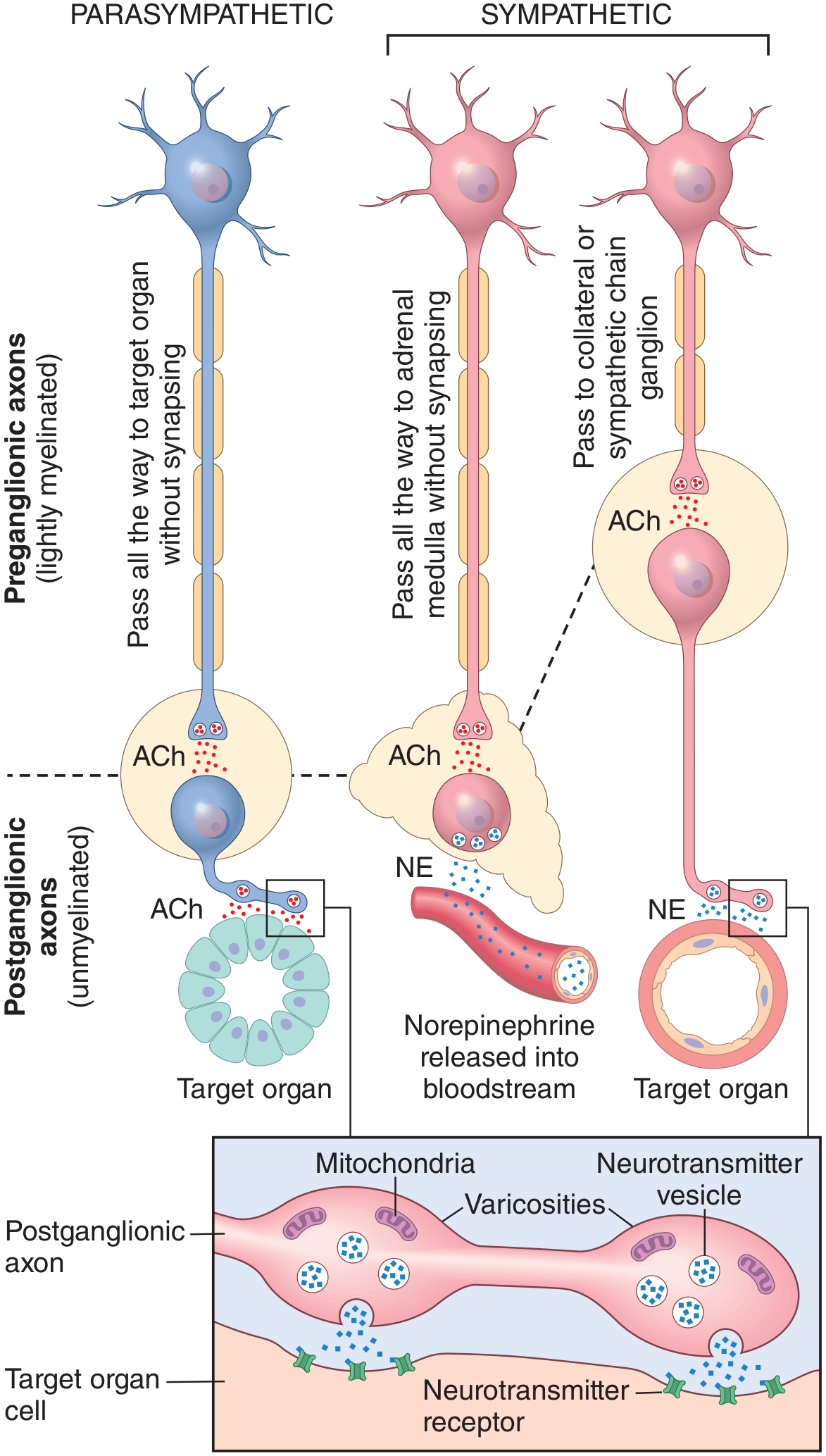
| Feature | Sympathetic | Parasympathetic |
|---|---|---|
| Outflow origin | Thoracolumbar (T1-L2) | Craniosacral (CN III, VII, IX, X; S2-S4) |
| Preganglionic fiber | Short, lightly myelinated | Long, lightly myelinated |
| Ganglion location | Paravertebral chain or prevertebral (far from target) | Near or within the target organ |
| Postganglionic fiber | Long, unmyelinated | Short, unmyelinated |
| Preganglionic neurotransmitter | ACh (nicotinic receptor) | ACh (nicotinic receptor) |
| Postganglionic neurotransmitter | Norepinephrine (adrenergic) - except sweat glands (cholinergic) | Acetylcholine (muscarinic receptor) |
| Response character | Diffuse, widespread - one preganglionic activates many postganglionic neurons | Discrete, organ-specific - limited distribution |
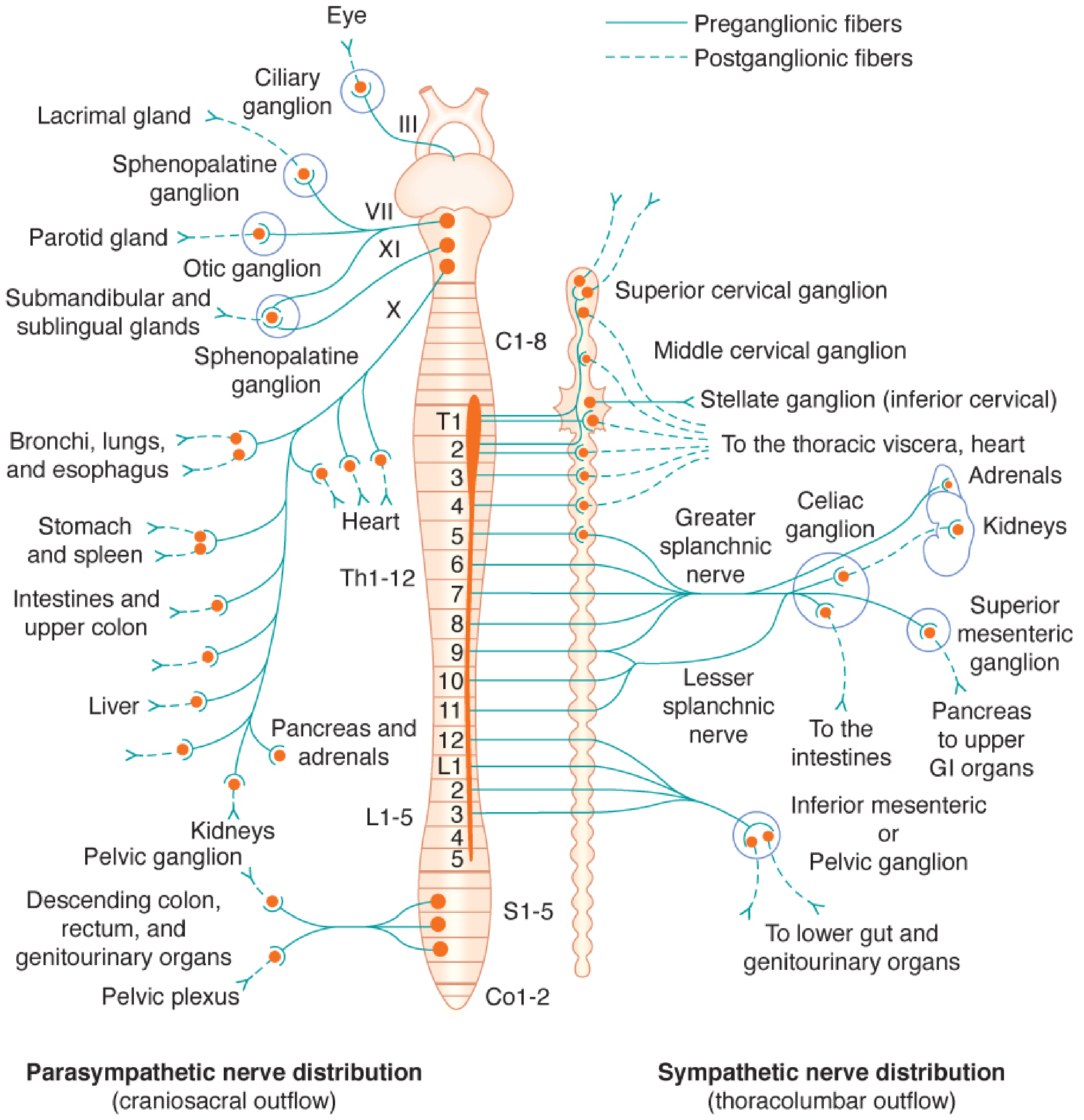
- Barash Clinical Anesthesia, 9e; Guyton and Hall Medical Physiology
2. Neurotransmitters and Receptors
Sympathetic receptors (adrenergic)
| Receptor | Location | Effect |
|---|---|---|
| α₁ | Most vascular smooth muscle, iris dilator | Vasoconstriction, pupil dilation |
| α₂ | Presynaptic (inhibitory), some vessels | ↓NE release, vasoconstriction |
| β₁ | Heart (SA node, AV node, ventricles) | ↑HR, ↑contractility, ↑AV conduction |
| β₂ | Bronchial, skeletal muscle vessels, uterus | Bronchodilation, vasodilation |
| β₃ | Adipose tissue | Lipolysis |
Parasympathetic receptors (cholinergic - muscarinic)
| Receptor | Location | Effect |
|---|---|---|
| M₁ | Gastric glands, CNS | ↑Gastric acid secretion |
| M₂ | Heart (SA/AV nodes) | ↓HR, ↓AV conduction |
| M₃ | Smooth muscle, glands, endothelium | Contraction (GI, bladder), secretion, NO release |
3. Organ-by-Organ Functions
(Data compiled from Guyton & Hall Table 61.2 and Costanzo Physiology)
| Organ / System | Sympathetic Effect | Parasympathetic Effect |
|---|---|---|
| Heart rate (SA node) | ↑ (β₁) - tachycardia | ↓ (M₂) - bradycardia |
| AV conduction | ↑ speed (β₁) | ↓ speed / block (M₂) |
| Cardiac contractility | ↑ (β₁) | ↓ atrial contractility |
| Arterioles (most) | Constriction (α₁) | Little/no innervation |
| Arterioles (skeletal muscle) | Dilation (β₂) / Constriction (α₁) | - |
| Bronchi | Dilation (β₂) | Constriction (M₃) |
| Pupil | Dilation - mydriasis (α₁, radial muscle) | Constriction - miosis (M₃, circular muscle) |
| Ciliary muscle (lens) | Slight relaxation → far vision | Contraction → near vision (accommodation) |
| Salivary glands | ↓ volume, ↑ viscous/enzyme-rich secretion | ↑ profuse, watery secretion |
| Lacrimal/nasal glands | Mild vasoconstriction | Copious secretion |
| Sweat glands | ↑ Sweating (cholinergic sympathetic) | No effect |
| GI motility | ↓ (relaxation of wall, α and β) | ↑ (contraction of wall, M₃) |
| GI sphincters | Contraction (α₁) | Relaxation (M₃) |
| Liver | Glycogenolysis → ↑ blood glucose | Glycogen synthesis |
| Pancreas | ↓ Insulin secretion (α₂), ↑ glucagon | ↑ Insulin + digestive enzyme secretion |
| Adrenal medulla | Releases epinephrine + NE into bloodstream | No innervation |
| Urinary bladder (detrusor) | Relaxation (β₂) → urine storage | Contraction (M₃) → micturition |
| Urinary sphincter | Contraction (α₁) → continence | Relaxation → voiding |
| Uterus | Variable (α: contraction; β₂: relaxation) | Variable |
| Male genitalia | Ejaculation | Erection |
| Skin (piloerection) | Hair erection (α₁) | No effect |
| Fat cells | Lipolysis (β₃) | - |
| Blood coagulation | ↑ (reduces clotting time) | - |
| Spleen | Contracts → mobilizes RBCs | - |
| Kidney (renin) | ↑ Renin release (β₁) | - |
4. The Fight-or-Flight Response (Sympathetic Mass Discharge)
The sympathetic system can produce a massive, coordinated output to all end organs simultaneously while parasympathetic output ceases. This was described by Walter Cannon (1915) as the fight-or-flight response:
- ↑ Heart rate and contractility
- ↑ Blood pressure
- ↑ Ventilation; bronchodilation
- Sweating, piloerection
- Liberation of glucose (glycogenolysis)
- ↓ Insulin secretion
- ↓ GI activity
- ↓ Blood clotting time
- Mobilization of RBCs from spleen
Under normal, non-stressful conditions, sympathetic output can also be discrete and organ-specific ("chemical coding") - different postganglionic neurons co-release different peptides (e.g., NPY for vasoconstriction, somatostatin for gut control).
"The fight-or-flight response is a primitive mechanism for survival. In some people, such a response can be triggered spontaneously with minimal provocation; each individual episode is then called a panic attack."
- Medical Physiology (Boron & Boulpaep)
5. The Rest-and-Digest Response (Parasympathetic)
In contrast to the broad sympathetic response, parasympathetic neurons function only in a discrete, organ-specific, and reflexive manner. Key examples:
- Baroreceptor reflex: vagal bradycardia in response to hypertension
- Micturition reflex: bladder contraction in response to distension
- Salivation: in response to smell/sight of food
- Vagovagal GI reflexes: colon contraction in response to food in stomach
- Bronchoconstriction: in response to lung receptor activation
- Pupillary light reflex: constriction in response to bright light
6. Reciprocal vs. Synergistic Control
Most organs receive dual innervation that works reciprocally:
| Example | Sympathetic | Parasympathetic |
|---|---|---|
| Heart rate | ↑ (β₁) | ↓ (M₂) |
| GI wall | Relaxation | Contraction |
| GI sphincters | Contraction | Relaxation |
| Pupil size | Dilation | Constriction |
| Male sexual response | Ejaculation | Erection |
When blood pressure falls, the vasomotor center simultaneously increases sympathetic activity to the SA node AND decreases parasympathetic activity - both changes increase heart rate synergistically. Neither system competes; they are orchestrated. - Costanzo Physiology, 7e
7. Key Summary: "SLUDD" vs. "Fight-or-Flight"
| State | Division Active | Mnemonic Effects |
|---|---|---|
| Stress / Emergency | Sympathetic | E's - Exercise, Excitement, Emergency, Embarrassment |
| Rest / Feeding | Parasympathetic | SLUDD - Salivation, Lacrimation, Urination, Defecation, Digestion |
Sources: Guyton and Hall Textbook of Medical Physiology, Medical Physiology (Boron & Boulpaep), Costanzo Physiology, 7e, Barash Clinical Anesthesia, 9e, Morgan and Mikhail's Clinical Anesthesiology, 7e
This is a shared conversation. Sign in to Orris to start your own chat.