
Signs of portal hypertension
portal hypertension signs clinical findings caput medusae splenomegaly
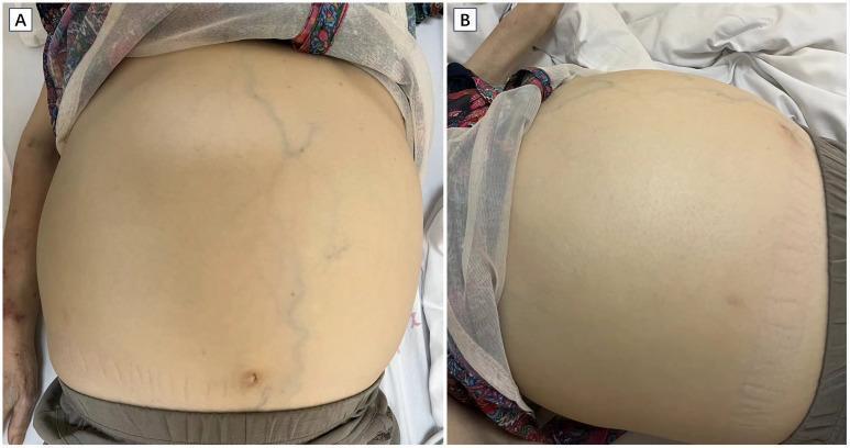
Two clinical photographs (A and B) show significant abdominal distension in a patient with massive splenomegaly. Image A, a frontal view, displays a globally distended abdomen with prominent caput medusae (engorged superficial veins) and visible striae in the lower quadrants. The patient's left upper extremity is visible, demonstrating marked muscle wasting consistent with cachexia, along with irregular, patchy purpura or skin hemorrhages. Image B provides a lateral-oblique perspective, further emphasizing the degree of abdominal protrusion and skin tension. These findings collectively illustrate the physical manifestations of chronic liver or hematological disease, specifically showing signs of portal hypertension and systemic nutritional depletion. Key educational concepts include the identification of splenomegaly-related distension, recognition of muscle wasting in chronic illness, and the visual assessment of vascular abnormalities on the abdominal wall.
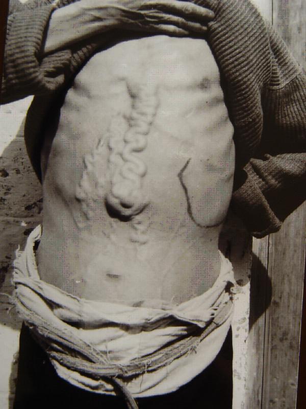
This clinical photograph displays the abdominal region of a patient with advanced schistosomiasis, illustrating severe portal hypertension. The most prominent feature is the presence of massively dilated and tortuous abdominal collateral veins (caput medusae) originating from the epigastric region and extending toward the umbilicus. A central, large, rope-like vein exhibits extreme serpentine morphology along the midline. On the left side of the abdomen (patient's left), an ink marking outlines the margin of a significant megalosplenia (enlarged spleen), extending well below the costal margin. The abdomen appears distended, suggestive of underlying ascites. These clinical signs—prominent venous collaterals and splenomegaly—are classic manifestations of late-stage hepatic schistosomiasis, where fibrotic changes in the liver obstruct portal blood flow, forcing the development of systemic-portal shunts and causing congestive splenomegaly. This image serves as an educational example of the physical examination findings associated with chronic hepatosplenic parasitic infections and secondary portal hypertensive syndrome.
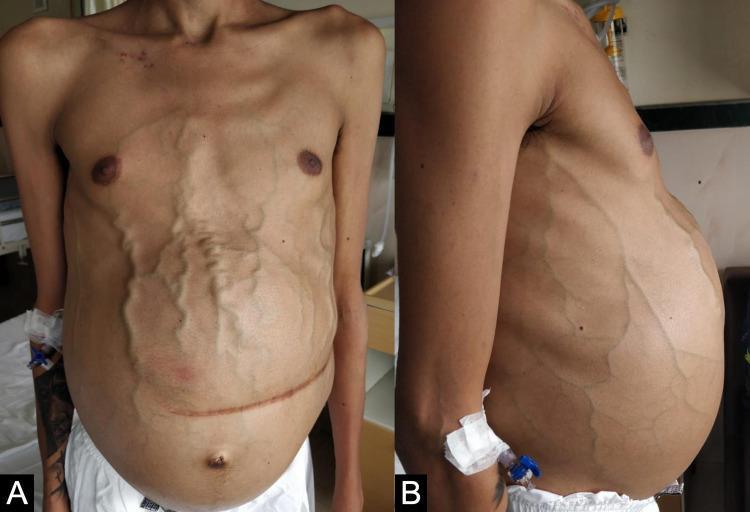
Clinical photograph of a male patient's torso in anterior (A) and lateral (B) views, demonstrating advanced clinical signs of portal hypertension and chronic liver disease. The abdomen is severely distended, consistent with tense ascites. Prominent, tortuous, and dilated superficial veins (caput medusae) are visible across the entire abdominal wall and flanks, radiating from the peri-umbilical region. A well-healed, horizontal surgical scar from a previous splenectomy is present in the mid-abdomen. Additional findings include a small umbilical hernia, bilateral gynecomastia, and significant sarcopenia characterized by muscle wasting in the pectoral region and visible bony landmarks of the clavicles and shoulders. An intravenous cannula is present in the left arm. These visual markers collectively illustrate the systemic manifestations of severe portal venous congestion, commonly associated with conditions such as Gaucher disease or advanced cirrhosis.
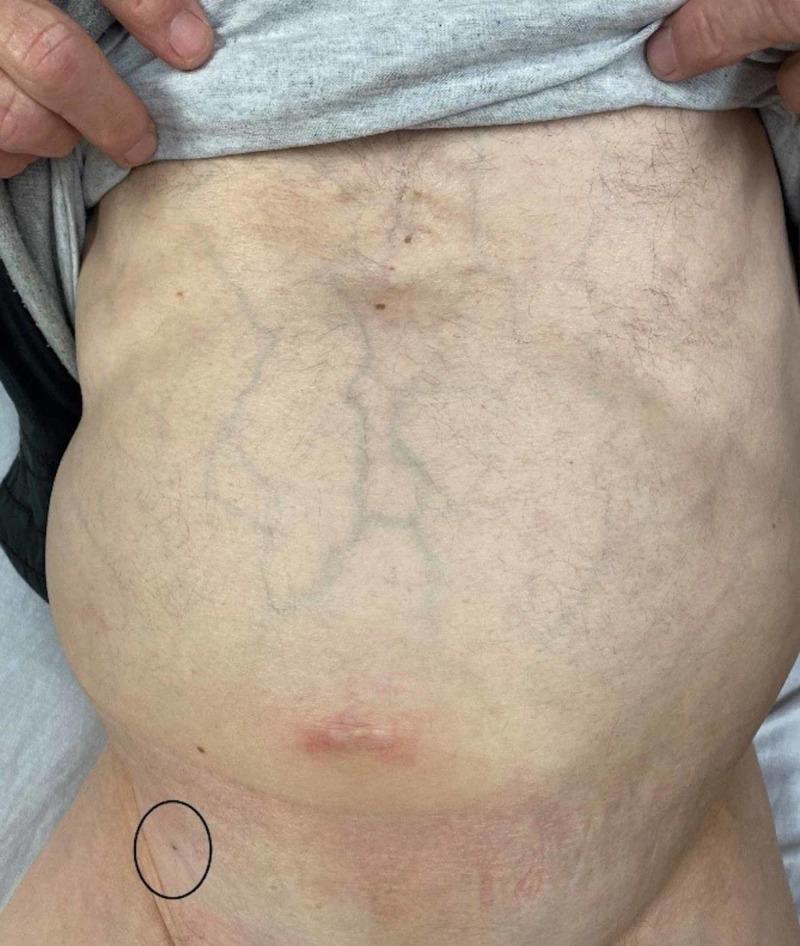
This clinical photograph shows a distended adult abdomen with several notable physical examination findings. Centrally, the abdomen displays prominent, dilated superficial veins, characteristic of caput medusae or collateral venous circulation often associated with portal hypertension. A red, linear scar or healing wound is visible in the lower midline (periumbilical region). A black circle in the patient's right lower quadrant highlights a small puncture site on the skin. This marked area represents a paracentesis site from three days prior, located medial to the right anterior inferior iliac spine. The skin within the circle shows a clean, dry puncture wound without evidence of active infection, such as surrounding erythema, swelling, or discharge. The overall clinical presentation illustrates a patient with likely ascites (suggested by the distention and venous patterns) and the follow-up appearance of a diagnostic or therapeutic paracentesis procedure. This image is relevant for gastroenterology and internal medicine education regarding the physical signs of chronic liver disease and post-procedural site assessment.
"portal hypertension" AND "clinical signs"
Signs of Portal Hypertension
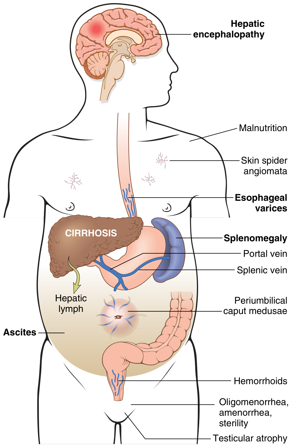
1. Portosystemic Collaterals (Varices)
| Site | Collateral Formed | Clinical Sign |
|---|---|---|
| Lower esophagus / gastric fundus | Esophageal & gastric varices | Hematemesis, melena (life-threatening) |
| Around the umbilicus | Paraumbilical vein dilation | Caput medusae (visible dilated veins radiating from umbilicus) |
| Rectum | Superior/inferior rectal anastomoses | Anorectal varices / hemorrhoids |
| Retroperitoneum | Retroperitoneal collaterals | Usually asymptomatic |
2. Ascites
- Raised splanchnic capillary pressure (Starling forces shift fluid into peritoneum)
- Splanchnic arterial vasodilation activating the RAAS and SNS, driving sodium and water retention
- Hypoalbuminemia reducing oncotic pressure
- Abdominal distension with bulging flanks
- Shifting dullness on percussion
- Fluid thrill in tense ascites
- Eversion or herniation of the umbilicus
- SAAG (serum-ascites albumin gradient) ≥1.1 g/dL confirms portal hypertensive ascites (97% specificity) - Washington Manual of Medical Therapeutics
3. Splenomegaly (Congestive)
- Thrombocytopenia (most common hematologic finding)
- Anemia
- Leukopenia
- Pancytopenia in severe cases
4. Cutaneous Signs (Hyperestrogenemia)
- Spider angiomas (spider naevi) - dilated arterioles with radiating vessels on the upper trunk, face, and arms; >5 is significant
- Palmar erythema - erythema of the thenar and hypothenar eminences (local vasodilation)
- Gynecomastia (in men)
- Testicular atrophy (in men)
- Oligomenorrhea / amenorrhea / sterility (in women)
5. Hepatic Encephalopathy
6. Other Abdominal / Systemic Signs
- Hepatomegaly (early disease) or a shrunken, nodular liver (advanced cirrhosis)
- Jaundice - reflecting underlying liver dysfunction
- Fetor hepaticus - a musty odor to breath from mercaptans
- Leukonychia (white nails) and Terry's nails - associated with hypoalbuminemia
- Dupuytren's contracture - fibrosis of the palmar fascia (especially in alcoholic cirrhosis)
- Parotid enlargement (alcoholic liver disease)
- Muscle wasting / cachexia - from protein catabolism
- Coagulopathy - bruising, petechiae from impaired clotting factor synthesis
7. Cardiopulmonary Manifestations
- Hyperdynamic circulation - raised cardiac output, low systemic vascular resistance, bounding pulse
- Hepatopulmonary syndrome - intrapulmonary vasodilation causing hypoxia; patients may have platypnea (dyspnea worse when sitting up, better when lying down) and orthodeoxia
- Portopulmonary hypertension - pulmonary arterial hypertension due to portal hypertension
- Hepatic hydrothorax - transudative pleural effusion (usually right-sided)
8. Pressure Thresholds Summary
| HVPG (mmHg) | Clinical Significance |
|---|---|
| >5 | Portal hypertension |
| >10 | "Clinically significant" - splenomegaly, ascites possible |
| >12 | Variceal bleeding risk; varices develop |
| >20 | High risk of treatment failure and death from variceal bleed |
Diagnostic Confirmation
- Imaging (ultrasound, CT, MRI): portal vein diameter >13 mm, splenomegaly, collaterals, ascites
- Endoscopy: visualizes esophagogastric varices directly
- HVPG measurement (transjugular): gold standard, rarely needed clinically
- Lab findings: thrombocytopenia, hypoalbuminemia, raised INR, elevated bilirubin