I now have all the library content needed. Let me compile the full Paper I answers.---
BJMC MD Preliminary Examination 2025
Paper I - Basics of Forensic Medicine, Basic Sciences & Allied Subjects
Complete Model Answers
QUESTION 1 (10 Marks)
Arterial Supply of the Heart + Autopsy Diagnosis of Myocardial Infarction
PART A: ARTERIAL SUPPLY OF THE HEART
Introduction
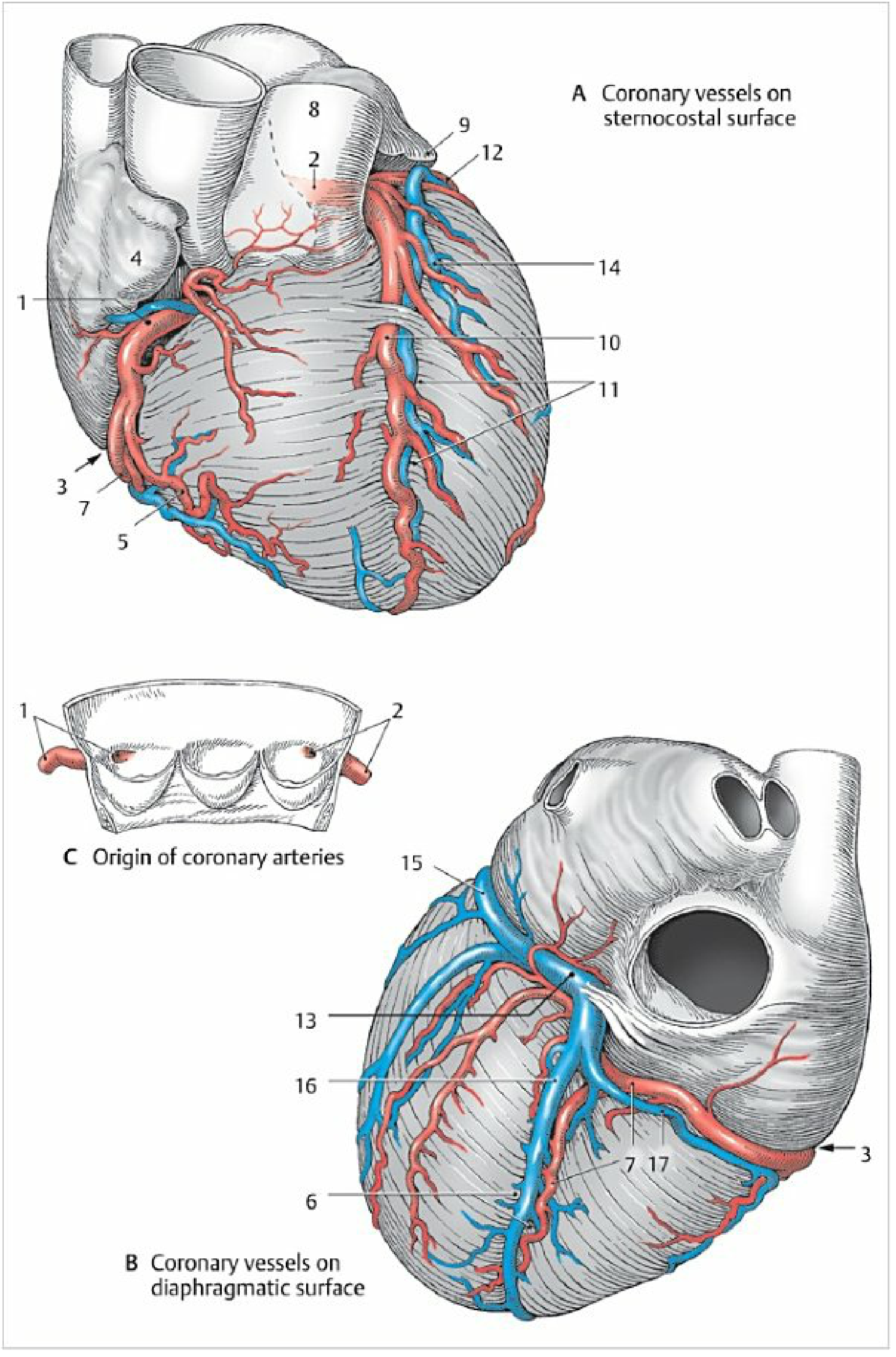
The heart is supplied by two main coronary arteries - the Right Coronary Artery (RCA) and the Left Coronary Artery (LCA) - both arising from the aortic sinuses (sinuses of Valsalva) just above the aortic valve. These are functionally end arteries; though small anastomoses exist, they are insufficient to establish collateral circulation acutely if a vessel is occluded.
Coronary artery anatomy - sternocostal (A), diaphragmatic (B) surfaces, and aortic origin (C) (Color Atlas of Human Anatomy, Vol. 2)
1. Right Coronary Artery (RCA)
- Origin: Right coronary (anterior) sinus of Valsalva; ostium ~2-3 mm diameter
- Course: Runs rightward and obliquely in the right atrioventricular (coronary) sulcus, initially covered by the right auricle
Branches:
| Branch | Territory Supplied |
|---|
| Right atrial branches | SA node (in ~60% of people) |
| Right marginal artery (acute marginal artery) | Lateral/acute margin of the right ventricle toward apex |
| Posterior Descending Artery (PDA) / Posterior Interventricular Artery | Inferior interventricular septum, adjacent ventricular walls |
| Right posterolateral artery | Posterior left ventricle |
| AV nodal artery | AV node (in ~90% of people) |
Territory: Right atrium, SA node (60%), AV node (90%), most of the right ventricle, posterior interventricular septum, diaphragmatic surface of the left ventricle (in right-dominant individuals).
2. Left Coronary Artery (LCA) / Left Main Coronary Artery (LMCA)
- Origin: Left coronary (posterior) sinus of Valsalva
- Course: Short main stem (~1 cm) courses posterolateral to the pulmonary trunk, passing between the left atrium and pulmonary artery before bifurcating into:
A. Left Anterior Descending Artery (LAD)
- Runs anterolaterally along the anterior interventricular sulcus toward the apex
- Branches: Diagonal branches (to LV free wall), septal perforators (to anterior 2/3 of interventricular septum)
- Territory: Anterior wall of left ventricle, anterior interventricular septum, apex, bundle of His, bundle branches
B. Left Circumflex Artery (LCx)
- Runs posteriorly in the left atrioventricular groove
- Branches: Obtuse marginal branches (to lateral LV wall), left posterolateral branches
- Territory: Left atrium, lateral and posterior left ventricular wall; SA node (in ~40%), AV node (in ~10%)
3. Coronary Dominance
Dominance is defined by which artery gives rise to the Posterior Descending Artery (PDA)
| Type | Frequency | PDA Origin |
|---|
| Right dominant | ~85% | RCA |
| Left dominant | ~8% | LCx |
| Codominant | ~7% | Both RCA + LCx |
4. Venous Drainage of the Heart
- Coronary Sinus (posterior atrioventricular sulcus) - receives ~2/3 of venous drainage
- Great cardiac vein (anterior interventricular vein) → drains LAD territory
- Middle cardiac vein → posterior interventricular sulcus
- Small cardiac vein → right coronary sulcus
- Anterior cardiac veins - drain directly into right atrium
- Thebesian veins (smallest cardiac veins) - empty directly into cardiac chambers
PART B: AUTOPSY DIAGNOSIS OF MYOCARDIAL INFARCTION
Introduction (Forensic Significance)
MI is a leading cause of sudden unexpected death. Autopsy diagnosis is challenging because gross and histological changes take time to develop and may be absent in the first few hours (the "silent window"). The forensic pathologist must correlate gross findings, histology, histochemistry, and ancillary tests.
Timeline of Morphological Changes in MI
| Time | Gross Changes | Histological Changes | Histochemical/Enzyme Changes |
|---|
| 0-4 hours | None visible (gross silent window) | No change on H&E; electron microscopy shows mitochondrial swelling and nuclear changes | Loss of glycogen (PAS stain); loss of NADH-diaphorase activity; loss of nitro-BT (NBT) staining |
| 4-12 hours | Subtle pallor; slight hemorrhagic mottling | Early coagulation necrosis; wavy myofibers; slight nuclear pyknosis | Loss of succinic dehydrogenase (SDH), LDH activity |
| 12-24 hours | Pale/yellowish area; soft texture; hemorrhagic margins | Coagulation necrosis with loss of nuclei and striations; early neutrophil infiltration begins | Loss of most oxidative enzymes |
| 1-3 days | Pale yellow-white center; soft, friable; hyperemic border | Dense neutrophil infiltration (PMN); myocyte ghosts; nuclear karyolysis | Elevated acid phosphatase in infiltrating cells |
| 4-7 days | Softest point (risk of rupture); yellow-white; depressed surface | Macrophage infiltration begins; granulation tissue at margins; neutrophils declining | |
| 1-3 weeks | Granulation tissue (vascular, pink-red) | Active fibroblast proliferation; neovascularization; collagen deposition begins | |
| >6 weeks | White fibrous scar; depressed, firm | Dense collagen scar; loss of myocytes; loss of vascularity | |
Gross Autopsy Findings
- Coronary arteries: Atherosclerotic plaques (stenosis >70% in culprit artery); look for fresh thrombus on ruptured plaque (present in >90% of acute MIs); assess all three vessels systematically by serial transverse cuts
- External heart examination: Increased heart weight (normal: 250-300 g female, 300-350 g male); wall thinning or bulging (aneurysm) in old infarcts
- Cross-sections of myocardium: Pale, yellow-white areas (acute); firm white scars (healed); note the territory affected:
- Anterior wall + septum = LAD territory
- Lateral wall = LCx territory
- Posterior wall + inferior septum = RCA territory
- Look for complications: Cardiac rupture (hemopericardium), mural thrombus, papillary muscle rupture, ventricular aneurysm
Histochemical Methods (Most Important for Early/Acute MI)
These are critical in the forensic context because they detect MI before gross or H&E changes appear (0-4 hours):
| Test | Principle | Positive Result | Negative (Infarcted) |
|---|
| Nitro-blue tetrazolium (NBT) / NADH-diaphorase | Reduces NBT to blue formazan in viable tissue with dehydrogenase enzymes | Blue-black (viable) | Pale/unstained (infarcted) - loss within 2-4 hours |
| Succinic dehydrogenase (SDH) | Mitochondrial enzyme; reduces NBT | Blue (viable) | Pale (infarcted) |
| Masson's Trichrome | Collagen (fibrosis) | Blue-green collagen | Identifies healing/healed infarcts |
| PAS (Periodic Acid-Schiff) | Glycogen in myocytes | Magenta (glycogen present) | Loss of glycogen in ischemic area |
| PTAH (Phosphotungstic Acid-Haematoxylin) | Cross striations | Normal striations | Loss of striations in early infarction |
Ancillary Tests at Autopsy
- Vitreous humor sampling: Potassium levels; glucose; may show biochemical evidence of ischemia
- Blood biochemistry (postmortem): Troponin I and T can be detected in postmortem serum/pericardial fluid - postmortem troponin elevation is a useful marker but must be interpreted with caution (postmortem redistribution occurs)
- Histology of conduction system: Section SA node, AV node, bundle of His to detect ischemia/necrosis affecting conduction
- Immunohistochemistry: Fibronectin, complement C9, myoglobin, and early ischemic markers detectable within 1-2 hours; myoglobin leaches out of infarcted cells
Key Forensic Points
- In sudden death within the first 2-4 hours of MI, the autopsy may be entirely negative on gross and H&E - histochemistry (NBT stain) is the only way to demonstrate infarction
- Always record the degree of coronary stenosis systematically - >75% cross-sectional area reduction is hemodynamically significant
- Fresh thrombus indicates an acute event vs. old organized thrombus = remote occlusion
- A healed infarct is a risk factor for sudden arrhythmic death even without new infarction
QUESTION 2 (10 Marks)
Circle of Willis Anatomy + Etiopathology of Intracranial Haemorrhage
(For the anatomy of the Circle of Willis, refer to the detailed answer provided earlier in this session. The following covers the etiopathology of intracranial haemorrhage.)
CIRCLE OF WILLIS - SUMMARY
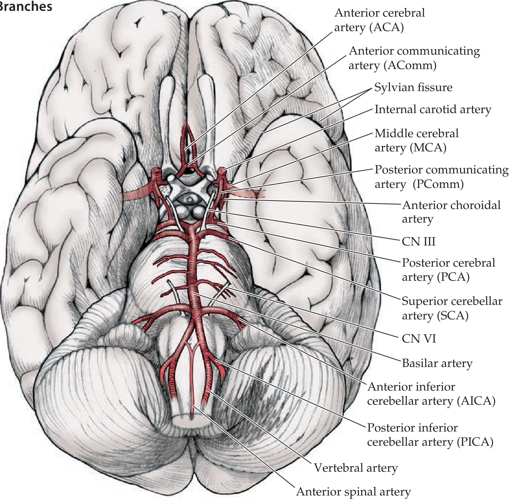
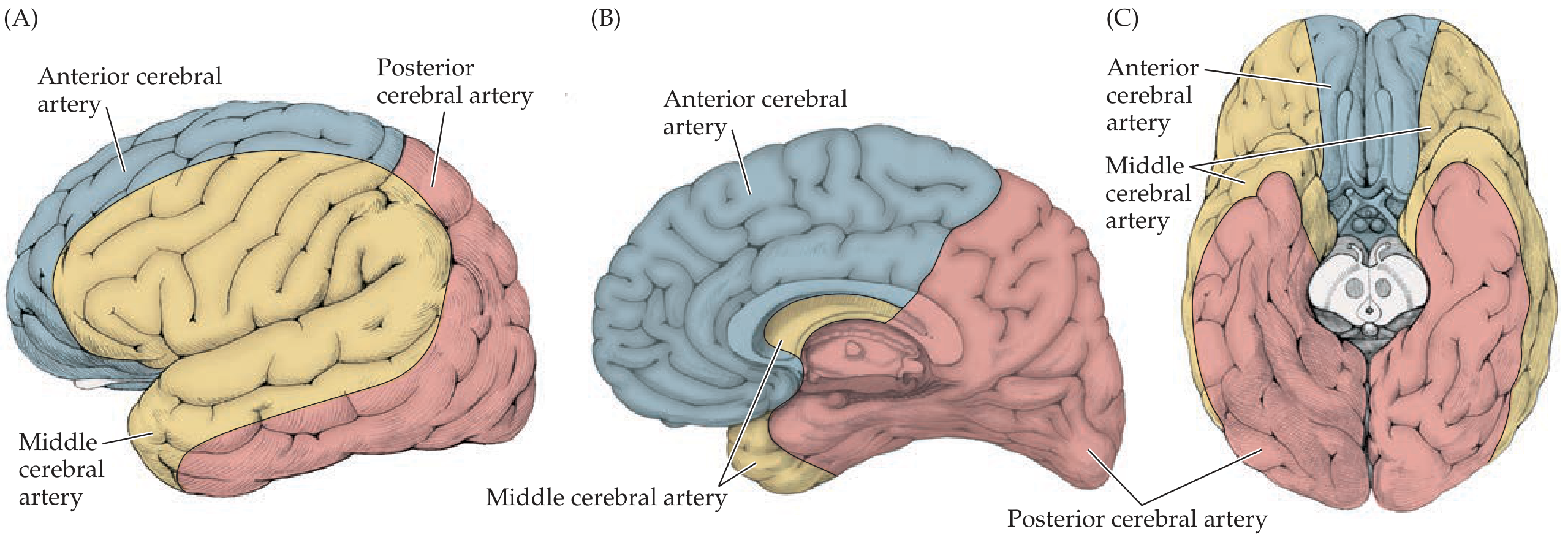
The Circle of Willis is an anastomotic hexagonal/heptagonal ring at the base of the brain formed by:
- Anterior part: ACA (bilateral) + Anterior communicating artery (AComm)
- Middle: ICA (bilateral)
- Posterior: PCA (bilateral, from basilar tip)
- Connecting links: Posterior communicating arteries (PComm, bilateral)
A complete, full-caliber ring is present in only ~34% of individuals. Its function is collateral flow redistribution during arterial occlusion.
ETIOPATHOLOGY OF INTRACRANIAL HAEMORRHAGE
Classification
| Type | Location | Common Cause |
|---|
| Extradural (Epidural) Haemorrhage (EDH) | Between skull and dura | Traumatic - rupture of middle meningeal artery |
| Subdural Haemorrhage (SDH) | Between dura and arachnoid | Traumatic - tearing of bridging veins |
| Subarachnoid Haemorrhage (SAH) | Subarachnoid space | Ruptured berry aneurysm (85%), AVM, trauma |
| Intracerebral Haemorrhage (ICH) | Within brain parenchyma | Hypertension (most common), AVM, amyloid angiopathy |
| Intraventricular Haemorrhage (IVH) | Within ventricles | Extension of ICH, neonatal (prematurity) |
1. Extradural Haemorrhage (EDH)
Mechanism: Trauma (usually temporal/parietal skull fracture) ruptures the middle meningeal artery (branch of maxillary artery, groove on inner temporal bone). Arterial bleeding accumulates between bone and dura, progressively stripping the dura.
Pathology:
- Biconvex/lenticular (lens-shaped) hematoma on imaging
- Lucid interval (loss of consciousness → recovery → deterioration) is classic but present in only ~30%
- Bilateral pupil dilation (CN III compression) = transtentorial herniation
2. Subdural Haemorrhage (SDH)
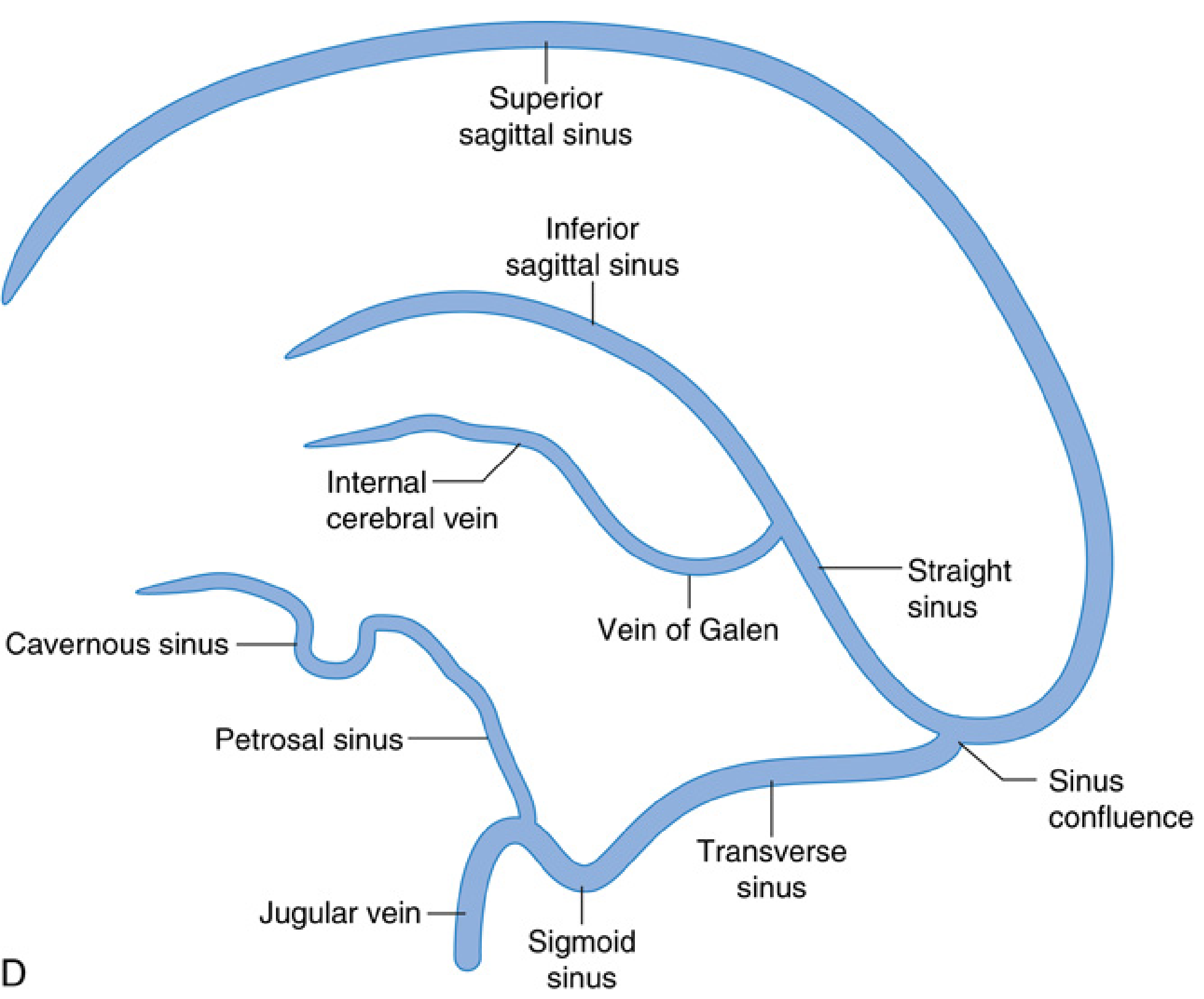
Mechanism: Acceleration-deceleration shear forces tear bridging veins (cortical veins draining into superior sagittal sinus). Venous bleed accumulates slowly in the subdural space.
Types and Pathology:
| Type | Onset | Appearance | Consistency |
|---|
| Acute | <3 days | Bright red | Liquid/clot |
| Subacute | 3-21 days | Mixed / dark red | Partial clot |
| Chronic | >21 days | Yellow-brown (xanthochromic) | Liquid membranes |
Predisposing factors: Elderly (cerebral atrophy - longer bridging vein stretch), alcoholism, anticoagulation, shaken baby syndrome.
3. Subarachnoid Haemorrhage (SAH)
Mechanism: Rupture of a berry (saccular) aneurysm in ~85% of non-traumatic cases. Berry aneurysms form at arterial bifurcations due to congenital weakness of the tunica media, compounded by hemodynamic stress.
Common sites (Circle of Willis):
- Anterior communicating artery (AComm) - most common (~35%)
- Posterior communicating artery (PComm) - ~30%
- MCA bifurcation
- Basilar tip
Pathophysiology of bleeding:
- Aneurysm wall ruptures (often triggered by sudden rise in BP - straining, coitus, exertion)
- Blood enters subarachnoid space under arterial pressure
- Sudden severe headache ("thunderclap" / "worst headache of life")
- Secondary complications: vasospasm (peak day 4-14), hydrocephalus, rebleeding
Autopsy finding: Blood in basal cisterns, around Circle of Willis; cerebral sulci filled with blood; possible herniation.
4. Hypertensive Intracerebral Haemorrhage (ICH)
Mechanism: Chronic hypertension causes:
- Lipohyalinosis and fibrinoid necrosis of small penetrating arteries (especially lenticulostriate arteries)
- Formation of Charcot-Bouchard microaneurysms (on vessels <200 µm diameter)
- Rupture causes hematoma that expands as it strips white matter tracts
Common sites (in order of frequency):
- Putamen/basal ganglia (~50%) - from lenticulostriate arteries
- Thalamus (~15%)
- Pons (~10-15%)
- Cerebellum (~10%)
- Lobar (subcortical) - more common in elderly, amyloid angiopathy
Pathology:
- Hematoma compresses surrounding tissue → cerebral edema → rising ICP
- Midline shift, transtentorial herniation, Duret hemorrhages in brainstem (secondary)
- Hemosiderin-laden macrophages at margins (after 5-7 days)
QUESTION 3 (10 Marks)
Modern Methods of Diagnosis of Brain Death
Definition
Brain death is the irreversible cessation of all functions of the entire brain, including the brainstem, while the heart continues to beat with mechanical support. It is the only form of death recognized as legally, ethically, and medically equivalent to biological death.
Prerequisites (Must Be Established Before Testing)
Before any brain death assessment, the following confounding factors must be excluded:
| Prerequisite | Threshold |
|---|
| Cause of coma known and irreversible (neuroimaging mandatory) | - |
| No hypothermia | Core temperature ≥ 36°C |
| No drug intoxication | Sedatives, opioids, barbiturates, neuromuscular blockers must be cleared |
| No severe metabolic derangement | Electrolytes, acid-base, glucose corrected |
| Hemodynamic stability | Systolic BP ≥ 100 mmHg |
| No residual effect of neuromuscular blockers | Confirm with peripheral nerve stimulator |
A minimum observation period (usually 6-24 hours from onset, longer if cardiac arrest) should elapse.
Step 1: Clinical Examination (Primary Method)
A. Test for Coma - Absent Motor Response
- Apply deep painful stimuli to both condyles at the TMJ, supraorbital notch, fingernail beds, sternal rubbing
- Brain dead: No grimacing, no motor response in extremities
- Note: Deep tendon reflexes and triple flexion (spinal cord reflexes) may persist and do not preclude brain death diagnosis; posturing responses (decorticate/decerebrate) exclude brain death
B. Brainstem Reflex Assessment
| Reflex | Test | Brain Dead Result |
|---|
| Pupillary light reflex | Bright light to each eye | Pupils fixed and dilated (4-9 mm); no constriction bilaterally |
| Corneal reflex | Saline drops or cotton swab on cornea | Absent bilaterally |
| Oculocephalic reflex (Doll's eye) | Rapid passive head turning | Absent - eyes move with head ("fixed") |
| Oculovestibular (caloric) reflex | 50 mL ice water into each ear (head at 30°); observe 1 min, wait 5 min between sides | No eye movement (intact = slow conjugate deviation toward cold side) |
| Gag reflex | Stimulation of posterior oropharynx | Absent |
| Cough reflex | Suction catheter to carina via ETT | No cough response |
Step 2: Apnea Test (Mandatory)
The apnea test confirms the absence of respiratory drive from the brainstem (medullary respiratory center).
Procedure:
- Pre-oxygenate with 100% O₂ for 10 minutes → PaO₂ ≥ 200 mmHg
- Obtain baseline ABG: normalize PaCO₂ to 35-45 mmHg
- Disconnect ventilator; deliver O₂ via insufflation catheter to carina at 6 L/min (or T-piece with CPAP)
- Observe for 8-10 minutes for any breathing effort (chest excursion, abdominal movement, gasping)
- Obtain ABG at end of apnea period
Positive result (confirms brain death):
No respiratory effort AND PaCO₂ ≥ 60 mmHg OR rise of ≥ 20 mmHg above normal baseline
Abort test if: Systolic BP drops <90 mmHg, O₂ saturation falls below 85%, or cardiac arrhythmia occurs → proceed to ancillary tests.
Step 3: Ancillary/Confirmatory Tests
Required when the clinical exam cannot be completed (e.g., severe facial trauma, inability to perform apnea test, persistent confounders). Not mandatory in most countries if the clinical exam is complete.
A. Electrophysiological Tests
| Test | Criteria for Brain Death | Notes |
|---|
| EEG (Electroencephalography) | Electrocerebral silence (isoelectric EEG): no electrical activity >2 µV during minimum 30-minute recording with ≥8 scalp electrodes (interelectrode distance ≥10 cm) | Most widely used; limitations: artifacts from ventilator/cardiac monitors; some comatose patients may have flat EEG with intact brainstem |
| Auditory Evoked Potentials (BAEP) | Absence of waves III-V (only wave I or I-II may persist from cochlear nerve) | Confirms brainstem death |
| Somatosensory Evoked Potentials (SSEP) | Bilateral absence of cortical N20-P22 responses after median nerve stimulation | Confirms cortical non-function |
B. Cerebral Blood Flow Studies (Most Reliable)
| Test | Criteria for Brain Death | Advantage |
|---|
| Cerebral Angiography (4-vessel) | No intracerebral filling above the level of the cavernous sinus (no flow beyond circle of Willis) | "Gold standard" CBF test; highly specific |
| Radionuclide Brain Scan (Tc-99m HMPAO SPECT) | "Hollow skull" sign - no uptake of tracer within the cranial vault (no perfusion); "hot nose" sign (blood flow diverted to face/scalp) | Non-invasive; widely available |
| CT Angiography (CTA) | Absence of contrast filling of intracranial vessels beyond circle of Willis | Fast; widely available in ICU settings |
| Transcranial Doppler (TCD) | Reverberating/oscillating flow pattern or systolic spikes only (indicating zero net perfusion or retrograde diastolic flow) | Bedside; non-invasive; requires adequate temporal window |
C. Other Tests
| Test | Finding in Brain Death |
|---|
| Cerebral Perfusion Pressure (ICP monitoring) | ICP equals or exceeds MAP → zero CPP |
| CT Brain | Diffuse cerebral edema, loss of grey-white differentiation, effaced cisterns, evidence of herniation |
Legal Framework (India)
Under the Transplantation of Human Organs and Tissues Act, 1994 (amended 2011), brain death must be certified by a board of four doctors:
- Registered Medical Practitioner in charge of the hospital/medical superintendent
- Authorized specialist (neurologist, neurosurgeon, or intensivist)
- Neurologist or neurosurgeon (as appropriate)
- Treating physician / anaesthesiologist
Two sets of tests must be conducted with a minimum gap of 6 hours between them. Both sets must confirm brain death. The certificate (Form 10) must be signed by all four doctors.
QUESTION 4 (10 Marks)
Pathological and Histochemical Changes in Injured Tissue
Introduction
When tissue is injured, a predictable and time-dependent sequence of cellular, biochemical, and structural changes occurs. From a forensic pathology perspective, the ability to determine the age of a wound is of paramount importance in reconstructing the sequence of events and determining whether wounds were inflicted ante-mortem, perimortem, or postmortem.
A. PATHOLOGICAL CHANGES IN INJURED TISSUE
1. Immediate Phase (0-2 Hours)
Vascular changes:
- Transient vasoconstriction (neurogenic, seconds to minutes) followed by vasodilation
- Increased vascular permeability (histamine, bradykinin, prostaglandins)
- Exudation of plasma proteins and fluid → edema
- Margination and emigration of neutrophils (PMNs) begins
Cellular changes:
- Disruption of cell membranes
- Mitochondrial swelling
- Cellular swelling (cloudy swelling)
- No inflammatory cells seen on histology yet
Gross: Edges of wound pink-red, soft; minimal swelling
2. Acute Inflammatory Phase (2-24 Hours)
- Neutrophil (PMN) infiltration is the hallmark
- PMNs begin at 2-4 hours, peak at 12-24 hours at wound edges
- Phagocytosis of debris and bacteria
- Fibrin deposition at wound edges
- Gross: Redness (rubor), swelling (tumor), heat (calor), pain (dolor) - the four cardinal signs of inflammation
3. Subacute/Reparative Phase (24 Hours - 7 Days)
- Monocyte/macrophage infiltration begins at 24-48 hours; macrophages predominate after 48-72 hours; PMNs decline
- Macrophages phagocytose dead cells, fibrin, and debris; secrete growth factors (PDGF, TGF-β, VEGF)
- Granulation tissue formation begins: proliferating fibroblasts + neovascularization
- Epithelialization begins at wound edges (migration of keratinocytes) within 12-24 hours in skin wounds
- Gross: Wound edges become pink/red, granular, vascular
4. Chronic Healing Phase (7 Days - 6 Weeks)
- Fibroblast proliferation and collagen synthesis (Type III collagen initially → remodeled to Type I)
- Progressive replacement of granulation tissue by fibrous scar
- Wound contraction (myofibroblasts)
- Re-epithelialization complete
- Gross: Scar formation; initially pink-raised (hypertrophic), eventually white and flat
B. HISTOCHEMICAL CHANGES IN INJURED TISSUE
Histochemical techniques detect enzymatic and biochemical changes that occur before gross or standard H&E histology can confirm a wound. This is the key forensic tool for wound age estimation.
Key Enzyme Changes
| Enzyme | Normal Finding | Change After Injury | Timing |
|---|
| Acid Phosphatase | Low in connective tissue | Increases early in wound edges; later elevated in macrophages and PMNs | Detectable within 30 min - 1 hour; rises progressively |
| Alkaline Phosphatase | Present in normal dermis | Decreases in wound area acutely; later increases in proliferating endothelial cells of new capillaries | Loss: 0-12 hours; Recovery: 24-72 hours |
| Succinic Dehydrogenase (SDH) | Active in mitochondria of healthy cells | Decreases in damaged cells | Loss within hours |
| Esterases (non-specific) | Present in tissue | Elevated in PMN-rich phase | 4-12 hours |
| 5'-Nucleotidase | Present in normal cells | Decreases in necrotic tissue | |
| Mast cell degranulation | Mast cells intact | Degranulation within minutes - release histamine/heparin | < 1 hour; detectable by Toluidine Blue, Giemsa stain |
Vital Reaction - Key Concept
Vital reaction = the tissue changes that occur only in living (ante-mortem) tissue and are absent in postmortem injuries
| Feature | Ante-mortem Wound | Postmortem Wound |
|---|
| Hemorrhage (tissue infiltration) | Present; blood is clotted and infiltrates tissue | Absent / minimal (no vascular pressure) |
| Vital bleeding | Present | Absent |
| PMN infiltration | Present (after 2-4 hours) | Absent |
| Histamine elevation | Present (mast cell degranulation) | Absent |
| Acid phosphatase elevation | Present | Absent |
| Serotonin elevation | Present | Absent |
| Vasodilation/edema | Present | Absent |
| Leukocyte margination | Present | Absent |
Special Histochemical Stains for Wound Aging
| Stain | Target | Use |
|---|
| Hematoxylin & Eosin (H&E) | General morphology | PMN infiltration (4-24 hrs), macrophages (24-48 hrs), fibroblasts (3-5 days) |
| Toluidine Blue / Giemsa | Mast cell granules | Mast cell degranulation within minutes to hours |
| Naphthalamide AS-D chloroacetate esterase (CAE) | PMNs and mast cells | Specific PMN identification from 2-4 hours |
| PAS (Periodic Acid Schiff) | Glycogen, basement membranes | Basement membrane regeneration in healing |
| Masson's Trichrome | Collagen | Fibrosis/scar from 5-7 days; mature scar after weeks |
| van Gieson | Collagen and muscle | Differentiating collagen (red) from muscle (yellow) |
| Immunohistochemistry (CD3, CD20, CD68) | T cells, B cells, macrophages | Precise inflammatory cell typing; useful in research |
| Fibronectin IHC | Extracellular matrix protein | Appears within 1-2 hours in wound; useful marker for very early ante-mortem injury |
C. BIOCHEMICAL MARKERS IN WOUND AGE ESTIMATION
| Marker | Change | Timing |
|---|
| Histamine | Elevated (mast cell release) | Minutes to hours |
| Serotonin | Elevated (platelet release) | Minutes to hours |
| Prostaglandins (PGE2, PGI2) | Elevated | Hours |
| Interleukin-1 (IL-1), IL-6, TNF-α | Elevated (inflammatory cytokines) | 2-12 hours |
| VEGF (Vascular Endothelial Growth Factor) | Elevated | 24+ hours (angiogenesis) |
| TGF-β | Elevated | 24+ hours (fibrosis) |
QUESTION 5 (10 Marks)
Pathophysiology of Erectile Dysfunction + Tests for Detection of Semen
PART A: PATHOPHYSIOLOGY OF ERECTILE DYSFUNCTION (ED)
Definition
Erectile dysfunction (ED) is the persistent inability to attain or maintain a penile erection sufficient for satisfactory sexual performance for a period of at least 6 months.
Normal Erection Physiology
- Psychogenic or tactile stimulation activates the parasympathetic sacral outflow (S2-S4)
- Non-adrenergic, non-cholinergic (NANC) neurons and endothelial cells release Nitric Oxide (NO)
- NO activates guanylyl cyclase → cGMP production → smooth muscle relaxation in the corpora cavernosa
- Relaxation of helicine arteries → increased blood flow into lacunar spaces
- The expanded corpora compress the emissary veins against the tunica albuginea → venoocclusive mechanism traps blood → rigid erection
- Detumescence: PDE-5 degrades cGMP → smooth muscle contracts → venous drainage restored
Etiology and Pathophysiology of ED
| Type | Mechanism | Key Pathology |
|---|
| Vasculogenic (most common, ~70%) | Reduced arterial inflow (arteriogenic) or failure of veno-occlusive mechanism (venogenic) | Atherosclerosis of pudendal/cavernosal arteries; venous leak from cavernosal fibrosis |
| Neurogenic | Disruption of autonomic pathways to corpora | Diabetes (peripheral neuropathy), spinal cord injury, pelvic surgery (prostatectomy, abdominoperineal resection), multiple sclerosis |
| Hormonal / Endocrine | Low testosterone reduces libido and NO synthase activity; hyperprolactinemia inhibits GnRH; hyperthyroidism/hypothyroidism | Hypogonadism, hyperprolactinoma, diabetes mellitus (also causes neuropathy and vasculopathy) |
| Psychogenic | Heightened adrenergic tone inhibits parasympathetic erection pathway; performance anxiety suppresses NO release | Anxiety, depression, relationship issues |
| Drug-induced | Anti-androgens, antihypertensives (beta-blockers, thiazides), SSRIs, antipsychotics, alcohol, opioids | Medication side effects |
| Structural/Local | Peyronie's disease (penile plaques), priapism-induced fibrosis | Corporeal fibrosis disrupts veno-occlusive mechanism |
PART B: TESTS FOR DETECTION OF SEMEN
Detection of semen is critically important in cases of:
- Sexual assault / rape
- Disputed paternity
- Sodomical assault
1. Naked Eye Examination
- Dry stains: white, stiff, starchy, fluorescent under Wood's lamp (long-wave UV 365 nm) - fluorescence due to flavin compounds
- Fresh semen: opalescent, viscous, characteristic odor (spermine + oxidation)
- Wood's lamp examination: Semen fluoresces blue-white; note - many other substances also fluoresce (saliva, urine, vaginal secretions)
2. Microscopic Examination (Definitive for Semen)
- Identification of spermatozoa (head + midpiece + tail) - definitive proof of semen
- Stains used: H&E, Papanicolaou, Christmas tree stain (Kernechtrot + Fast green) - heads stain red, tails green
- Spermatozoa survive in:
- Vagina: up to 5 days (motile up to 12-24 hours)
- Cervix: up to 5-7 days
- Anus/rectum: 24-48 hours
- Clothing (dry): weeks to months
3. Chemical Tests
| Test | Principle | Positive Result | Notes |
|---|
| Acid Phosphatase (AP) test | Seminal vesicles secrete very high concentrations of AP (500-1000x serum levels) | Purple color (using sodium alpha-naphthyl phosphate + Fast Blue B dye) | Screening test; high sensitivity but low specificity (also present in vaginal secretion at lower levels); activity decreases with time |
| Florence test | Potassium triiodide (Florence solution) reacts with choline in semen | Brown rhomboid crystals (choline periodide) | Non-specific; also positive with some other body fluids |
| Barberio's test | Picric acid reacts with spermine in semen | Yellow needle-shaped crystals (spermine picrate) | Specific for spermine but can be positive in prostatic secretions |
4. Immunological Tests (Modern)
| Test | Principle | Notes |
|---|
| PSA (Prostate-Specific Antigen) / p30 | PSA is secreted exclusively by the prostate into semen; detected by ELISA or lateral flow immunoassay | Highly sensitive and specific for semen; detectable even in azoospermic males; standard forensic test in modern labs |
| ELISA for seminal vesicle-specific antigen (SVSA / Sg proteins) | Sg3 and Sg5 proteins specific to seminal vesicle secretions | Highly specific |
5. DNA Profiling
- Short Tandem Repeat (STR) analysis on sperm cells or epithelial cells from the seminal fluid
- Identifies the contributor of the semen with very high precision
- Sperm can be separated from vaginal cells by differential extraction
- Possible even from mixed stains and degraded samples
6. ABO Blood Group Typing
- ~80% of individuals are secretors - they secrete ABO blood group antigens into all body fluids including semen
- ABO grouping of seminal stain can be compared with the accused's blood group
- Less reliable than DNA profiling; largely superseded
QUESTION 6 (10 Marks)
Mechanisms of Death in Pressure on the Neck
Introduction
Compression of the neck is encountered in homicidal strangulation, suicidal and accidental ligature strangulation, manual strangulation, hanging (suicidal/accidental/judicial), and traumatic neck compression. Death may result through multiple mechanisms acting simultaneously, and their relative contribution varies with the method and degree of compression.
Anatomical Structures Relevant to Neck Compression
| Structure | Role in Mechanism of Death |
|---|
| Carotid arteries (common, internal) | Cerebral blood supply |
| Jugular veins | Cerebral venous drainage |
| Vertebral arteries | Posterior cerebral + brainstem blood supply |
| Carotid sinus (at bifurcation, C4) | Baroreceptor - vagal reflex |
| Carotid body | Chemoreceptor |
| Larynx (thyroid cartilage, cricoid, hyoid) | Airway |
| Trachea | Airway |
| Vagus nerve | Cardiac parasympathetic |
| Cervical spinal cord | Neural control of respiration and circulation |
Mechanisms of Death
1. Venous Obstruction / Asphyxia (Primary Mechanism)
Pressure required: Very low (~2 kg applied to neck)
Mechanism:
- Jugular veins (thin-walled, superficial) are compressed first
- Venous outflow from the brain is blocked while arterial inflow continues
- Cerebral congestion and raised intracranial pressure (ICP)
- Progressive hypoxia of brain tissue
- Petechial hemorrhages in conjunctivae, sclera, skin of face (tardieu spots) due to elevated venous pressure
- Loss of consciousness occurs in 10-15 seconds
This is the most important and earliest mechanism in strangulation.
2. Arterial Occlusion (Cerebral Ischemia)
Pressure required: Moderate (~5 kg for carotid; ~20 kg for vertebral arteries)
Mechanism:
- Compression of carotid arteries stops cerebral blood supply
- Vertebral arteries require greater force but may be compressed in hyperextension
- Complete arterial occlusion causes unconsciousness in 4-5 seconds
- Carotid artery dissection may occur with tearing of the intima, leading to thrombus formation and delayed cerebral infarction (can occur hours to days after the assault - the person may appear to recover but die later)
3. Vagal Inhibition / Reflex Cardiac Arrest
Mechanism:
- Sudden pressure on the carotid sinus (at the carotid bifurcation, C4 level) stimulates baroreceptors
- Reflex afferents via the glossopharyngeal nerve (CN IX) → nucleus tractus solitarius → dorsal vagal nucleus
- Vagal efferents cause sudden severe bradycardia or asystole (cardiac arrest)
- Death can be instantaneous before asphyxial changes develop
- This explains why some victims of strangulation die very quickly with minimal external marks and absence of asphyxial signs (petechiae, congestion)
- Forensically important because little external evidence of violence may be present
4. Airway Obstruction (Asphyxia)
Mechanism:
- Compression of the larynx and trachea directly occludes the airway
- Fracture of the hyoid bone or thyroid/cricoid cartilages causes mucosal edema, hemorrhage, and airway collapse
- Requires relatively greater force than venous obstruction
- More prominent in:
- Ligature strangulation where the ligature is placed at the level of the larynx
- Manual strangulation with thumbs pressing on the larynx
- Traumatic compression (e.g., a heavy object on the neck)
5. Spinal Cord Injury
Mechanism:
- In judicial hanging and suicidal hanging with a long drop, the sudden jerk fractures/dislocates the cervical spine (classically C2-C3, "hangman's fracture" = bilateral fractures of C2 pedicles)
- Transection or severe compression of the cervical spinal cord causes:
- Immediate respiratory arrest (C3-C5 - phrenic nerve origin)
- Sudden loss of all motor control
- Death may be instantaneous due to brainstem concussion/laceration
6. Combined / Synergistic Mechanism
In practice, death from neck pressure involves multiple simultaneous mechanisms:
Neck Compression
│
├── Jugular vein occlusion → cerebral venous congestion → ICP → asphyxia
├── Carotid occlusion → cerebral ischemia → anoxic death
├── Carotid sinus stimulation → vagal cardiac arrest
├── Airway obstruction → hypoxia
└── (In hanging) Spinal cord transection → respiratory arrest
Autopsy Findings
| Finding | Significance |
|---|
| Petechial hemorrhages in conjunctivae, face, scalp | Venous obstruction (elevated venous pressure) |
| Tardieu spots (subpleural, subepicardial petechiae) | Asphyxia |
| Hyoid bone fracture | Direct laryngeal compression (common in manual strangulation in older victims) |
| Thyroid cartilage fracture | Manual/ligature strangulation |
| Carotid intimal tears / hemorrhage | Manual strangulation; may cause delayed death |
| Ligature mark / bruise pattern | Indicates method and material |
| Pale face / congested face | Arterial vs. venous mechanism predominance |
| Cervical spine fracture | Long-drop hanging |
| Pulmonary edema / congestion | Asphyxia |
QUESTION 7 (10 Marks)
Anatomy of Tooth + Gustafson's Method
PART A: ANATOMY OF THE TOOTH
Gross Structure
A tooth consists of two main parts:
1. Crown - the portion above the gumline (visible)
2. Root - the portion embedded in the alveolar socket; teeth may have 1-3 roots depending on type
Histological Structure
| Layer | Location | Composition | Key Facts |
|---|
| Enamel | Outer crown | Hardest substance in body (~96% hydroxyapatite); avascular; produced by ameloblasts (destroyed after eruption - no regeneration) | Most radio-dense; survives fire, decomposition, acids better than other tissues |
| Dentine | Beneath enamel (crown) and cementum (root) | 70% inorganic (hydroxyapatite), 30% organic (collagen + water); contains dentinal tubules running from pulp to periphery; produced by odontoblasts | Secondary dentine deposited throughout life → narrows pulp cavity progressively (key in Gustafson's method) |
| Cementum | Outer root surface | Bone-like mineralized tissue (~45-50% inorganic); attaches tooth to periodontal ligament via Sharpey's fibers | Cementum apposition continues throughout life → incremental lines visible (key in Gustafson's method) |
| Pulp | Central cavity of crown + root canals | Soft connective tissue: odontoblasts, nerves (CN V branches), blood vessels (via apical foramen) | Contains nociceptors; regresses/narrows with age due to secondary dentine deposition |
Supporting Structures (Periodontium)
- Periodontal ligament (PDL): Dense fibrous connective tissue suspending the root in the alveolar socket; Sharpey's fibers insert into cementum and alveolar bone
- Alveolar bone: Surrounds and supports the root
- Gingiva (gum): Mucosal tissue covering alveolar bone
Types of Teeth (Dental Formula)
| Type | Number (adult) | Number (primary) | Function |
|---|
| Incisors | 8 (4 upper, 4 lower) | 8 | Cutting |
| Canines | 4 | 4 | Tearing |
| Premolars | 8 | 0 | Crushing |
| Molars | 12 (including wisdom) | 8 | Grinding |
Permanent dental formula: 2I 1C 2P 3M / 2I 1C 2P 3M × 2 = 32 teeth
PART B: GUSTAFSON'S METHOD OF AGE ESTIMATION FROM TEETH
Introduction
Gustafson (1950) developed a systematic method of estimating age from teeth by scoring six progressive regressive changes that occur in teeth with aging. This method can be applied to a single extracted tooth (most commonly a canine or incisor) even from decomposed, burned, or skeletonized remains.
Gustafson's Six Parameters
Each parameter is scored from 0 to 3 (0 = no change, 1 = beginning, 2 = marked, 3 = extensive):
| # | Parameter | Change | Method of Assessment |
|---|
| 1. Attrition (A) | Progressive wear of the occlusal/incisal surface through use | 0 = no wear; 1 = enamel only; 2 = enamel + dentine; 3 = pulp exposed | Gross examination of the crown surface |
| 2. Periodontosis (P) | Recession of the periodontal ligament and alveolar bone from the cemento-enamel junction (CEJ) | 0 = no recession; 1 = up to 1/3 root; 2 = up to 2/3 root; 3 = >2/3 root | Radiograph + examination |
| 3. Secondary Dentine (D) | Deposition of secondary/tertiary dentine on the pulp chamber walls, progressively obliterating it | 0 = normal pulp; 1 = slight reduction; 2 = extensive reduction; 3 = complete obliteration | Longitudinal ground section (histology); radiograph |
| 4. Cementum Apposition (C) | Progressive deposition of cementum on the root surface with age | 0 = normal; 1 = slight thickening; 2 = moderate; 3 = extensive | Ground section (histology) |
| 5. Root Resorption (R) | Resorption of the root apex | 0 = no resorption; 1 = slight; 2 = moderate; 3 = considerable root shortened | Radiograph; ground section |
| 6. Transparency of Root Dentine (T) | The dentinal tubules progressively calcify with age, causing the root dentine to become optically transparent when examined under transmitted light | 0 = no transparency; 1 = transparent to apical 1/3; 2 = up to 2/3; 3 = entire root transparent | Ground longitudinal section, 0.3 mm thick, transmitted light |
Transparency of root dentine (parameter T) is considered the most reliable single indicator of age.
Scoring and Age Calculation
- Score each of the 6 parameters (0-3)
- Calculate the Total Score (S) = sum of all six scores (range 0-18)
- Apply Gustafson's regression formula:
Age = 11.43 + 4.56 × S
Example: S = 8 → Age = 11.43 + (4.56 × 8) = 11.43 + 36.48 = ~48 years
Standard error of estimate: ±3.6 years (acceptable for forensic purposes)
Modifications and Limitations
| Modification | Author | Contribution |
|---|
| Bang and Ramm (1970) | Simplified - used only root transparency | Correlated transparency length with age more precisely |
| Maples (1978) | Regression formula revision | Improved accuracy |
| Johanson (1971) | Added more intermediate scores (0, 0.5, 1, 1.5, 2, 2.5, 3) | Better precision |
Limitations:
- Population-specific variation (dietary habits, tooth wear patterns differ across populations)
- Requires an extracted tooth (or post-extraction study)
- Accuracy reduces at extreme ages (<15 years, >70 years)
- Individual variation in rates of dental aging
- Does not work well with primary (deciduous) teeth
- May be affected by dental disease (caries, periodontitis, bruxism) giving falsely elevated scores
SHORT NOTES
Q8: HEAT STROKE (5 Marks)
Definition: A life-threatening form of heat illness characterized by a core body temperature >40°C (104°F) with associated central nervous system dysfunction (altered mental status, confusion, seizures, coma), resulting from failure of thermoregulatory homeostasis.
Classification
| Type | Classic Heat Stroke | Exertional Heat Stroke |
|---|
| Population | Elderly, chronically ill | Young healthy adults (athletes, military) |
| Mechanism | Passive exposure to high ambient temperature | Vigorous exercise in hot environment; endogenous heat overwhelms dissipation |
| Onset | Gradual (days) | Rapid (hours) |
| Sweating | Absent (anhidrosis) | Often present |
Pathophysiology
- Heat production exceeds dissipation → core temperature rises
- Above 40°C: hypothalamic set point overcome; thermoregulatory failure
- Direct cellular thermal injury (protein denaturation, lipid membrane disruption)
- Cytokine release (IL-1, IL-6, TNF-α) → systemic inflammatory response
- Multi-organ failure: brain (cerebellar Purkinje cell damage), liver (centrizonal necrosis), kidneys (acute tubular necrosis), DIC, rhabdomyolysis
Features
- Hot, dry skin (classic type); flushed face
- Core temp >40°C (rectal)
- CNS: confusion, delirium, seizures, coma
- Cerebellar ataxia (Purkinje cell sensitivity to heat)
- Tachycardia, hypotension
Management
- Immediate cooling is the priority: cold water immersion, ice packs to axillae/groin/neck, evaporative cooling + fans
- Target: reduce core temperature to <38.5°C within 30-60 minutes
- IV fluids, monitor electrolytes
- No antipyretics (hypothalamus not the problem)
Autopsy (Forensic)
- Cerebral edema; cerebellar degeneration
- Hepatic necrosis (pericentral)
- Renal cortical pallor/congestion (ATN)
- Petechial hemorrhages in serosal surfaces
Q9: TRANSFORMATION OF PUBIC SYMPHYSIS (5 Marks)
Todd (1920) and McKern & Stewart (1957) described age-related morphological changes in the pubic symphyseal face used for skeletal age estimation.
The pubic symphyseal face undergoes progressive, predictable changes from adolescence to old age. Todd's 10-phase system (later simplified) describes:
| Age Phase | Pubic Face Characteristics |
|---|
| Phase 1 (18-19 yrs) | Ridged and furrowed surface (billowing); horizontal ridges and furrows prominent |
| Phase 2 (20-21 yrs) | Beginning fusion of ridges at dorsal margin; ventral beveling begins |
| Phase 3 (22-24 yrs) | Further dorsal fusion; ridges still partially present |
| Phase 4 (25-26 yrs) | Dorsal plateau formed; ridges mostly fused; ventral rampart developing |
| Phase 5 (27-30 yrs) | Complete dorsal and ventral rims formed; face becoming smooth (granular texture) |
| Phase 6 (30-35 yrs) | Complete delimitation; smooth, flat oval face; defined margins |
| Phase 7 (35-39 yrs) | Some rarefaction (porosity) begins; slight lipping of margins |
| Phase 8 (39-44 yrs) | Lipping more pronounced; increased porosity |
| Phase 9 (45-50 yrs) | Irregular, eroded face; significant lipping and osteophyte formation |
| Phase 10 (>50 yrs) | Erosion and disintegration of symphyseal face; osteophytes, pitting, irregular margins |
Key features: Billowing → smooth face formation → rim formation → erosion/porosity → disintegration
Forensic significance: Used in skeletal remains to estimate age at death (biological profile); accuracy approximately ±5-10 years in middle-aged individuals.
Q10: SNAKE VENOM (5 Marks)
Snake venom is a modified saliva produced in parotid/venom glands - a complex mixture of proteins (enzymes), polypeptides, glycoproteins, and metalions that is toxic when injected.
Classification of Snake Venoms
| Type | Examples | Primary Component | Mechanism |
|---|
| Neurotoxic (neuroparalytic) | Cobra (Naja), Krait (Bungarus) | Neurotoxins (alpha and beta bungarotoxin, cobratoxin) | Alpha-toxins: Post-synaptic - bind to nicotinic acetylcholine receptors (like curare) blocking neuromuscular junction. Beta-toxins: Pre-synaptic - destroy motor nerve terminals, prevent ACh release |
| Haemotoxic / Cytotoxic | Viper (Russell's viper, Daboia russelii), Pit vipers | Phospholipases A2, hyaluronidase, proteases, kinins | Local tissue necrosis; coagulation cascade activation → DIC (consumptive coagulopathy); haemolysis; renal failure |
| Cardiotoxic | Cobras (also neurotoxic) | Cardiotoxin polypeptides | Depolarize cardiac cell membranes → arrhythmia |
| Myotoxic | Sea snakes (Enhydrina) | Phospholipase A2 myotoxins | Rhabdomyolysis → myoglobinuria → renal failure |
Major Components and Their Actions
- Phospholipase A2: Destroys cell membranes; haemolysis; presynaptic neurotoxicity; myotoxicity
- Proteases (SVMPs - Snake Venom Metalloproteinases): Destroy fibrinogen, fibronectin, collagen → haemorrhage, DIC
- Hyaluronidase ("spreading factor"): Breaks down hyaluronic acid in connective tissue → aids venom spread
- L-amino acid oxidase: Produces H₂O₂ → cytotoxic
- Acetylcholinesterase: Present in elapid venoms
Clinical Features
- Elapid (cobra, krait): Minimal local reaction; progressive descending paralysis; ptosis → external ophthalmoplegia → bulbar palsy → respiratory failure; death from respiratory paralysis
- Viper: Severe local swelling, pain, necrosis; systemic coagulopathy (bleeding from all sites, haematuria, haematemesis); shock; renal failure
Treatment
- Antivenom (polyvalent - IV) - mainstay; must be given early
- Supportive: airway management, neostigmine (for neurotoxic bites), fresh frozen plasma/blood for coagulopathy, dialysis for renal failure
Forensic Significance
- Suicide: uncommon; cobra bite most used
- Homicide: rare; venom can be injected with a syringe (simulate bite)
- Detection: ELISA for specific venom proteins in blood/wound fluid; skin biopsy of bite site shows venom
Q11: CONCUSSION OF BRAIN (5 Marks)
Definition: Concussion is a mild traumatic brain injury (mTBI) - a trauma-induced, transient and reversible alteration in brain function resulting from rapid acceleration-deceleration of the head or rotational forces, typically without macroscopic structural damage.
Mechanism
- Sudden rotational acceleration/deceleration of the brain within the skull
- Diffuse axonal stretching and shearing (especially in the reticular activating system and corpus callosum)
- Ionic flux: K⁺ efflux and Ca²⁺ influx disrupts neuronal membrane potential
- Metabolic depression: reduced cerebral glucose metabolism; relative ischemia despite normal blood flow
Clinical Features
- Loss of consciousness: Typically brief (<30 minutes); may be absent in mild cases
- Post-traumatic amnesia (PTA): Retrograde (events before impact) and anterograde (events after)
- Headache, dizziness, nausea, vomiting
- Confusion ("being dazed"), disorientation
- Visual disturbances, photophobia, phonophobia
- Cognitive impairment: slowed processing, memory difficulties
Grading (AAN Classification)
| Grade | Features |
|---|
| Grade 1 (mild) | No LOC; confusion/disorientation <15 minutes; transient |
| Grade 2 (moderate) | No LOC; confusion/disorientation >15 minutes |
| Grade 3 (severe) | Any loss of consciousness (brief seconds or prolonged minutes) |
Pathology
- Macroscopic: Usually normal CT/MRI (distinguishes it from contusion/DAI)
- Microscopic: Diffuse axonal injury (DAI) detectable on MRI (DWI, GRE, susceptibility-weighted sequences); beta-amyloid precursor protein (β-APP) accumulation in axons on IHC
- Biochemical: Elevated serum GFAP (glial fibrillary acidic protein) and UCH-L1 (ubiquitin carboxy-terminal hydrolase L1) - validated biomarkers
Forensic Significance
- Second impact syndrome: A second concussion before recovery from the first → catastrophic cerebral swelling; potentially fatal (especially in young athletes)
- Chronic Traumatic Encephalopathy (CTE): Cumulative concussions → progressive tau protein deposition → dementia, behavioral changes; linked to contact sport athletes
- Lucid interval: Must be distinguished from extradural haemorrhage (which also has a lucid interval but with progressive deterioration - different mechanism)
- Post-concussion syndrome: symptoms persisting >3 months; medicolegal implications in personal injury claims
Sources: Sabiston Textbook of Surgery (8e), Color Atlas of Human Anatomy Vol.2, Neuroanatomy through Clinical Cases (3e), Bradley & Daroff's Neurology in Clinical Practice, Harrison's Principles of Internal Medicine (22e), Miller's Anesthesia (10e)
---Your credits are exhausted. Your daily credits reset tomorrow, or you can purchase a top-up for more.